. 2003 Sep;23(18):6373-84.
doi: 10.1128/MCB.23.18.6373-6384.2003.

The human I-mfa domain-containing protein, HIC, interacts with cyclin T1 and modulates P-TEFb-dependent transcription

Affiliations

The human I-mfa domain-containing protein, HIC, interacts with cyclin T1 and modulates P-TEFb-dependent transcription

Tara M Young et al. Mol Cell Biol. 2003 Sep.

Abstract

Positive transcription elongation factor b (P-TEFb) hyperphosphorylates the carboxy-terminal domain of RNA polymerase II, permitting productive transcriptional elongation. The cyclin T1 subunit of P-TEFb engages cellular transcription factors as well as the human immunodeficiency virus type 1 (HIV-1) transactivator Tat. To identify potential P-TEFb regulators, we conducted a yeast two-hybrid screen with cyclin T1 as bait. Among the proteins isolated was the human I-mfa domain-containing protein (HIC). HIC has been reported to modulate expression from both cellular and viral promoters via its C-terminal cysteine-rich domain, which is similar to the inhibitor of MyoD family a (I-mfa) protein. We show that HIC binds cyclin T1 in yeast and mammalian cells and that it interacts with intact P-TEFb in mammalian cell extracts. The interaction involves the I-mfa domain of HIC and the regulatory histidine-rich region of cyclin T1. HIC also binds Tat via its I-mfa domain, although the sequence requirements are different. HIC colocalizes with cyclin T1 in nuclear speckle regions and with Tat in the nucleolus. Expression of the HIC cDNA modulates Tat transactivation of the HIV-1 long terminal repeat (LTR) in a cell type-specific fashion. It is mildly inhibitory in CEM cells but stimulates gene expression in HeLa, COS, and NIH 3T3 cells. The isolated I-mfa domain acts as a dominant negative inhibitor. Activation of the HIV-1 LTR by HIC in NIH 3T3 cells occurs at the RNA level and is mediated by direct interactions with P-TEFb.


Figures
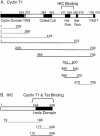
FIG. 1.
FIG. 1.
Mapping of interaction in yeast. HIC interacts with the His-rich region of cyclin T1, while cyclin T1 and Tat interact with the C-terminal I-mfa domain of HIC. (A) Top, schematic representation of cyclin T1. Bottom, the indicated cyclin T1 truncations depicted were tested for their ability to interact with the HIC construct isolated from the library screen (aa 19 to 246; nucleotides 645 to 4152). Yeast cells were transformed with Gal4AD-HIC and plasmids expressing Gal4BD-cyclinT1ΔPEST or the truncations illustrated. (B) Top, schematic representation of HIC. Bottom, HIC (aa 19 to 246) truncations were tested for their ability to interact with cyclin T1 and Tat. Yeast cells were transformed with plasmids expressing Gal4AD-HIC or its truncations and with plasmids expressing Gal4BD-cyclin T1 or Gal4BD-Tat. Interactions were determined as described for Table 1. Solid lines, truncations that interacted; dotted lines, truncations that did not interact.
FIG. 2.
FIG. 2.
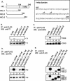
HIC interacts with P-TEFb in vivo, and this interaction is dependent upon the I-mfa domain. (A) Schematic representation of HIC and its truncations, HICΔ1 to -Δ3. The sequence of the I-mfa region (aa 165 to 246) is shown in the box, and the C termini of the Δ2 and Δ3 proteins are marked (/). (B) HIC interacts with cyclin T1 in vivo. COS cells were transfected with plasmids expressing FLAG-HIC (lane 2), FLAG-HICΔ1 (lane 3), FLAG-HICΔ2 (lane 4), FLAG-HICΔ3 (lane 5), or the FLAG tag alone (lane 1), and cell extracts prepared at 24 h posttransfection were subjected to immunoprecipitation (IP) followed by Western blotting (WB). Top, complexes immunoprecipitated with anti-FLAG antibody were resolved in a sodium dodecyl sulfate-polyacrylamide gel, transferred to nitrocellulose, and probed with anti-cyclin T1 antibody. Bottom, as a control, 10% of the inputs used for the immunoprecipitation reactions were analyzed. (C) HIC coimmunoprecipitates with CDK9 in vivo. Transfected COS cell extracts were analyzed as for panel B except that the blot was probed with anti-CDK9 antibody. (D) HIC interacts with Tat in vivo. COS cells cotransfected with HA-Tat plasmid and the FLAG-HIC constructs were analyzed as for panel B except that the blot was probed for Tat with anti-HA antibody. (E) As for panel D, except that Tat-containing complexes were immunoprecipitated with anti-HA antibody and probed for HIC with anti-FLAG antibody.
FIG. 3.
FIG. 3.
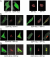
HIC colocalizes with Tat and cyclin T1. COS7 cells were transfected or cotransfected with plasmids expressing HIC proteins carrying the EGFP fluorescent tag (green) and cyclin T1 (T1) or Tat proteins carrying the RFP fluorescent tag (red). Live cells were viewed at 24 h posttransfection by confocal microscopy. (A) Cells were transfected singly with the plasmids indicated. Single fluorescence, green or red, is shown in each panel. (B to D) Cells were cotransfected with the plasmids indicated. The green, red, and dual fluorescences are shown from left to right. Yellow signifies colocalization of the two proteins.
FIG. 4.
FIG. 4.
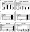
Effects of HIC on Tat transactivation. HIC inhibits Tat transactivation in CEM cells (A and B) but stimulates Tat transactivation in HeLa, COS, and 3T3 cells (C to F). (A and B) CEM cells were electroporated with 4 μg of HIV-1 LTR-firefly luciferase reporter plasmid, 1 μg of CMV-Renilla luciferase reporter plasmid, 0.5 μg of pcDNA3.1-HA-Tat or pcDNA3.1, and 10 μg of pcDNA3.1-HIC expression vector or pcDNA3.1 empty vector. Cells were harvested at the times indicated and assayed for luciferase by using the dual luciferase reporter assay. Cells used for electroporation either were grown at low density and were actively dividing (A) or were grown at a high density and were not actively dividing (B). Data represent Tat transactivation, calculated as the relative firefly/Renilla luciferase activity normalized to the value obtained without Tat, at each time point, in the absence and presence of HIC. Data for each time point are averages from two to four experiments with standard errors. (C) HeLa cells were cotransfected with 100 ng of HIV-1 LTR-firefly luciferase reporter plasmid, 20 ng of CMV-Renilla luciferase reporter plasmid, 5 ng of pcDNA3.1-HA-Tat expression vector or pcDNA3.1 empty vector, and 2 μg of pcDNA3.1-HIC expression vector or pcDNA3.1 empty vector. (D) COS cells were cotransfected as for panel C except that 20 ng of RSV-Renilla luciferase reporter plasmid was used to control for transfection efficiency. (E) NIH 3T3 cells were cotransfected with 100 ng of HIV-1 LTR-firefly luciferase reporter plasmid, 20 ng of CMV-Renilla luciferase reporter plasmid, 5 ng of pcDNA3.1-HA-Tat, 100 ng of pcDNA3.1-HA-cyclinT1 (T1), and 2 μg of pcDNA3.1-HIC expression vector. The total amount of DNA was kept constant in each sample by the addition of the pcDNA3.1 empty vector. In panels C to E, transactivation was measured at 24 h in the presence or absence of HIC, and the data represent averages from three experiments with standard errors. (F) NIH 3T3 cells were cotransfected as for panel E (shaded bars), except that 100 ng of PCNA-firefly luciferase was used in place of HIV-1 firefly luciferase (solid bars). The total amount of DNA was kept constant in each sample by the addition of the pcDNA3.1 empty vector. Transactivation was measured at 24 h and expressed as firefly/Renilla luciferase activity. Data represent the averages from two experiments.
FIG. 5.
FIG. 5.
The I-mfa domain inhibits Tat transactivation. (A) Schematic representation of HIC and its truncations, HIC-N and HIC-Imfa. (B) The HIC I-mfa domain interacts with P-TEFb in vitro. Total HeLa cell extract was incubated with the indicated GST fusion proteins or with GST itself, as described by Hoque et al. (17). Bound proteins were examined by Western blotting (WB) with antibody directed against cyclin T1 (top panel) or CDK9 (bottom panel). (C) HIC I-mfa-P-TEFb complexes in cell extracts. COS cells were transfected with the indicated pcDNA3.1 expression vectors, and immunoprecipitation-Western analysis was conducted as described for Fig. 2B. Immunoprecipitates prepared with anti-Flag antibodies were probed with antibody against cyclin T1 (top panel), CDK9 (middle panel), or the Flag epitope (bottom panel). The second and fourth panels show Western blots of cell extract (without immunoprecipitation, equivalent to 10% of the input) probed with anti-cyclin T1 and anti-CDK9 antibodies, respectively. (D) HeLa, COS, 293, and NIH 3T3 cells were transfected with 100 ng of HIV-1 LTR-firefly luciferase, 20 ng of CMV-Renilla luciferase, 5 ng of pcDNA3.1-HA-Tat, and 2 μg of pFLAG-HIC-N or pFLAG-HIC-Imfa. The total amount of DNA was kept constant in each sample by the addition of pQW3.1FLAG empty vector. Transactivation was measured at 24 h and expressed as firefly/Renilla luciferase activity normalized to the value obtained with Tat alone. Data represent the averages from three experiments with standard errors.
FIG. 6.
FIG. 6.
HIC increases reporter gene RNA levels. NIH 3T3 cells were cotransfected as described for Fig. 4. RNA was isolated from nuclear and cytoplasmic fractions and subjected to RNase protection assays with a probe for firefly luciferase RNA. The control lane lacked cellular RNA but was digested with RNases A and T1. The probe lane contains 10% of undigested probe RNA. The arrow indicates the position of protected luciferase probe.
FIG. 7.
FIG. 7.
Stimulation of gene expression by HIC in the absence of Tat. (A) Schematic representation of HIV-1 LTR-firefly luciferase construct furnished with five upstream Gal4 sites (top) and the Gal4 binding domain-cyclin T1 fusion protein (bottom). (B) NIH 3T3 cells were transfected with 100 ng of Gal4BD-HIV-1 LTR-firefly luciferase reporter plasmid, 20 ng of CMV-Renilla luciferase reporter plasmid, 100 ng of Gal4-cyclin T1 (Gal4-T1) expression vector or pcDNA3.1 empty vector, 2 μg of pcDNA3.1-HIC expression vector or pcDNA3.1 empty vector, and 5 ng of pcDNA3.1-HA-Tat expression vector or pcDNA3.1 empty vector. Data represent activation by Gal4-cyclin T1 at 24 h, calculated as the relative firefly/Renilla luciferase activity normalized to the value obtained without Gal4-cyclin T1, HIC, or Tat.

Similar articles

Cited by

References

    1. Barboric, M., R. M. Nissen, S. Kanazawa, N. Jabrane-Ferrat, and B. M. Peterlin. 2001. NF-kappaB binds P-TEFb to stimulate transcriptional elongation by RNA polymerase II. Mol. Cell 8:327-337. - PubMed
    1. Bourgeois, C. F., Y. K. Kim, M. J. Churcher, M. J. West, and J. Karn. 2002. Spt5 cooperates with human immunodeficiency virus type 1 Tat by preventing premature RNA release at terminator sequences. Mol. Cell. Biol. 22:1079-1093. - PMC - PubMed
    1. Chen, C. M., N. Kraut, M. Groudine, and H. Weintraub. 1996. I-mf, a novel myogenic repressor, interacts with members of the MyoD family. Cell 86:731-741. - PubMed
    1. Chun, R. F., O. J. Semmes, C. Neuveut, and K. T. Jeang. 1998. Modulation of Sp1 phosphorylation by human immunodeficiency virus type 1 Tat. J. Virol. 72:2615-2629. - PMC - PubMed
    1. Darbinian, N., B. E. Sawaya, K. Khalili, N. Jaffe, B. Wortman, A. Giordano, and S. Amini. 2001. Functional interaction between cyclin T1/cdk9 and Puralpha determines the level of TNFalpha promoter activation by Tat in glial cells. J. Neuroimmunol. 121:3-11. - PubMed

Publication types

MeSH terms