. 2003 Sep 1;22(17):4465-77.
doi: 10.1093/emboj/cdg428.

TLP, a novel modulator of TGF-beta signaling, has opposite effects on Smad2- and Smad3-dependent signaling

Affiliations

TLP, a novel modulator of TGF-beta signaling, has opposite effects on Smad2- and Smad3-dependent signaling

Angelina Felici et al. EMBO J. .

Abstract

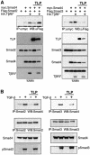
Transforming growth factor-beta (TGF-beta) is a multifunctional cytokine signaling to the nucleus through cell surface transmembrane receptor serine/threonine kinases and cytoplasmic effectors, including Smad proteins. We describe a novel modulator of this pathway, TLP (TRAP-1-like protein), which is 25% identical to the previously described Smad4 chaperone, TRAP-1, and shows identical expression patterns in human tissues. Endogenous TLP associates with both active and kinase-deficient TGF-beta and activin type II receptors, but interacts with the common-mediator Smad4 only in the presence of TGF-beta/activin signaling. Overexpression of TLP represses the ability of TGF-beta to induce transcription from SBE-Luc, a Smad3/4-specific reporter, while it potentiates transcription from ARE-Luc, a Smad2/4-specific reporter. Consistent with this, TLP inhibits the formation of Smad3/4 complexes in the absence of effects on phosphorylation of Smad3, while it affects neither Smad2 phosphorylation nor hetero-oligomerization. We propose that TLP might regulate the balance of Smad2 and Smad3 signaling by localizing Smad4 intracellularly, thus contributing to cellular specificity of TGF-beta transcriptional responses in both normal and pathophysiology.


Figures
None
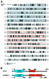
Fig. 1. Sequence similarity and domain organization of human TLP and TRAP-1. (A) Alignment of TRAP-1 and TLP amino acid sequence. Identical and homologous residues are shaded black and gray, respectively. Numbers represent amino acid positions. Blue and red lines denote the hypothetical CNH and CLH domains respectively. (B) Domain organization of human TLP and TRAP-1: blue box, CNH domain; red box, CLH domain. Numbers represent amino acid residue positions.
None
Fig. 2. TLP and TRAP-1 show similar tissue distribution. (A) TLP and TRAP-1 are ubiquitously expressed in similar patterns. A single TLP transcript of approximately 5.5 kb was detected in all human tissues examined, with the exception of peripheral blood leukocytes which displayed two distinct transcripts of 5.5 and 5.8 kb. (B) TLP and TRAP-1 expression in human cell lines: HepG2, hepatocarcinoma; Colo357, pancreatic carcinoma; MDA468, breast carcinoma; BxPC3, pancreatic carcinoma; HeLa, cervix adenocarcinoma; SKN-SH, neuroblastoma.
None
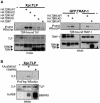
Fig. 3. TLP interacts with TGF-β and activin receptors in vivo. (A) TLP and TRAP-1 display different binding affinities for TβRs. COS-1 cells were transiently transfected with Xpr.TLP (left panels) or GFP.TRAP-1 (right panels) in combination with the indicated HA-TβRs. Thirty hours after transfection, cells were serum starved for 12 h and cell lysates were immunoprecipitated with anti-HA antibody and blotted as indicated. Lysates of cells untransfected or transfected with TLP or TRAP-1 cDNA alone were analyzed as negative controls. Top panels, immunoprecipitated complexes; bottom panels, unfractionated extracts showing expression levels of the transfected proteins. (B) TLP associates with ActRs but not with BMPRII. COS-1 cells were transiently transfected with Xpr.TLP and Flag.ActRIIB or BMPRII receptors as indicated. Lysates were subjected to immunoprecipitation with anti-Flag and blotted with anti-Xpr antibody. In the two leftmost lanes, lysates of cells untransfected or transfected with TLP cDNA alone were analyzed as negative controls. Expression levels of receptors and TLP were confirmed by western blot analysis of total cell extracts using anti-Xpr and anti-Flag antibodies, respectively. WT, wildtype; KD, kinase-deficient (K277R) mutant; asterisk, mutationally activated (T204D) mutant.
None
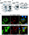
Fig. 4. TLP interacts with TGF-β/activin receptor complexes in a signal-independent fashion. (A) In contrast to TRAP-1, TLP associates with ActRIIB constitutively. COS-1 cells were transiently transfected with Xpr.TLP (left panel) or with GFP.TRAP-1 (right panel) in combination with the indicated epitope-tagged activin receptors type II and type I. Lysates were immunoprecipitated with anti-Flag and blotted as indicated. (B) TLP associates with TGF-β receptor complex constitutively. Left panels: COS-1 cells transfected with Xpr.TLP and the indicated HA-TβRs type II and type I were immuno precipitated with anti-HA and blotted with anti-Xpr. Bottom left panels: control western blots for the immunoprecipitations. Leftmost lane (A and B): lysate of cells transfected with either TLP or TRAP-1 cDNA alone, negative control. Right panels: endogenous TLP associates with TβRII constitutively. HaCaT cells treated with TGF-β for 1 h, followed by treatment with cross-linker for 20 min, were immunoprecipitated with anti-TβRII and blotted with anti-TLP specific rabbit polyclonal antibody (749). Comparable levels of TβRII in the immunoprecipitates are shown. Leftmost lanes: lysate of cells untreated (–) or treated (+) was immunoprecipitated in the presence of TβRII specific blocking peptide (sc-400P) as negative control. (C) TLP colocalizes with TGF-β receptor complex at the plasma membrane and submembrane vesicular domains in a ligand-independent fashion. COS-1 cells were transiently transfected with Xpr.TLP and either HA.TβRII.KD and HA.TβRI.KD in the absence of TGF-β (top panels) or HA.TβRII.WT and HA.TβRI.WT, and treated with TGF-β for 20 min (bottom panels). TLP and TβRs were visualized by the monoclonal anti-Xpr antibody (green) and by the polyclonal anti-HA antibody (red), respectively. DAPI staining (blue) highlights the location of nuclei. Colocalization of TLP and TβRs appears as yellow. Areas marked by a rectangle are enlarged and shown as insets.
None
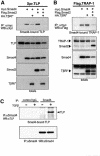
Fig. 5. TLP associates with Smad4 in vivo. (A) TLP and Smad2 form mutually exclusive complexes with Smad4. COS-1 cells that co- expressed myc.Smad4 and Xpr.TLP were transfected with or without HA.TβRI* to activate TGF-β pathway in either the absence or the presence of Flag.Smad2. Lysates were immunoprecipitated with myc and probed with Xpr antibody. (B) Effect of TGF-β signaling and Smad2 expression on TRAP-1/Smad4 association. COS-1 cells that co-expressed myc.Smad4 with Flag.TRAP-1 were transfected with or without HA.TβRI* in either the absence or the presence of Flag.Smad2 as competitor. Cells treated as in (A) were immunoprecipitated with myc and probed with Flag antibody. Leftmost lane: lysate of cells transfected with (A) TLP cDNA or (B) TRAP-1 cDNA alone was analyzed as negative control. (C) TGF-β-dependent interaction between endogenous TLP and Smad4. Lysates of HaCaT cells untreated (–) or treated (+) with TGF-β for 1 h were subjected to Smad4 immunoprecipitation and immunoblotting with a TLP antiserum (749) (top panel). Equal levels of Smad4 in the immunoprecipitates are shown (bottom panels). Two leftmost lanes: lysates immunoprecipitated with an irrelevant IgG were analyzed as negative control.
None
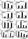
Fig. 6. TLP inhibits Smad-3 and activates Smad-2 dependent transcription in TGF-β/activin reporter assays while having no effect on BMP signaling. (A and C) Effect of TLP on TGF-β-induced transcription of SBE4 and ARE reporters. Hep3B cells were transiently transfected with a control vector or increasing amounts of TLP along with either (A) SBE4 or (C) ARE reporter and FAST1. (B and D) Effect of Smad2 and Smad3 on TGF-β-induced transcription of SBE4 and ARE. Hep3B cells were transiently transfected with a control vector or with Smad2- and Smad3-expressing vectors as indicated, along with either (B) SBE4 or (D) ARE reporter and FAST1. Luciferase activity in cells treated with 5 ng/ml TGF-β1 (black bars) or left untreated (open bars) is shown. (EG) TLP blocks both TGF-β and activin-dependent Smad3-specific transcription while having no effect on activation of the BMP-specific BRE reporter. (E and F) The indicated expression vectors were cotransfected with BRE reporter and Hep3B cells were treated with 100 ng/ml BMP2 (black bars) or left untreated (open bars). (G) Hep3B cells were transiently transfected with a control vector or TLP (0.2 µg/well) along with SBE4. The cells were treated with 5 ng/ml TGF-β1 or 25 ng/ml activin A (black bars) or left untreated (open bars). (H) TLP blocks TGF-β-mediated transcription of human Smad7 promoter. The Smad7 promoter construct was cotransfected with increasing amounts of TLP in Hep3B cells treated with 5 ng/ml TGF-β (black bars) or left untreated (open bars). Luciferase activity was normalized to β-galactosidase activity and plotted as mean ± SD of triplicates from a representative experiment.
None
Fig. 7. TLP has no effect on TGF-β-induced phosphorylation of Smad2 and Smad3. (A) Increasing concentrations of TLP have no effect on the extent of Smad2 and Smad3 phosphorylation induced by 30 min stimulation with TGF-β. Hep3B cells were transiently transfected with increasing amounts of Xpr.TLP (7–600 ng/well) along with either Flag.Smad2 (left panel) or Flag.Smad3 (right panel). After 18 h of serum starvation, cells were treated with 5 ng/ml TGF–β1 for 30 min (+) or left untreated (–). Smad phosphorylation was examined by anti-pSmad2 or anti-pSmad3 immunoblotting of equivalent protein lysates (35 µg) prepared from plates transfected as indicated. Total levels of TLP, Smad2 and Smad3 expression are also shown (bottom panels). Occasionally, expression of exogenous TLP at medium-high concentrations appeared not to be dose dependent. This may reflect degradation of TLP, suggesting a strict control of TLP intracellular level above the physiologic threshold. (B) TLP has no effect on the kinetics of TGF-β-induced Smad2/3 phosphorylation. Hep3B cells were transiently transfected with a control vector (top panels) or with Xpr.TLP (middle panels), serum-starved for 18 h and treated with 5 ng/ml TGF-β1 for the indicated times. Phosphorylation of endogenous Smad2 and Smad3 was examined on equivalent protein lysates (70 µg) as described in (A). TLP expression is shown in the bottom panels.
None
Fig. 8. Overexpression of TLP blocks TGF-β-induced formation of Smad3/4 complexes while it does not alter Smad2/4 complex levels. (A) TLP effects on the formation of exogenous Smad2/4 and Smad3/4 complexes. COS-1 cells that co-expressed myc.Smad4 and Flag.Smad2 (left panel) or Flag.Smad3 (right panel) were transfected with HA.TβRI* along with a negative control vector (lanes 2 and 3) or with Xpr.TLP (lanes 4 and 5). Cells treated as in Figure 5A were immunoprecipitated with myc antibody and probed with anti-Flag. Leftmost lane: lysate of cells transfected with Flag.Smad2 (left panel) or Flag.Smad3 (right panel) cDNA alone was analyzed as negative control. Protein expression was confirmed by immunoblotting total cell lysates for the indicated tagged proteins (bottom panels). (B) TLP effects on the formation of endogenous Smad2/4 and Smad3/4 complexes. HaCaT cells, left untreated or stimulated with TGF-β (2.5 ng/ml) for 40 min, were treated with cross-linker for 20 min. The amount of endogenous Smad4 bound to Smad2 and Smad3 was determined by immunoprecipitation with anti-Smad2 and anti-Smad3 followed by immunoblotting with anti-Smad4, as indicated (top panels). Equal immunoprecipitations were confirmed by blotting anti-Smad2 or anti-Smad3 of the corresponding immunoprecipitates (second top panels). Steady state levels of endogenous Smad4 were analysed by anti-Smad4 blotting of total lysates (second bottom panels). Immunoblotting of total lysates with specific anti-pSmad2 and anti-pSmad3 antibodies shows endogenous activated Smad2 and Smad3 concentrations (bottom panels).
None
Fig. 9. Downregulation of endogenous TLP specifically blocks Smad3-dependent transcription. (A) siRNA-mediated TLP downregulation inhibits TGF-β-induced activation of Smad3/4 transcriptional response. HaCaT cells were transiently transfected with the indicated amounts of either control or TLP-specific siRNA followed by transfection with either SBE4 (left panel) or BRE (right panel). Luciferase activity was normalized for both transfection efficiency and protein concentration. (B) siRNA-dependent reduction of endogenous TLP protein levels. Aliquots of total lysates from HaCaT cells transfected as in (A) were tested in immunoblotting for endogenous TLP (top panel) and actin (bottom panel) expression.
None
Fig. 10. Proposed model for TLP opposite effects on Smad2- and Smad3-dependent transcriptional response to TGF-β. A model integrating defined early events in TGF-β signaling and a proposed mechanism of action for TLP is shown. Steps 1 and 2: TLP constitutive binding to TβRs, lack of association of TLP with BMPRs and association of TLP with Smad4 upon signal activation. Step 3: positioning of TLP downstream of Smad2 and Smad3 phosphorylation (described to occur on post-plasma membrane vesicles and here symbolized by red dots on Smad2 and blue dots on Smad3). Steps 4 and 5: TLP inhibition of Smad3/Smad4 complex formation and TLP effects on Smad-dependent transcription following deregulation of endogenous TLP levels. Although TLP is shown bound to Smad4 on the TβR complex throughout, our current observations do not address whether endocytic vesicle formation is required for TLP/Smad4 association. TLP, green; Smad4, blue; SARA, pink; Smad2, yellow; Smad3, red; Smad1, purple.

Similar articles

Cited by

References

    1. Altschul S.F., Madden,T.L., Schaffer,A.A., Zhang,J., Zhang,Z., Miller,W. and Lipman,D.J. (1997) Gapped BLAST and PSI-BLAST: a new generation of protein database search programs. Nucleic Acids Res., 25, 3389–3402. - PMC - PubMed
    1. Caplan S., Hartnell,L.M., Aguilar,R.C., Naslavsky,N. and Bonifacino,J.S. (2001) Human Vam6p promotes lysosome clustering and fusion in vivo. J. Cell Biol., 154, 109–122. - PMC - PubMed
    1. Charng M.J., Zhang,D., Kinnunen,P. and Schneider,M.D. (1998) A novel protein distinguishes between quiescent and activated forms of the type I transforming growth factor beta receptor. J. Biol. Chem., 273, 9365–9368. - PubMed
    1. Chen R.H., Miettinen,P.J., Maruoka,E.M., Choy,L. and Derynck,R. (1995) A WD-domain protein that is associated with and phos phorylated by the type II TGF-beta receptor. Nature, 377, 548–552. - PubMed
    1. Datta P.K. and Moses,H.L. (2000) STRAP and Smad7 synergize in the inhibition of Transforming growth factor β signaling. Mol. Cell. Biol., 20, 3157–3167. - PMC - PubMed

Publication types

MeSH terms

Substances