. 2003 Sep 1;22(17):4346-55.
doi: 10.1093/emboj/cdg439.

Gangliosides are receptors for murine polyoma virus and SV40

Affiliations

Gangliosides are receptors for murine polyoma virus and SV40

Billy Tsai et al. EMBO J. .

Abstract

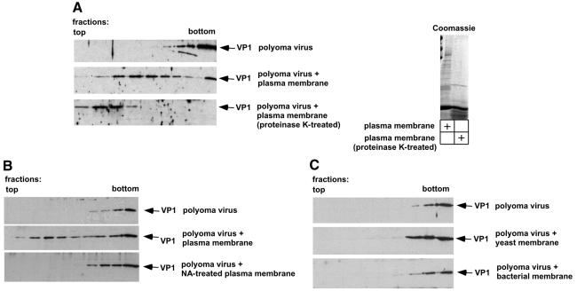
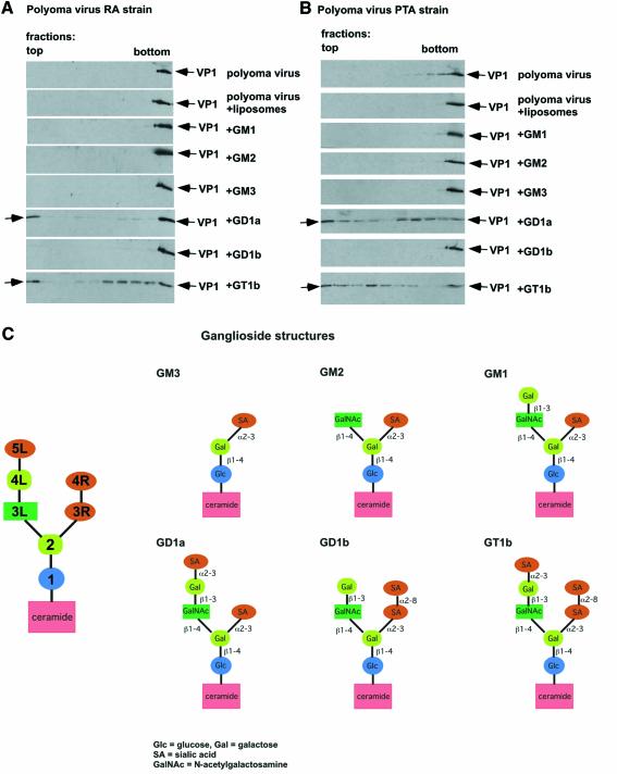
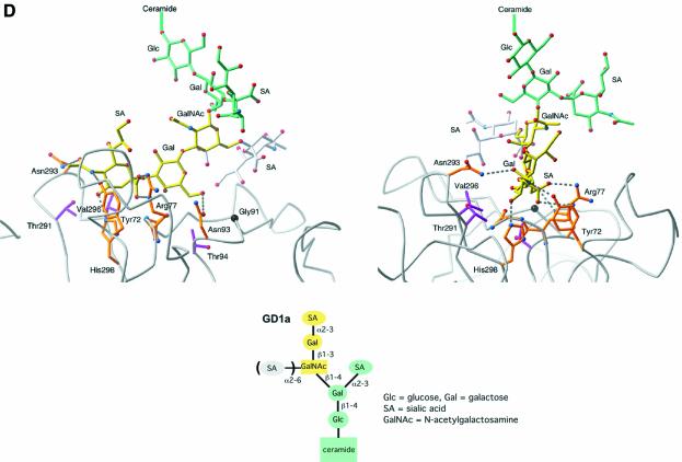
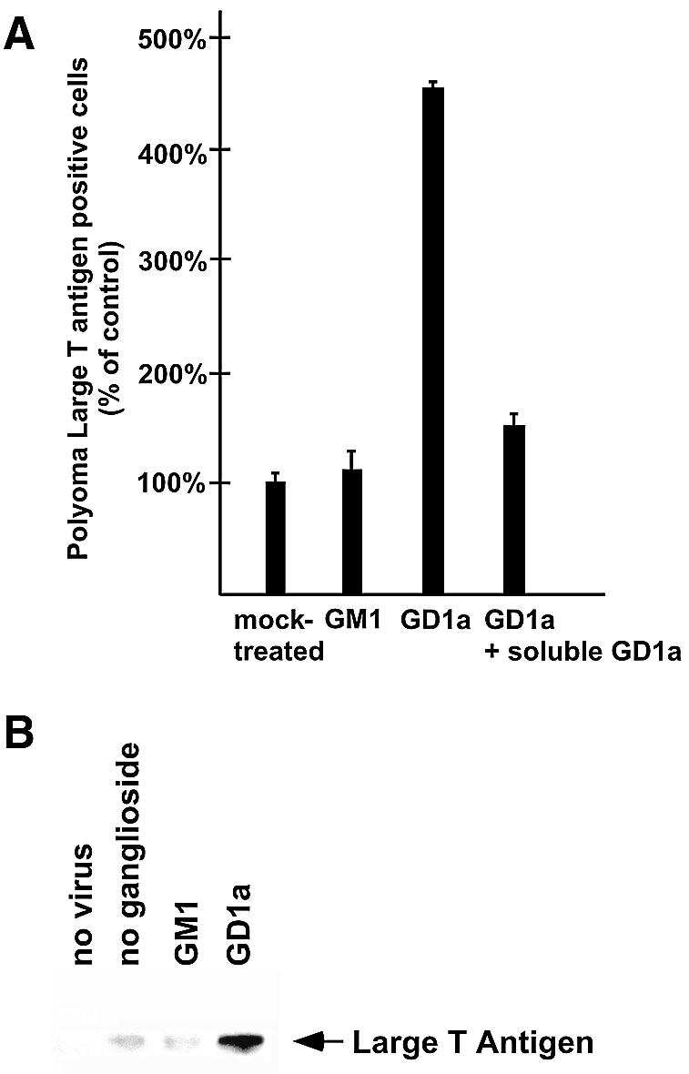
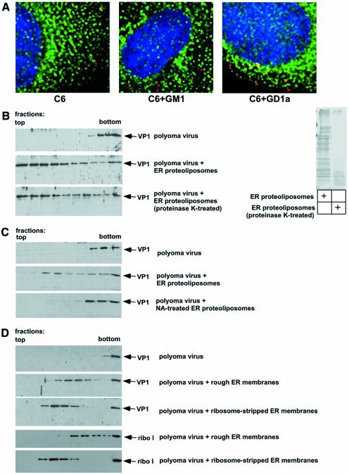
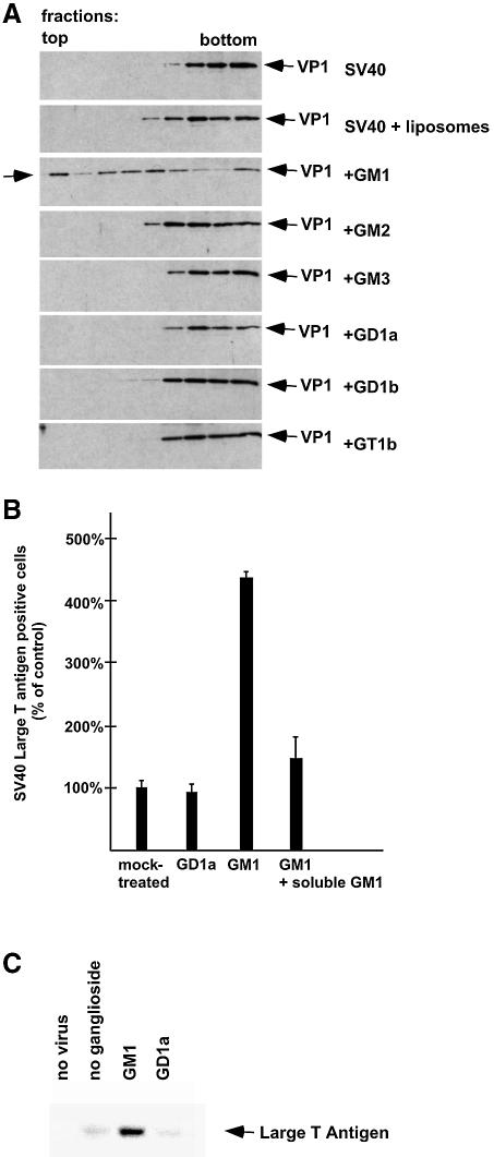
Polyoma virus (Py) and simian virus 40 (SV40) travel from the plasma membrane to the endoplasmic reticulum (ER) from where they enter the cytosol and then the nucleus to initiate infection. Here we demonstrate that specific gangliosides can serve as plasma membrane receptors for these viruses, GD1a and GT1b for Py and GM1 for SV40. Binding and flotation assays were used to show that addition of these gangliosides to phospholipid vesicles allowed specific binding of the respective viruses. The crystal structure of polyoma VP1 with a sialic acid-containing oligosaccharide was used to derive a model of how the two terminal sugars (sialic acid-alpha2,3-galactose) in one branch of GD1a and GT1b are recognized by the virus. A rat cell line deficient in ganglioside synthesis is poorly infectible by polyoma and SV40, but addition of the appropriate gangliosides greatly facilitates virus uptake, transport to the ER and infection. Lipid binding sites for polyoma are shown to be present in rough ER membranes, suggesting that the virus travel with the ganglioside(s) from the plasma membranes to the ER.


Figures

None
Fig. 1. Py binds to a protease-resistant receptor at the plasma membrane. (A) Purified Py (strain RA) was incubated in the absence of membranes or with plasma membranes pre-treated with or without proteinase K. The samples were floated in a discontinuous sucrose gradient. Fractions were collected from the top of the gradient and analyzed by SDS–PAGE and immunoblotting with a Py VP1 antibody (left panels). The proteins in the non-proteolyzed and proteolyzed plasma membranes were analyzed by SDS–PAGE followed by staining with Coomassie blue (right panel). (B) Purified Py (strain RA) was incubated in the absence of membranes or with plasma membranes pre-treated with or without α2,3-neuraminidase (NA). The analysis was performed as in (A). (C) Purified Py was incubated alone, or with yeast or bacterial membranes. The analysis was performed as in (A).
None
Fig. 2. The Py receptor is a ganglioside. (A) Py (RA strain) was incubated in the absence of membranes or with liposomes containing different gangliosides. The samples were floated in a discontinuous sucrose gradient and fractions were analyzed by immunoblotting with antibodies to the polyoma VP1 protein (see Figure 1A). (B) As in (A) except that the more tumorigenic Py PTA strain was used. (C) Structures of the various gangliosides used in (A) and (B). (D) Modeling of interactions between GD1a and polyoma VP1 coat protein. Two orthogonal views are shown. The backbone structure of VP1 is shown as a gray ribbon, with residues involved in carbohydrate contacts shown in ball-and-stick representation (orange for residues with polar interactions, magenta for residues with hydrophobic contacts). VP1 was crystallized with a branched disialylated oligosaccharide (yellow and white) (Stehle and Harrison, 1997). The yellow portion of this oligosaccharide is identical to the longer branch of GD1a (except GlcNAc, which does not contact VP1, is replaced with GalNAc in GD1a), and thus we expect that this portion of GD1a will engage VP1 in the same manner. The remaining three carbohydrate residues of GD1a are attached to the O4 position of GalNAc, and in their most likely conformation they face away from VP1 and are unlikely to engage in contacts (light blue). The second sialic acid binding site of VP1 that accomodates the branched sialic acid (white) is probably not occupied by GD1a, and we therefore expect GD1a to bind equally well to RA and PTA Py strains.
None
Fig. 3. Binding of Py to ganglioside GD1a is required for infection. (A) Quantification of Py LTAg expression. C6 glioma cells lacking gangliosides more complex than GM3 (see Figure 2C) were incubated with or without gangliosides GM1 or GD1a as indicated. The cells were washed and incubated with Py (RA strain) in the absence or presence of GD1a. Expression of Py LTAg was monitored after immunostaining in a fluorescence microscope. The data are presented as the percentage of total nuclei (duplicates of about 500 nuclei) that were Py LTAg positive, normalized to 100%. (B) As in (A) except expression of Py LTAg in C6 cells was analyzed by SDS–PAGE followed by immunoblotting with Py LTAg antibodies.
None
Fig. 4. Transport of Py to the ER by ganglioside GD1a. (A) C6 glioma cells lacking gangliosides more complex than GM3 (see Figure 2C) were incubated with GM1 or GD1a and then infected with Texas Red-labeled Py (RA strain). After 4 h the cells were fixed and immunostained for the ER luminal protein BiP using Oregon Green-labeled secondary antibodies (green). Nuclei were labeled with DAPI. Yellow points (white arrow head) indicate co-localization. (B) Purified Py (strain RA) was incubated in the absence or presence of ER proteoliposomes pre-treated or not with proteinase K. The samples were floated in a sucrose gradient and fractions were analyzed by immunoblotting with antibodies to the Py VP1 protein (left panels). The proteins in the non-proteolyzed or proteolyzed membranes were analyzed by SDS–PAGE followed by staining with Coomassie blue (right panel). (C) Purified Py (strain RA) was incubated in the absence or presence of ER proteoliposomes pre-treated or not with α2,3-neuraminidase (NA). The samples were analyzed as in (A). (D) Purified Py (strain RA) was incubated in the presence or absence of rough ER membranes or ribosome-stripped ER membranes. The samples were analyzed as in (A). An antibody against ribophorin I was used to monitor ER membranes in the fractions.
None
Fig. 5. SV40 binds to ganglioside GM1 to infect cells. (A) SV40 was incubated in the absence or presence of liposomes containing different gangliosides, as indicated. The samples were floated in a sucrose gradient and fractions were analyzed by immunoblotting with antibodies directed against Py VP1, VP2 and VP3 proteins. (B) Quantification of SV40 LTAg expression. C6 glioma cells lacking gangliosides more complex than GM3 (see Figure 2C) were incubated with or without gangliosides GM1 or GD1a, as indicated. The cells were washed and incubated with SV40 in the absence or presence of GM1. Expression of SV40 LTAg was monitored after immunostaining in a fluorescence microscope. The data are presented as the percentage of total nuclei (duplicates of about 500 nuclei) that were SV40 LTAg positive, normalized to 100%. (C) As in (B) except expression of SV40 LTAg in C6 cells was analyzed by SDS–PAGE followed by immunoblotting with SV40 LTAg antibodies.

Similar articles

Cited by

References

    1. Anderson H.A., Chen,Y. and Norkin,L.C. (1996) Bound simian virus 40 translocates to caveolin-enriched membrane domains, and its entry is inhibited by drugs that selectively disrupt caveolae. Mol. Biol. Cell, 7, 1825–1834. - PMC - PubMed
    1. Anderson H.A., Chen,Y. and Norkin,L.C. (1998) MHC class I molecules are enriched in caveolae but do not enter with simian virus 40. J. Gen. Virol., 79, 1469–1477. - PubMed
    1. Atwood W.J. and Norkin,L.C. (1989) Class I major histocompatibility proteins as cell surface receptors for simian virus 40. J. Virol., 63, 4474–4477. - PMC - PubMed
    1. Barouch D.H. and Harrison,S.C. (1994) Interactions among the major and minor coat proteins of polyomavirus. J. Virol., 68, 3982–3989. - PMC - PubMed
    1. Bauer P.H., Cui,C., Stehle,T., Harrison,S.C., DeCaprio,J.A. and Benjamin,T. (1999) Discrimination between sialic acid-containing receptors and pseudoreceptors regulates polyomavirus spread in the mouse. J. Virol., 73, 5826–5832. - PMC - PubMed

Publication types

MeSH terms