. 2003 Aug;14(8):3414-26.
doi: 10.1091/mbc.e02-09-0581. Epub 2003 Apr 17.

Association of chromatin proteins high mobility group box (HMGB) 1 and HMGB2 with mitotic chromosomes

Affiliations

Association of chromatin proteins high mobility group box (HMGB) 1 and HMGB2 with mitotic chromosomes

Coralie Pallier et al. Mol Biol Cell. 2003 Aug.

Abstract

High mobility group box (HMGB) 1 and 2 are two abundant nonhistone nuclear proteins that have been found in association with chromatin. Previous studies based on immunofluorescence analysis indicated that HMGB1 dissociates from chromosomes during mitosis. In the present work, HMGB1 and 2 subcellular localization was reinvestigated in living cells by using enhanced green fluorescent protein- and Discosome sp. red fluorescent protein-tagged proteins. Contrary to previous reports, HMGB1 and 2 were shown to be present under two forms in mitotic cells, i.e., free and associated with the condensed chromatin, which rapidly exchange. A detailed analysis of HMGB2 interaction with mitotic chromosomes indicated that two sites encompassing HMG-box A and B are responsible for binding. Importantly, this interaction was rapidly inactivated when cells were permeabilized or exposed to chemical fixatives that are widely used in immunodetection techniques. A comparable behavior was also observed for two proteins of the HMG-nucleosome binding (HMGN) group, namely, HMGN1 and HMGN2.


Figures

Figure 1.
Figure 1.
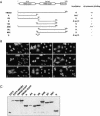
Cloning of human HMGB cDNAs. HMGB1 and HMGB2 cDNAs were amplified by PCR from reverse transcribed polyadenylated RNAs. The position of the primers used to amplify HMGB2 is indicated by arrows on the premessenger. Numbered open boxes represent exons. Whereas a single cDNA was detected for HMGB1 (lane 1), two cDNAs (hmgb 2.1 and hmgb 2.2) were amplified for HMGB2 (lane 2). Sequence analysis indicated that hmgb1 and hmgb 2.1 corresponded to the expected mature mRNA, whereas hmgb 2.2 arose from a partly spliced RNA which contained an intron between exon 3 and 4 (open boxes).
Figure 2.
Figure 2.
HMGB proteins interact with human and mouse chromosomes. (A) Living HeLa cells expressing EGFP or histone H1, HMGB2, HMGB1 fused to EGFP (green) or to DsRed (red) were observed by fluorescent microscopy. DNA was stained with Hoechst 33342 (blue). Whereas EGFP does not interact with mitotic chromosomes, H1-EGFP strictly colocalized with condensed chromatin during mitosis. HMGB2 binding to mitotic chromosomes was observed independently of the N- or C-terminal position of EGFP. HMGB2 binding was also observed in the context of DsRed fusions. Similar observations were done for HMGB1 tagged with either EGFP or DsRed to its N or C termini. (B) HeLa cells were transfected by an expression vector encoding HMGB1-EGFP. Binding was observed during early to late phases of mitosis. Similar observations were done for HMGB2 (our unpublished data). (C) Mouse 3T3 cells were transfected by an expression vector encoding HMGB1-EGFP (green). DNA was stained by Hoechst 33342 (blue). In living cells, HMGB1 localized to the nucleus and was not excluded from the nucleolus. It colocalized to most heterochromatin containing regions (yellow arrowheads). In paraformaldehyde-fixed cells, HMGB1-EGFP detached from Hoechst-bright spots (red arrowheads) and remained in the nucleolus.
Figure 3.
Figure 3.
HMGB2 fused to EGFP or to DsRed interact with mitotic chromosomes 8 wk after transfection. HeLa cells were transfected by pHMGB2-EGFP or pDsRed-HMGB2, grown for 3 wk in the presence of geneticin and then for 5 wk in the absence of selection. Fluorescent foci were cloned from 3 to 5 times. Positive foci contained cells expressing undetectable to high level of the fluorescent protein (top, low magnification). In all instances, HMGB2-EGFP (g) interacts with mitotic chromosomes. Similar results were obtained with cloned HeLa cells expressing DsRed-HMGB2 (d) (bottom, high magnification). DNA was stained with Hoechst 33342 (h). Note that DsRed-HMGB2 colocalized exclusively with the condensed chromosomes, whereas HMGB2-EGFP was also detected in the cytoplasm.
Figure 6.
Figure 6.
Expression and localization of HMGB deletion mutants. (A) Schematic representation of HMGB2 and truncated forms. The nuclear (N) or cytoplasmic localization (C) as well as their binding to mitotic chromosomes is indicated. (B) Cellular localization of HMGB2 and truncated forms fused to EGFP (g) in HeLa cells. Cellular DNA was counterstained with Hoechst 33342 (h). (C) Expression of HMGB2, HMGB1 and various deletion mutants of HMGB2 fused to EGFP. Ten micrograms of total protein extract was subjected to SDS-PAGE and analyzed by Western blot with an antibody directed against EGFP.
Figure 4.
Figure 4.
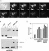
EGFP does not alter known properties of HMGB1. (A) Leakage of HMGB1-EGFP from permeabilized cells. HeLa cells expressing HMGB1-EGFP were permeabilized with NP-40 directly on the microscope, and sequential images were collected before and after detergent addition. Less than 1.5 min is sufficient to lose the entire complement of HMGB1 upon permeabilization. (B) HMGB1 and HMGB1-EGFP diffuse similarly upon cell permeabilization. HeLa cells expressing EGFP, EGFP-H1, or HMGB1-EGFP were incubated in the presence of PBS plus NP40 for 5 min. An equivalent volume of soluble (S) and insoluble (chromatin bound) (P) proteins was analyzed by Western blot by using an antibody directed against EGFP. The membrane was then reprobed with an antibody directed against human HMGB1. A comparable amount of total proteins was also analyzed (T). HMGB1 and HMGB1-EGFP were exclusively detected in the soluble fraction, indicating that both proteins similarly diffuse out of the cells.(C) The fusion to EGFP does not impair the transactivational activity of HMGB1. HeLa cells were transfected with fixed amounts of the reporter plasmid pTHCR and pSGD9, and increasing amounts of either pHMGB1 or pEGFP-HMGB1. Both HMGB1 and HMGB1-EGFP enhance HOXD9 transcriptional activity in transient cotransfection assays.
Figure 5.
Figure 5.
The cytoplasmic and chromosome associated forms of HMGB1 can rapidly exchange. (A) FLIP on a cytoplasmic region of a mitotic cell expressing HMGB1-EGFP (a). The area indicated by a circle was repeatedly bleached for 200 ms, and cells were imaged between bleach pulses. The loss of fluorescence was fast and complete in the entire cell, including the condensed chromosomes. There is interchange between chromosomal and cytoplasmic pools of HMGB1-EGFP, and a rapid dissociation of the protein from condensed chromatin. FLIP on mitotic cells expressing EGFP-H1 (b). A cytoplasmic region (cell on the left) and a region on condensed chromosomes (cell on the right) of two mitotic cells were repeatedly bleached for 200 ms, and cells were imaged between bleach pulses. Bleaching in the cytoplasm did not lead to loss of fluorescence on chromosomes, whereas bleaching on chromosomes lead to a slow loss of fluorescence in the regions near the bleached spot. (B) FLIP quantitation in living HeLa cells expressing EGFP-H1 or HMGB1-EGFP.
Figure 7.
Figure 7.
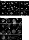
Two regions are responsible for HMGB2 binding to mitotic chromosomes. (A) Analysis of the chromosome binding properties of HMGB2 truncated forms fused to EGFP (g) in living HeLa cells. Cells were observed in culture medium 72 h after transfection. DNA was counterstained by Hoechst 33342 (h). (B) Analysis of the chromosome binding properties of HMGB2 derivatives fused to EGFP (g) after chromosome spread. HeLa cells were arrested in mitosis by a 16-h treatment in the presence of colchicine. DNA was counterstained by Hoechst 33342 (h). Mitotic cells were then subjected to a hypotonic shock and cytocentrifuged on microscopic slides. Binding to mitotic chromosomes was assessed immediately either in the absence of mounting medium (EGFP and mutant B) or in the presence of PBS plus 20% glycerol. Note that Hoechst staining was barely detectable in the absence of mounting medium. Unbound proteins rapidly diffuse in the mounting medium, whereas bound proteins were observed for up to 1 h.
Figure 8.
Figure 8.
HMGB interaction with mitotic chromosomes is disrupted by paraformaldehyde. HeLa cells expressing EGFP or histone H1, HMGB1, and HMGB2 fused to EGFP were incubated for 10 min in the presence of PBS containing 4% paraformaldehyde. As soon as 1 min after the beginning of the treatment, HMGB proteins dissociate from the condensed chromatin, whereas H1-EGFP stayed stably associated even after prolonged incubation. Note that HMGB proteins were detected in the vicinity of telophase chromosomes even in fixed cells.
Figure 9.
Figure 9.
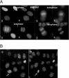
HMGN1-EGFP binding to mitotic chromosomes is observed in living cells, not in paraformaldehyde-treated cells. (A) HeLa cells were transfected by an expression vector encoding HMGN1 fused to the N terminus of EGFP (g). DNA was counterstained by Hoechst 33342 (h). Living cells were observed 72 h after transfection by fluorescent microscopy. HMGN1 was associated with mitotic chromosomes from early to late phases of mitosis. (B) Binding of HMGN1 to mitotic chromosomes was disrupted when the cells were treated for 10 min by PBS containing 4% paraformaldehyde (white arrow). Note that HMGN1 concentrated in nucleolar areas in paraformaldehyde-treated cells.

Similar articles

Cited by

References

    1. Aidinis, V., Bonaldi, T., Beltrame, M., Santagata, S., Bianchi, M.E., and Spanopoulou, E. (1999). The RAG1 homeodomain recruits HMG1 and HMG2 to facilitate recombination signal sequence binding and to enhance the intrinsic DNA-bending activity of RAG1-RAG2. Mol. Cell. Biol. 19, 6532–6542. - PMC - PubMed
    1. Baird, G.S., Zacharias, D.A., and Tsien, R.Y. (2000). Biochemistry, mutagenesis, and oligomerization of DsRed, a red fluorescent protein from coral. Proc. Natl. Acad. Sci. USA 97, 11984–11989. - PMC - PubMed
    1. Bianchi, M.E. (1995). The HMG-box domain. In: DNA-Protein: Structural Interactions, ed. D. Lilley, Oxford: Oxford University Press, 177–200.
    1. Bianchi, M.E., and Beltrame, M. (2000). Upwardly mobile proteins. Workshop: the role of HMG proteins in chromatin structure, gene expression and neoplasia. EMBO Rep. 1, 109–114. - PMC - PubMed
    1. Boonyaratanakornkit, V., Melvin, V., Prendergast, P., Altmann, M., Ronfani, L., Bianchi, M.E., Taraseviciene, L., Nordeen, S.K., Allegretto, E.A., and Edwards, D.P. (1998). High-mobility group chromatin proteins 1 and 2 functionally interact with steroid hormone receptors to enhance their DNA binding in vitro and transcriptional activity in mammalian cells. Mol. Cell. Biol. 18, 4471–4487. - PMC - PubMed

Publication types