. 2003 Aug 18;162(4):703-17.
doi: 10.1083/jcb.200304140.

Trafficking of prion proteins through a caveolae-mediated endosomal pathway

Affiliations

Trafficking of prion proteins through a caveolae-mediated endosomal pathway

Peter J Peters et al. J Cell Biol. .

Abstract

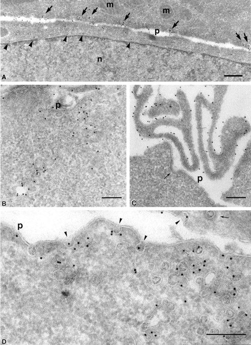
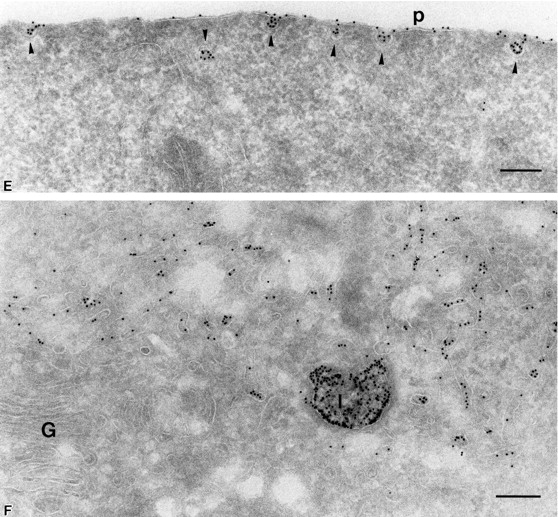
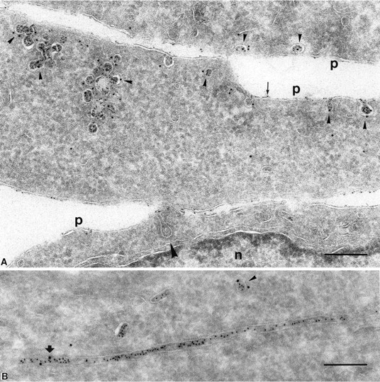
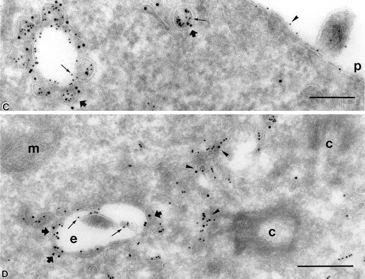
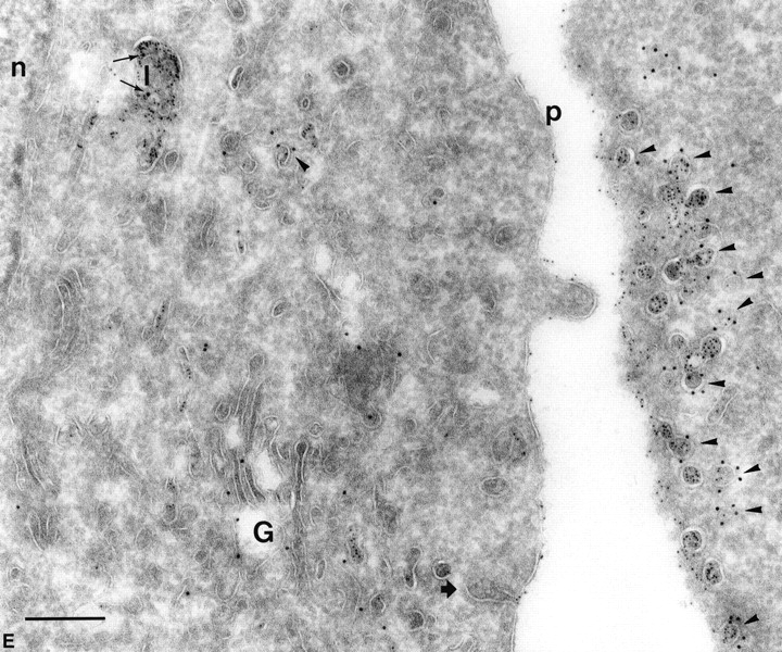
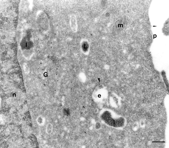
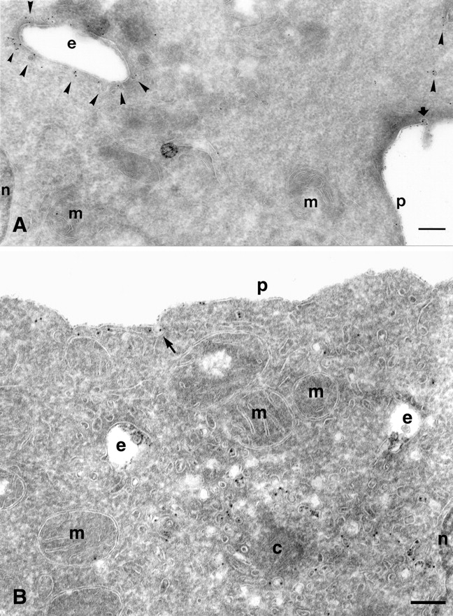
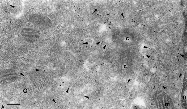
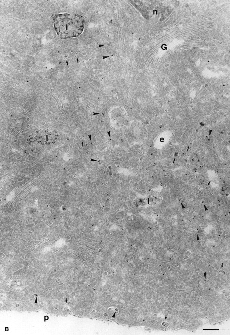
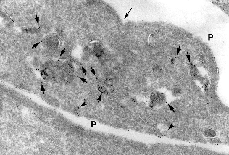
To understand the posttranslational conversion of the cellular prion protein (PrPC) to its pathologic conformation, it is important to define the intracellular trafficking pathway of PrPC within the endomembrane system. We studied the localization and internalization of PrPC in CHO cells using cryoimmunogold electron microscopy. At steady state, PrPC was enriched in caveolae both at the TGN and plasma membrane and in interconnecting chains of endocytic caveolae. Protein A-gold particles bound specifically to PrPC on live cells. These complexes were delivered via caveolae to the pericentriolar region and via nonclassical, caveolae-containing early endocytic structures to late endosomes/lysosomes, thereby bypassing the internalization pathway mediated by clathrin-coated vesicles. Endocytosed PrPC-containing caveolae were not directed to the ER and Golgi complex. Uptake of caveolae and degradation of PrPC was slow and sensitive to filipin. This caveolae-dependent endocytic pathway was not observed for several other glycosylphosphatidyl inositol (GPI)-anchored proteins. We propose that this nonclassical endocytic pathway is likely to determine the subcellular location of PrPC conversion.


Figures

Figure 1.
Figure 1.
At steady state, PrP C is enriched in lamellae and caveolae both at the plasma membrane and inside the cell. (A–C) PrPC was immunogold labeled on ultrathin cryosections of CHO/30C3 cells (steady-state distribution of PrPC) using the R1 recombinant antibody Fab fragment against PrPC. Labeling was observed on the ER (A, arrowheads), plasma membrane caveolae-like structures (A, arrows), caveolae-like structures that appeared as flask-shaped invaginations on the plasma membrane (p) and interconnecting chains of caveolae-like structures deeper into the cytoplasm (B), and microvilli at the leading edge of the cell (C). Clathrin-coated pits (C, thin arrow) did not label. (D and E) Caveolin-1 was immunogold labeled on ultrathin cryosections of CHO/30C3 cells using anti-caveolin-1–gold labeling. (D) Caveolae-like structures and interconnecting chains of caveolae-like structures deeper into the cytoplasm labeled with a caveolin-1 antibody under steady-state conditions. (E) Protein A–gold uptake in an SHaPrPC-expressing cell, after a 10-min pulse. Gold labeling was observed on the plasma membrane and was enriched in caveolae (arrowheads). (F) After a 10-min pulse and 50-min chase, gold labeling was observed in small vesicles near the Golgi complex and lysosomes. G, Golgi complex; l, lysosomes; m, mitochondria; n, nucleus; p, plasma membrane. Bar, 200 nm (applies to all panels).
Figure 1.
Figure 1.
At steady state, PrP C is enriched in lamellae and caveolae both at the plasma membrane and inside the cell. (A–C) PrPC was immunogold labeled on ultrathin cryosections of CHO/30C3 cells (steady-state distribution of PrPC) using the R1 recombinant antibody Fab fragment against PrPC. Labeling was observed on the ER (A, arrowheads), plasma membrane caveolae-like structures (A, arrows), caveolae-like structures that appeared as flask-shaped invaginations on the plasma membrane (p) and interconnecting chains of caveolae-like structures deeper into the cytoplasm (B), and microvilli at the leading edge of the cell (C). Clathrin-coated pits (C, thin arrow) did not label. (D and E) Caveolin-1 was immunogold labeled on ultrathin cryosections of CHO/30C3 cells using anti-caveolin-1–gold labeling. (D) Caveolae-like structures and interconnecting chains of caveolae-like structures deeper into the cytoplasm labeled with a caveolin-1 antibody under steady-state conditions. (E) Protein A–gold uptake in an SHaPrPC-expressing cell, after a 10-min pulse. Gold labeling was observed on the plasma membrane and was enriched in caveolae (arrowheads). (F) After a 10-min pulse and 50-min chase, gold labeling was observed in small vesicles near the Golgi complex and lysosomes. G, Golgi complex; l, lysosomes; m, mitochondria; n, nucleus; p, plasma membrane. Bar, 200 nm (applies to all panels).
Figure 2.
Figure 2.
PrP C is internalized into nonclassical endosomes via caveolae, and this internalization is triggered by surrogate ligand protein A–gold. (A) Endocytosed protein A–gold is found in caveolae of SHaPrPC-expressing CHO/30C3 cells. Cells were incubated at 37°C for 10 min with protein A–gold (5 nm) and chased for 50 min before fixation. Ultrathin cryosections were labeled with anti–caveolin-1 (10-nm gold). Protein A–gold (5 nm) detecting PrPC labeling was observed on the plasma membrane (thin arrow) and was enriched in caveolae (arrowheads) and caveolin-1–positive (10-nm gold) structures. Clathrin-coated pit (large arrowhead) did not label for either PrPC or caveolin-1. (B) Tubule and vesicular structures (small arrowhead) that contained endocytosed protein A–gold (5 nm) labeled for caveolin-1 (10 nm, large arrow). (C) Caveolae (caveolin-1, 10 nm, thick arrows) were present on endocytic structures that were enriched for endocytosed protein A–gold (5-nm gold, small gold, thin arrows). Protein A–gold (5 nm) was also present on the plasma membrane (arrowhead). Panel D depicts an endocytic structure loaded with endocytosed protein A–gold (5 nm, thin arrows) with fused caveolae (thick arrows) labeled for caveolin-1 (10-nm gold) surrounded by caveolae (arrowheads) at the pericentriolar region of the cell that also labeled for caveolin-1 (10 nm) and contained endocytosed protein A–gold (5 nm). Compared with C, the endocytic structure shown contained caveolae that have less caveolar appearance, as if they were fused with the endosome. (E) Endocytosed protein A–gold (5 nm) was present exclusively on the plasma membrane, on plasma membrane–associated caveolae (caveolin-1, 10 nm, arrowheads), and in lysosomes (thin arrows show caveolin-1 label, 10-nm gold). The Golgi complex, labeled for caveolin-1 in a nonclustered fashion, did not contain endocytosed protein A–gold (5 nm). Clathrin-coated pit (thick arrow), nuclear envelope–ER, and clathrin-coated vesicles (not depicted) did not exhibit endocytosed protein A–gold. c, centriole; e, endocytic structure; G, Golgi complex; l, lysosome; m, mitochondria; n, nucleus; p, plasma membrane. Bar, 200 nm (applies to all panels).
Figure 2.
Figure 2.
PrP C is internalized into nonclassical endosomes via caveolae, and this internalization is triggered by surrogate ligand protein A–gold. (A) Endocytosed protein A–gold is found in caveolae of SHaPrPC-expressing CHO/30C3 cells. Cells were incubated at 37°C for 10 min with protein A–gold (5 nm) and chased for 50 min before fixation. Ultrathin cryosections were labeled with anti–caveolin-1 (10-nm gold). Protein A–gold (5 nm) detecting PrPC labeling was observed on the plasma membrane (thin arrow) and was enriched in caveolae (arrowheads) and caveolin-1–positive (10-nm gold) structures. Clathrin-coated pit (large arrowhead) did not label for either PrPC or caveolin-1. (B) Tubule and vesicular structures (small arrowhead) that contained endocytosed protein A–gold (5 nm) labeled for caveolin-1 (10 nm, large arrow). (C) Caveolae (caveolin-1, 10 nm, thick arrows) were present on endocytic structures that were enriched for endocytosed protein A–gold (5-nm gold, small gold, thin arrows). Protein A–gold (5 nm) was also present on the plasma membrane (arrowhead). Panel D depicts an endocytic structure loaded with endocytosed protein A–gold (5 nm, thin arrows) with fused caveolae (thick arrows) labeled for caveolin-1 (10-nm gold) surrounded by caveolae (arrowheads) at the pericentriolar region of the cell that also labeled for caveolin-1 (10 nm) and contained endocytosed protein A–gold (5 nm). Compared with C, the endocytic structure shown contained caveolae that have less caveolar appearance, as if they were fused with the endosome. (E) Endocytosed protein A–gold (5 nm) was present exclusively on the plasma membrane, on plasma membrane–associated caveolae (caveolin-1, 10 nm, arrowheads), and in lysosomes (thin arrows show caveolin-1 label, 10-nm gold). The Golgi complex, labeled for caveolin-1 in a nonclustered fashion, did not contain endocytosed protein A–gold (5 nm). Clathrin-coated pit (thick arrow), nuclear envelope–ER, and clathrin-coated vesicles (not depicted) did not exhibit endocytosed protein A–gold. c, centriole; e, endocytic structure; G, Golgi complex; l, lysosome; m, mitochondria; n, nucleus; p, plasma membrane. Bar, 200 nm (applies to all panels).
Figure 3.
Figure 3.
PIPLC treatment of SHaPrPC-expressing CHO/30C3 cells inhibited PrPC-dependent protein A–gold uptake. Cells expressing PrPC were treated with PIPLC for 2 h and incubated with protein A–gold (5 nm; 10-min pulse and 50-min chase during the last hour of enzyme digestion) before fixation. Sections were labeled for PrPC using the R1 Fab. Only a very small fraction of the protein A–gold (small gold, arrowhead) was endocytosed. Only a few (arrow) protein A–gold particles, but no PrPC labeling (10-nm gold), were observed on the plasma membrane. However, abundant labeling for PrPC was seen in the Golgi. e, endocytic structure; G, Golgi complex; l, lysosome; n, nucleus; m, mitochondria; p, plasma membrane. Bar, 200 nm.
Figure 4.
Figure 4.
Caveolae-mediated uptake of PrP C is cholesterol dependent. (A) PrPC-dependent protein A–gold internalization in SHaPrPC- expressing CHO/30C3 cells was temperature sensitive. Cells were preincubated with protein A–gold (5 nm) at 4°C before fixation. Ultrathin cryosections were labeled with an antibody against caveolin-1 (10-nm gold). Abundant protein A–gold was only seen on the plasma membrane and on plasma membrane–associated caveolae (thick arrow). Caveolin-1–positive endocytic structures (arrowheads) and caveolae-1 intracellular caveolae (arrowheads) did not exhibit any protein A–gold labeling (compare with Fig. 5). (B) Effect of filipin treatment on protein A–gold uptake and the fate of caveolae. Cells were treated for 24 h with filipin followed by a 10-min incubation in media that also contained protein A–gold (5 nm) and a 50-min chase at 37°C. Sections were labeled with anti–caveolin-1 (15 nm). A significant number of protein A–gold (small size) binding on the membrane was seen but no enrichment in caveolae (arrow). The uptake of protein A–gold is sharply reduced; endocytic structures contain protein A–gold (small size) at relatively low levels. No evidence of caveolae and no label for caveolin-1 (large gold) can be seen on these endocytic structures. Many caveolin-1–positive (large gold) vesicles/tubules are present in the pericentriolar region devoid of small protein A–gold. c, centriole; e, endocytic structure; m, mitochondria; n, nucleus; p, plasma membrane. Bars, 200 nm.
Figure 5.
Figure 5.
Endocytosed protein A–gold was found in intracellular caveolae of SHaPrPC-expressing CHO/30C3 cells that did not contain the transferrin receptor. Cells were (A) incubated at 37°C for 10 min with 5-nm protein A–gold or (B) incubated at 37°C for 10 min with 5-nm protein A–gold and chased for 50 min before fixation. Ultrathin cryosections were labeled with antitransferrin antibodies (10-nm gold). (A) Protein A–gold (5 nm) detecting PrPC labeling enriched in pericentriolar caveolae that did not colocalize with transferrin receptor–positive structures (10-nm gold, arrowheads). Panel B shows an identical result after a 50-min chase but with an accumulation of endocytosed protein A–gold in late endocytic multivesicular bodies. Transferrin receptor–positive labeling (10-nm gold) is found in endocytic structures that show an identical morphological appearance to PrPC-containing structures. c, centriole; e, endocytic structure; G, Golgi complex; l, lysosomes; m, mitrochondria; n, nucleus; p, plasma membrane. Bars, 200 nm (also apply to Fig. 6).
Figure 5.
Figure 5.
Endocytosed protein A–gold was found in intracellular caveolae of SHaPrPC-expressing CHO/30C3 cells that did not contain the transferrin receptor. Cells were (A) incubated at 37°C for 10 min with 5-nm protein A–gold or (B) incubated at 37°C for 10 min with 5-nm protein A–gold and chased for 50 min before fixation. Ultrathin cryosections were labeled with antitransferrin antibodies (10-nm gold). (A) Protein A–gold (5 nm) detecting PrPC labeling enriched in pericentriolar caveolae that did not colocalize with transferrin receptor–positive structures (10-nm gold, arrowheads). Panel B shows an identical result after a 50-min chase but with an accumulation of endocytosed protein A–gold in late endocytic multivesicular bodies. Transferrin receptor–positive labeling (10-nm gold) is found in endocytic structures that show an identical morphological appearance to PrPC-containing structures. c, centriole; e, endocytic structure; G, Golgi complex; l, lysosomes; m, mitrochondria; n, nucleus; p, plasma membrane. Bars, 200 nm (also apply to Fig. 6).
Figure 6.
Figure 6.
Endocytosed protein A–gold accumulated in lysosomes of SHaPrPC-expressing CHO/30C3 cells that contained the classical marker for late endocytic structures. Cells were incubated at 37°C for 10 min with 5-nm protein A–gold and chased for 50 min before fixation. Ultrathin cryosections were labeled with anti–LGP-120 antibodies (15-nm gold). PrPC labeling (protein A–gold, 5 nm) accumulated in LGP-120–positive structures. P, plasma membrane.

Similar articles

Cited by

References

    1. Anderson, R.G. 1998. The caveolae membrane system. Annu. Rev. Biochem. 67:199–225. - PubMed
    1. Arnold, J.E., C. Tipler, L. Laszlo, J. Hope, M. Landon, and R.J. Mayer. 1995. The abnormal isoform of the prion protein accumulates in late-endosome-like organelles in scrapie-infected mouse brain. J. Pathol. 176:403–411. - PubMed
    1. Blochberger, T.C., C. Cooper, D. Peretz, J. Tatzelt, O.H. Griffith, M.A. Baldwin, and S.B. Prusiner. 1997. Prion protein expression in Chinese hamster ovary cells using a glutamine synthetase selection and amplification system. Protein Eng. 10:1465–1473. - PubMed
    1. Borchelt, D.R., M. Scott, A. Taraboulos, N. Stahl, and S.B. Prusiner. 1990. Scrapie and cellular prion proteins differ in their kinetics of synthesis and topology in cultured cells. J. Cell Biol. 110:743–752. - PMC - PubMed
    1. Bouillot, C., A. Prochiantz, G. Rougon, and B. Allinquant. 1996. Axonal amyloid precursor protein expressed by neurons in vitro is present in a membrane fraction with caveolae-like properties. J. Biol. Chem. 271:7640–7644. - PubMed

Publication types