. 2003 Sep;77(17):9652-61.
doi: 10.1128/jvi.77.17.9652-9661.2003.

Cell cycle regulation by Kaposi's sarcoma-associated herpesvirus K-bZIP: direct interaction with cyclin-CDK2 and induction of G1 growth arrest

Affiliations

Cell cycle regulation by Kaposi's sarcoma-associated herpesvirus K-bZIP: direct interaction with cyclin-CDK2 and induction of G1 growth arrest

Yoshihiro Izumiya et al. J Virol. 2003 Sep.

Abstract

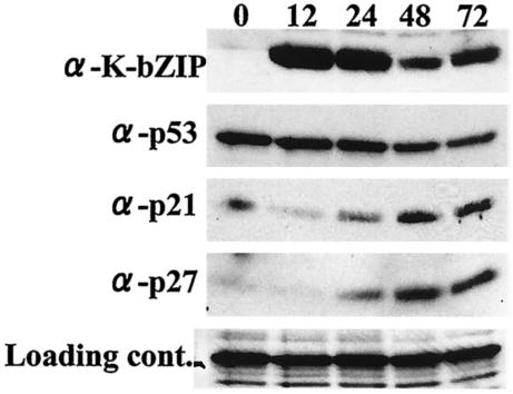
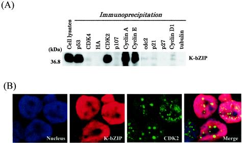
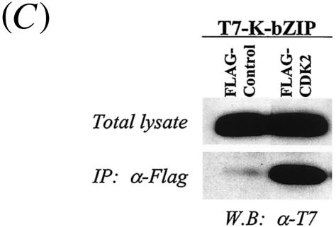
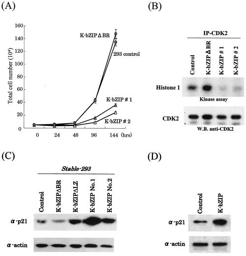
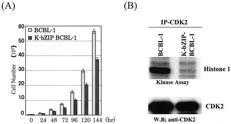
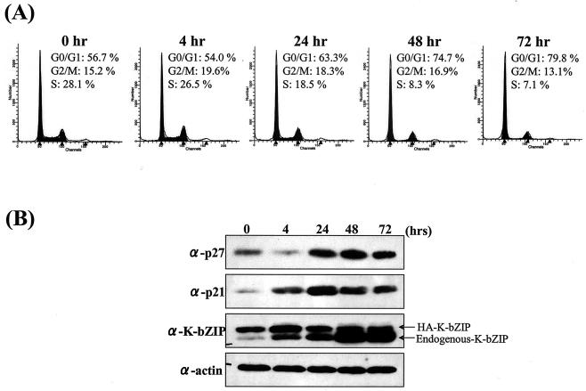
In order to cope with hostile host environments, many viruses have developed strategies to perturb the cellular machinery to suit their replication needs. Some herpesvirus genes protect cells from undergoing apoptosis to prolong the lives of infected cells, while others, such as Epstein-Barr virus Zta, slow down the G(1)/S transition phase to allow ample opportunity for transcription and translation of viral genes before the onset of cellular genomic replication. In this study, we investigated whether Kaposi's sarcoma-associated herpesvirus (KSHV) K-bZIP, a homologue of the Epstein-Barr virus transcription factor BZLF1 (Zta), plays a role in cell cycle regulation. Here we show that K-bZIP physically associates with cyclin-CDK2 and downmodulates its kinase activity. The association can be detected in the natural environment of KSHV-infected cells without artificial overexpression of either component. With purified protein, it can be shown that the interaction between K-bZIP and cyclin-CDK2 is direct and that K-bZIP alone is sufficient to inhibit CDK2 activity. The interacting domain of K-bZIP has been mapped to the basic region. The result of these associations is a prolonged G(1) phase, accompanied by the induction of p21 and p27 in a naturally infected B-cell line. Thus, in addition to the previously described transcription and genome replication functions, a new role of K-bZIP in KSHV replication is identified in this report.


Figures

FIG. 1.
FIG. 1.
Effect of KSHV reactivation on p53, p21, and p27 levels. After TPA treatment, BCP1 cells were lysed with EBC buffer. Protein extract (100 μg/lane) from each time point (in hours) was loaded. K-bZIP was detected with anti-K-bZIP rabbit serum. p53, p21, and p27 antibodies were purchased commercially. Nonspecific reaction of antibody was used as a loading control. Protein expression levels of both p21 and p27 were increased after viral reactivation.
FIG. 2.
FIG. 2.
Coimmunoprecipitation assay. (A) Coimmunoprecipitation assay with the KSHV-positive BCBL-1 cell line. BCBL-1 cells induced to viral lytic replication with TPA (48 h) were harvested with EBC buffer, and the same amounts of lysates (500 μg) were precipitated with antibodies against different molecules and then immunoblotted with anti-K-bZIP rabbit serum. (B) Colocalization of K-bZIP with CDK2. Confocal analysis was performed with anti-K-bZIP rabbit serum and anti-CDK2 mouse monoclonal antibody 48 h after TPA induction. K-bZIP (red) and CDK2 (green) were detected with Alexa Fluor 555-conjugated goat F(ab′)2 anti-rabbit immunoglobulin G and Alexa Fluor 488-conjugated goat F(ab′)2 anti-mouse immunoglobulin G. The nucleus was counterstained with To-Pro-3 (blue). This panel is representative of 10 different fields. (C) Association between K-bZIP and CDK2-cyclins in 293T cells. 293T cells were cotransfected with the indicated plasmids. Cell lysates were precipitated with Flag antibody-conjugated agarose, and coimmunoprecipitation of K-bZIP was detected with anti-T7 antibody. The expression of T7-tagged K-bZIP in total lysates is shown in the same blots as a control. IP, immunoprecipitation; W.B, Western blotting; α, anti.
FIG. 2.
FIG. 2.
Coimmunoprecipitation assay. (A) Coimmunoprecipitation assay with the KSHV-positive BCBL-1 cell line. BCBL-1 cells induced to viral lytic replication with TPA (48 h) were harvested with EBC buffer, and the same amounts of lysates (500 μg) were precipitated with antibodies against different molecules and then immunoblotted with anti-K-bZIP rabbit serum. (B) Colocalization of K-bZIP with CDK2. Confocal analysis was performed with anti-K-bZIP rabbit serum and anti-CDK2 mouse monoclonal antibody 48 h after TPA induction. K-bZIP (red) and CDK2 (green) were detected with Alexa Fluor 555-conjugated goat F(ab′)2 anti-rabbit immunoglobulin G and Alexa Fluor 488-conjugated goat F(ab′)2 anti-mouse immunoglobulin G. The nucleus was counterstained with To-Pro-3 (blue). This panel is representative of 10 different fields. (C) Association between K-bZIP and CDK2-cyclins in 293T cells. 293T cells were cotransfected with the indicated plasmids. Cell lysates were precipitated with Flag antibody-conjugated agarose, and coimmunoprecipitation of K-bZIP was detected with anti-T7 antibody. The expression of T7-tagged K-bZIP in total lysates is shown in the same blots as a control. IP, immunoprecipitation; W.B, Western blotting; α, anti.
FIG. 3.
FIG. 3.
In vitro interaction and interacting domain of K-bZIP. (A) Domains of K-bZIP and GST-K-bZIP mutants I to V are indicated in the panel. The results of GST pulldown assays for in vitro-translated (IVT), 35S-labeled full-length CDK2, cyclin A, and cyclin E are shown. FL, full length; TA, transactivation domain; BR, basic region; LZ, leucine zipper domain. (B) Inhibition of CDK2-cyclin kinase activity by K-bZIP. CDK2-cyclin kinase activity was measured by in vitro kinase assay with histone 1 as a substrate. The kinase activity of half of the reaction was measured by Quantity One. Relative kinase activity is shown. Protein amounts used in this assay are indicated at the bottom of the panel. The Coomassie-stained gel of the other half of the reaction is shown as a loading control. BSA, bovine serum albumin.
FIG. 4.
FIG. 4.
(A) K-bZIP influence on cell cycle division. For determination of cell growth rate, 5 × 10⁴ cells of Flag-293-K-bZIPwt 1, Flag-293-K-bZIPwt 2, Flag-K-bZIPΔBR, and Flag-293-vector were seeded in 60-mm plates and cultured in 10% fetal bovine serum-DMEM. Cell numbers were counted at the indicated time points postseeding. Triplicate plates were prepared for each cell clone. (B) Inhibition of CDK2 kinase activity in Flag-293-K-bZIPwt cell lines. CDK2 kinase activity was determined by in vitro kinase assay after immunoprecipitation (IP) of CDK2 from stable cell lines. Histone 1 was used as a substrate for the assay. The amounts of the CDK2 were measured by immunoblotting. W.B., Western blotting. (C) Upregulation of p21 in Flag-293-K-bZIPwt and Flag-K-bZIPΔLZ cell lines. Protein expression levels of p21 in stably K-bZIP-expressing cell lines were measured. Total cell lysates were prepared from the stable cell lines. Actin served as an internal control for the amounts of protein on the membrane. (D) Upregulation of p21 in transiently transfected HeLa cells. HeLa cells were transfected pFlag-empty vector or pFlag-K-bZIPwt. At 72 h posttransfection, total cell lysates were obtained, and 50 μg of protein was loaded in each lane and probed with anti-p21 antibody. Actin served as an internal control for the amount of protein on the membrane.
FIG. 5.
FIG. 5.
(A) K-bZIP slows down cell cycle division. A total of 5 × 10⁴ K-bZIP-BCBL-1 cells or parental BCBL-1 cells were seeded in 60-mm plates and cultured in 15% fetal bovine serum-RPMI 1640. Cell numbers were counted at the indicated time points. Triplicate plates were seeded for each cell clone. (B) Inhibition of CDK2 kinase activity in K-bZIP-BCBL-1 cells. CDK2 kinase activity was determined by the in vitro kinase assay after induction of K-bZIP expression (72 h). CDK2 was immunoprecipitated from K-bZIP-BCBL-1 or parental BCBL-1 cells. Histone 1 was used as a substrate for the assay. The amounts of CDK2 were measured by immunoblotting. W.B, Western blotting.
FIG. 6.
FIG. 6.
KSHV reactivation and K-bZIP expression prolong G0/G1 phase. (A) Cell cycle analysis of TPA-treated K-bZIP-BCBL-1 cells. Cell cycle analysis after induction by TPA was carried out with flow cytometry. Cell cycle analysis was performed with ModFit LT. The percentage of cells in G0/G1 phase is indicated. (B) Upregulation of p21 and p27 in K-bZIP-BCBL-1 cells after induction. Protein extract (100 μg/lane) from each time point (in hours) was loaded. Protein expression levels of p21, p27, and total K-bZIP in HA-K-bZIP-BCBL-1 cells were measured by immunoblotting. Actin served as an internal control for the amount of protein on the membrane. α, anti.

Similar articles

Cited by

References

    1. Adams, P. D., W. R. Sellers, S. K. Sharma, A. D. Wu, C. M. Nalin, and W. G. Kaelin, Jr. 1996. Identification of a cyclin-cdk2 recognition motif present in substrates and p21-like cyclin-dependent kinase inhibitors. Mol. Cell. Biol. 16:6623-6633. - PMC - PubMed
    1. Advani, S. J., R. R. Weichselbaum, and B. Roizman. 2001. cdc2 cyclin-dependent kinase binds and phosphorylates herpes simplex virus 1 UL42 DNA synthesis processivity factor. J. Virol. 75:10326-10333. - PMC - PubMed
    1. Alkan, S., D. S. Karcher, A. Ortiz, S. Khalil, M. Akhtar, and M. A. Ali. 1997. Human herpesvirus-8/Kaposi's sarcoma-associated herpesvirus in organ transplant patients with immunosuppression. Br. J. Haematol. 96:412-414. - PubMed
    1. Bartek, J., and J. Lukas. 2001. Pathways governing G1/S transition and their response to DNA damage. FEBS Lett. 490:117-122. - PubMed
    1. Bonin, L. R., and J. K. McDougall. 1997. Human cytomegalovirus IE2 86-kilodalton protein binds p53 but does not abrogate G1 checkpoint function. J. Virol. 71:5861-5870. - PMC - PubMed

Publication types

MeSH terms

LinkOut - more resources