. 2003 Aug;23(16):5882-95.
doi: 10.1128/MCB.23.16.5882-5895.2003.

Activation of heat shock genes is not necessary for protection by heat shock transcription factor 1 against cell death due to a single exposure to high temperatures

Affiliations

Activation of heat shock genes is not necessary for protection by heat shock transcription factor 1 against cell death due to a single exposure to high temperatures

Sachiye Inouye et al. Mol Cell Biol. 2003 Aug.

Abstract

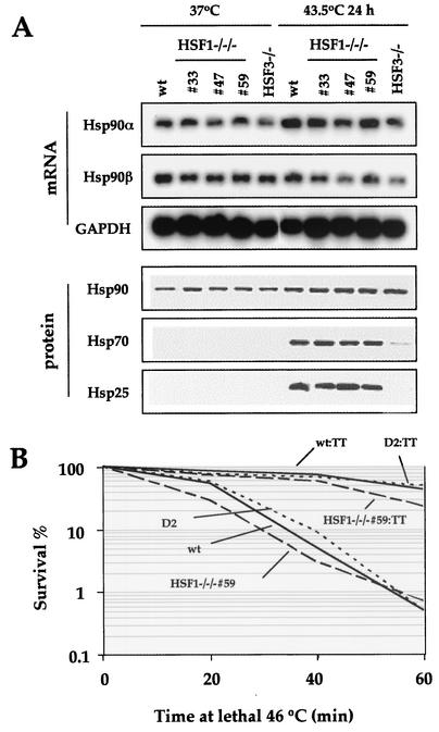
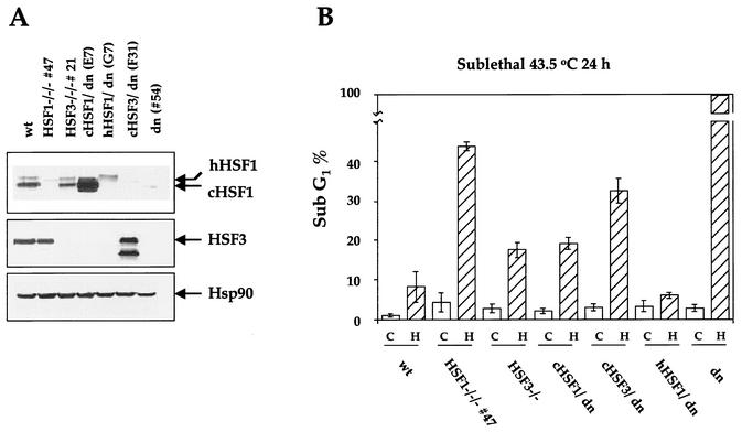
Heat shock response, which is characterized by the induction of a set of heat shock proteins, is essential for induced thermotolerance and is regulated by heat shock transcription factors (HSFs). Curiously, HSF1 is essential for heat shock response in mammals, whereas in avian HSF3, an avian-specific factor is required for the burst activation of heat shock genes. Amino acid sequences of chicken HSF1 are highly conserved with human HSF1, but those of HSF3 diverge significantly. Here, we demonstrated that chicken HSF1 lost the ability to activate heat shock genes through the amino-terminal domain containing an alanine-rich sequence and a DNA-binding domain. Surprisingly, chicken and human HSF1 but not HSF3 possess a novel function that protects against a single exposure to mild heat shock, which is not mediated through the activation of heat shock genes. Overexpression of HSF1 mutants that could not bind to DNA did not restore the susceptibility to cell death in HSF1-null cells, suggesting that the new protective role of HSF1 is mediated through regulation of unknown target genes other than heat shock genes. These results uncover a novel role of vertebrate HSF1, which has been masked under the roles of heat shock proteins.


Figures

FIG. 1.
FIG. 1.
Chicken HSF1 has little potential to activate heat shock genes in response to heat shock. (A) The phylogenetic tree generated in Clustal W (43) for members of the HSF family. The molecular tree was constructed by the neighbor-joining method. Gaps were excluded from all phylogenetic analyses. The numerals show bootstrap values (1,000 bootstrap replicates were performed). The unrooted tree was drawn with the program TreeView (31). The bar represents 0.1 substitutions per site. The amino acid sequences used in the tree construction are human HSF1 (hHSF1, SP accession no. Q00613), mouse HSF1 (mHSF1, SP accession no. P38532), chicken HSF1 (cHSF1, SP accession no. P38529), zebrafish HSF1 (Zebra HSF1, DDBJ accession no. AB062117), and Xenopus HSF1 (Xenopus HSF1, SP accession no. P41154), human HSF2 (hHSF2, SP accession no. Q03933), mouse HSF2 (mHSF2, SP accession no. P38533), and chicken HSF2 (cHSF2, SP accession no. P38530), chicken HSF3 (cHSF3, SP accession no. P38531), human HSF4b (hHSF4, SP accession no. Q9ULV5) and mouse HSF4b (mHSF4, SP accession no. Q9R0L1), and HSF from Saccharomyces cerevisiae (ScHSF, SP accession no. P10961), D. melanogaster (DmHSF, SP accession no. P22813), and Caenorhabditis elegans (CeHSF, PIR accession no. T27125). (B) HSF3-null cells were transfected with an expression vector for cHSF1, hHSF1, D. melanogaster HSF, or cHSF3. Clones grown in the presence of zeocine were selected for Western blot analysis with HSF1-specific antiserum αHSF1β and antiserum for HA or HSF3-specific antiserum αHSF3γ (26). Representative clones are shown. Arrows in the upper and lower columns indicate endogenous cHSF1 and cHSF3, respectively. An asterisk shows an unmodified form of HSF3 (42). Arrowheads indicate ectopically expressed HSFs. (C) Heat shock response in HSF3-null cells expressing the indicated HSFs. RNAs were extracted from cells untreated or treated with heat shock at 45°C for 60 min, and Northern blot analysis was performed with cDNA for Hsp70 or glyceraldehyde-3-phosphate dehydrogenase (GAPDH). (D) Schematic representation of wild-type and mutant HSF1 loci together with the targeting vector, mHSF1-neo. Exons are represented by solid boxes, and the DNA-binding domain is encoded by exons 2 and 3. The 3′ probe for Southern blot analysis (solid bar) and primer sites (arrowheads) are indicated. Restriction enzyme sites are: Bg, BglII; Hp, HpaI; S, SalI. (E) Southern blot analysis of DNA prepared from wild-type, HSF1+/−, and HSF1−/− ES cells. DNA was digested with BglII and hybridized with the 32P-labeled 3′ probe or neo probe. (F) HSF1−/− ES cells were transfected with expression vector pcHSF1-Hyg or phHSF1-Hyg, and clones which expressed a high level of cHSF1 or hHSF1 were selected. HSF1 protein levels in each representative clone were shown by Western blot analysis with αHSF1β antiserum. The lower column represents a longer exposure. (G) Examination of heat shock response by Northern blot analysis. HSF1−/− ES cells expressing cHSF1 or hHSF1 as well as wild-type and HSF1−/− ES cells were heat shocked at 43°C for 30 min. RNAs were extracted from these cells, and Northern blot analysis was performed with 32P-labeled cDNAs for mouse Hsp70-1 and human β-actin.
FIG. 2.
FIG. 2.
Amino-terminal sequence is responsible for inability of cHSF1 to induce heat shock gene activation. (A) Schematic representation of chimeras between chicken and human HSF1 and induction of Hsp70 expression. The expression vector for each chimeric HSF1 was stably transfected into HSF3-null DT40 cells. Total RNA was extracted from cells treated with and without heat shock at 45°C for 60 min, and Hsp70 and GAPDH expression was examined by Northern blot analysis. Relative positions of functional domains are indicated: DBD, DNA binding domain; HR, hydrophobic heptad repeat; RD, regulatory domain; AD, activation domain. The h370/c chimera represents a fusion protein of human HSF1 (amino acids 1 to 370) and chicken HSF1 (amino acids 333 to 491). (B) Extracts were prepared from cell lines expressing each chimeric HSF1. Western blot analysis was performed with the HSF1 antiserum αHSF1β. (C) Alignment of chicken and human HSF1s. There are unique sequences rich in alanine residues in chicken HSF1 (underlined). The position of the h20/c substitution is indicated. DBD, DNA binding domain.
FIG. 3.
FIG. 3.
Substitution of amino-terminal sequence of cHSF1 with that of hHSF1 does not alter the DNA binding activity, heat-induced phosphorylation, and nuclear translocation of cHSF1. (A) Wild-type DT40 cells, HSF3−/− cells, and HSF3−/− cells having cHSF1, hHSF1, and chimeric h20/c or c126/h were heat shocked at 45°C for 60 min. Northern blot analysis was performed. (B) The cells shown in panel A were heat shocked at 45°C for 60 min, and whole-cell extracts were prepared. The gel shift assay was performed with the 32P-labeled heat shock element probe. (C) Western blot analysis of the whole-cell extracts isolated in panel B was performed with HSF1 antibody. (D) Cells were treated as described for panel B. Cytoplasmic (c) and nuclear (n) extracts were prepared, and Western blot analysis of the extracts was performed with HSF1 antibody.
FIG. 4.
FIG. 4.
cHSF1-deficient cells are highly sensitive to high temperatures. (A) Wild-type DT40 (wt), double-null (54 cells), HSF1-null (59 cells), and HSF3-null (21 cells) cells were incubated at 43°C for 24 h. Representative cell cycle distributions of the indicated cell cultures are shown. Cells were pulse labeled for 10 min and stained with fluorescein isothiocyanate-conjugated antibromodeoxyuridine to detect bromodeoxyuridine incorporation (vertical axis) and propidium iodide to detect total DNA (horizontal axis). (B) Western blot analysis of HSF1 protein in cells of each indicated genotype as well as HSF1-null 59 cells reexpressing cHSF1 (D2). (C) The cells shown in panel B were incubated at 43.5°C for 24 h, and representative cell cycle distributions are shown. (D) Wild-type (wt) and HSF1-null 59 (HSF1−/−/−) cells were incubated for 24 h at the indicated temperatures. Proportions of sub-G1 fractions are shown. Means and standard deviations of three independent experiments are shown. (E) Cells were incubated at 43.5°C for the indicated periods, and proportions of sub-G1 fractions are shown as in panel D.
FIG. 5.
FIG. 5.
Expression of Hsp90 and induced thermotolerance in HSF1-null cells. (A) Expression of heat shock proteins, including Hsp90, in three independently generated cHSF1-null clones (33, 47, and 59). Wild-type, HSF1-null, and HSF3-null cells were incubated at 43.5°C for 24 h. Accumulations of Hsp90α and Hsp90β mRNAs were examined by Northern blot analysis. Accumulations of Hsp90, Hsp70, and Hsp25 proteins were determined by Western blot analysis. (B) cHSF1-null cells acquire induced thermotolerance. cHSF1-null cells (HSF1−/−/− 59) and cHSF1-restored cells (D2) as well as wild-type cells (wt) grown at 37°C were incubated at a lethal 46°C temperature for the indicated periods. The numbers of surviving cells were counted by a colony formation assay, and percent survival is shown. Some cells were treated at a sublethal 43°C for 30 min and then allowed to recover for 2 h before lethal heat shock (wt:TT, HSF1−/−/− 59:TT, and D2:TT). Means of three independent experiments are shown.
FIG. 6.
FIG. 6.
HSF1-null cells are sensitive to radiation and UV. (A) Wild-type (wt) cells, HSF1-null 59 cells, and cHSF1-restored 59 cells (D2) were exposed to 600 rads of irradiation and then incubated for 24 h. Proportions of sub-G1 fractions in control and irradiated cells are shown. (B) Cells were exposed to UV at 10 J/m2 and then incubated for 24 h. Means and standard deviations of three independent experiments are shown.
FIG. 7.
FIG. 7.
Mammalian HSF1 as well as chicken HSF1 is a survival factor in DT40 cells. (A) Double-null cells were stably transfected with an expression vector for cHSF1, hHSF1, or cHSF3. Protein levels of HSF1, HSF3, and Hsp90 were determined by Western blot analysis in wild-type cells (wt), HSF1-null cells (line 47), HSF3-null cells (line 21), double null cells (line 54), and null cells reexpressing cHSF1(E7), hHSF1 (G7), or cHSF3 (F31). (B) Proportions of sub-G1 fractions in the cells described in A are shown before (open bar) and after (hatched bar) incubation at 43.5°C for 24 h. All of the double-null cells died after the treatment. Note that the reexpression of hHSF1 completely restored cell survival after heat treatment.
FIG. 8.
FIG. 8.
Hypersensitivity of HSF1-null mouse embryonic fibroblast cells to high temperatures is restored by ectopic expression of chicken HSF1. (A) Northern blot analysis of heat shock genes in HSF1+/+, HSF1+/−, and HSF1−/− mouse embryonic fibroblast cells before and after heat shock at 43°C for 30 min. (B) Sixteen hours after the HSF1+/+ (open bars) and HSF1−/− (gray bars) mouse embryonic fibroblast cells were plated on culture dishes, the dishes were maintained in an incubator at each indicated temperature. Numbers of attached cells were counted at 9 h after the incubation. Means and standard deviations of three independent experiments are shown. (C) HSF1+/+ (diamonds) and HSF1−/− (squares) mouse embryonic fibroblast cells were incubated at 41.5°C for the indicated periods, and numbers of attached cells were counted. (D) Morphology of MEF cells incubated at 41.5°C under a Axiovert 200 microscope (Zeiss). (E) Mouse embryonic fibroblast cells were incubated in the presence of adenovirus expressing LacZ (Ad-LacZ) or chicken HSF1 (Ad-cHSF1). Western blot analysis of HSF1 was performed with these extracts. The level of endogenous HSF1 in HSF1+/+ mouse embryonic fibroblast cells was too low to be detected. (F) Twenty-four hours after the mouse embryonic fibroblast cells were plated, cells were maintained in the presence or absence of adenovirus (106/ml) for 24 h. Then the cells were incubated at 42°C for the indicated periods. Protein levels of Hsp90, Hsp70, Hsp27, and actin were examined by Western blot analysis with extracts from these cells. (G) Numbers of attached cells were counted at the indicated times after incubation at 42°C. Means and standard deviations of three independent experiments are shown.
FIG. 9.
FIG. 9.
DNA binding activity is necessary for human HSF1 to restore resistance to high temperatures. (A) Schematic representation of mutant human HSF1. The arginine at amino acid position 71 in human HSF1 is shown to contact DNA directly in S. cerevisiae HSF (20) and was mutated to alanine (hHSF1R71A) or glycine (hHSF1R71G). The conserved domains in other vertebrate HSFs are indicated: DBD, DNA binding domain; HR-A/B, amino-terminal hydrophobic repeat; HR-C, carboxyl-terminal hydrophobic repeat. (B) HSF1 protein levels were determined by Western blotting with extracts from wild-type DT40, double null cells (dn), and double null cells expressing hHSF1, hHSF1R71G, or hHSF1R71A. (C) Gel filtration analysis of extracts isolated from double null cells expressing hHSF1 (G7) and hHSF1R71G (R71G) before and after heat shock at 45°C for 30 min. The predicted elution positions of monomeric and trimeric forms of HSF1 are indicated at the bottom (26). (D) The cell extracts prepared in panel C were subjected to the gel mobility shift assay with the 32P-labeled ideal HSE oligonucleotide. Binding reaction mixes were incubated in the presence of anti-HSF2 antiserum to wipe out HSF2 binding activity. (E) Northern blot analysis of cells before and after heat shock at 45°C for 60 min. (F) Proportions of sub-G1 fractions in cells are shown before (open bar) and after (hatched bar) incubation at 43.5°C for 24 h. (G) HSF1 protein levels were determined by Western blotting with extracts from wild-type DT40 cells, HSF1-null cells (line 59), and HSF1-null cells expressing hHSF1 (hHSF1/line 59) and hHSF1R71G (R71G/line 59). (H) Proportions of sub-G1 fractions in cells are shown before (open bar) and after (hatched bar) incubation at 43.5°C for 24 h. Means and standard deviations of three independent experiments are shown.

Similar articles

Cited by

References

    1. C. N. Adra, P. H. Boer, and M. W. McBurney. 1987. Cloning and expression of the mouse pgk-1 gene and the nucleotide sequence of its promoter. Gene 60:65-74. - PubMed
    1. Ahn, S. G., P. C. Liu, K. Klyachko, R. I. Morimoto, and D. J. Thiele. 2001. The loop domain of heat shock transcription factor 1 dictates DNA-binding specificity and responses to heat stress. Genes Dev. 15:2134-2145. - PMC - PubMed
    1. Aschoff, J., and U. von Saint Paul. 1973. Brain temperature as related to gross motor activity in the unanesthetized chicken. Physiol. Behav. 10:529-533. - PubMed
    1. Baler, R., G. Dahl, and R. Voellmy. 1993. Activation of human heat shock genes is accompanied by oligomerization, modification, and rapid translocation of heat shock transcription factor HSF1. Mol. Cell. Biol. 13:2486-2496. - PMC - PubMed
    1. Beere, H. M., B. B. Wolf, K. Cain, et al. 2000. Heat-shock protein 70 inhibits apoptosis by preventing recruitment of procaspase-9 to the Apaf-1 apoptosome. Nat. Cell Biol. 2:469-475. - PubMed

Publication types

MeSH terms

LinkOut - more resources