. 2003 Aug;23(16):5790-802.
doi: 10.1128/MCB.23.16.5790-5802.2003.

HSP27 is a ubiquitin-binding protein involved in I-kappaBalpha proteasomal degradation

Affiliations

HSP27 is a ubiquitin-binding protein involved in I-kappaBalpha proteasomal degradation

Arnaud Parcellier et al. Mol Cell Biol. 2003 Aug.

Abstract

HSP27 is an ATP-independent chaperone that confers protection against apoptosis through various mechanisms, including a direct interaction with cytochrome c. Here we show that HSP27 overexpression in various cell types enhances the degradation of ubiquitinated proteins by the 26S proteasome in response to stressful stimuli, such as etoposide or tumor necrosis factor alpha (TNF-alpha). We demonstrate that HSP27 binds to polyubiquitin chains and to the 26S proteasome in vitro and in vivo. The ubiquitin-proteasome pathway is involved in the activation of transcription factor NF-kappaB by degrading its main inhibitor, I-kappaBalpha. HSP27 overexpression increases NF-kappaB nuclear relocalization, DNA binding, and transcriptional activity induced by etoposide, TNF-alpha, and interleukin 1beta. HSP27 does not affect I-kappaBalpha phosphorylation but enhances the degradation of phosphorylated I-kappaBalpha by the proteasome. The interaction of HSP27 with the 26S proteasome is required to activate the proteasome and the degradation of phosphorylated I-kappaBalpha. A protein complex that includes HSP27, phosphorylated I-kappaBalpha, and the 26S proteasome is formed. Based on these observations, we propose that HSP27, under stress conditions, favors the degradation of ubiquitinated proteins, such as phosphorylated I-kappaBalpha. This novel function of HSP27 would account for its antiapoptotic properties through the enhancement of NF-kappaB activity.


Figures

FIG. 1.
FIG. 1.
HSP27 enhances proteasome activation while inhibiting apoptosis. (A) U937 cells either were left untreated or were treated for 4 h with 100 μM etoposide (VP16) in the absence or presence of acetyl-calpastatin (25 μM), MG132 (25 μM), or lactacystin (25 μM) before measurement of the ability of cell lysates to cleave the substrate Suc-LLVY-AMC (black bars; AU, arbitrary units) and the percentage of apoptotic cells after nuclear chromatin staining with Hoechst 33342 (gray bars). (B) Control-transfected (white bars) and HSP27-transfected (black bars) cells either were left untreated or were treated for 4 h (U937 cells) or 24 h (MEF cells) with 100 μM etoposide (VP16) or 20 ng of ΤNF-α/ml in the absence or presence of 25 μM MG132 before measurement of the ability of cell lysates to cleave the substrate Suc-LLVY-AMC. Results are expressed as percentages (100% is the activity in VP16-treated, HSP27-transfected cells). Insets show Western blot analyses of HSP27 expression in control- and HSP27-transfected cells. (C) Kinetic analysis of Suc-LLVY-AMC cleavage activity (black bars) and apoptosis induction (gray bars) in control- and HSP27-transfected U937 cells treated with 100 μM etoposide for the indicated times. (D and E) Cells were treated with etoposide (100 μM, 4 h) in the presence of decreasing concentrations of MG132 or lactacystin, as indicated. Then, chromatin staining with Hoechst 33342 was used to measure the percentage of control-transfected and HSP27-transfected U937 apoptotic cells (D) or the percentage of cell survival induced by HSP27 overexpression (E). The percentage of proteasome inhibition (Suc-LLVY-AMC cleavage) is also indicated (E). Data are the means and standard deviations for three independent experiments.
FIG. 2.
FIG. 2.
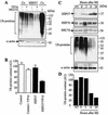
HSP27 enhances the degradation of ubiquitinated proteins in stressed cells. (A) Control-transfected (Co) and HSP27-transfected U937 cells either were left untreated or were treated with 100 μM etoposide (VP16) for 4 h in the absence or presence of 25 μM MG132. Protein ubiquitination (Ub-proteins) was monitored by Western blotting with an antiubiquitin antibody. α-actin served as the loading control. (B) Densitometry analysis of Ub-proteins in three independent experiments (mean and standard deviation) similar to that shown in panel A. AU, arbitrary units. (C) U937 cells either were left untreated (NT) or were exposed for 1 h to 42°C (HS) and then were incubated at 37°C for the indicated times. The indicated proteins were studied by Western blotting. α-Actin served as the loading control. (D) Densitometry analysis of Ub-proteins shown in panel C.
FIG. 3.
FIG. 3.
HSP27 associates with ubiquitin. (A) Cytoplasmic extracts obtained from control-transfected and HSP27-transfected U937 cells, either left untreated or treated with 100 μM etoposide (VP16) for 4 h, were incubated with a ubiquitin-agarose matrix. The presence of the indicated proteins in the input material was analyzed by Western blotting (left panels). Pull-down analysis of ubiquitin-agarose was performed in the absence or presence of 50 μg of free ubiquitin (free-Ub) before elution of bound proteins and Western blot analysis of HSPs (right panels). One representative experiment of three independent experiments is shown. (B) Western blot analysis of GST-monoubiquitin (GST-Ub) and GST-polyubiquitin (GST-polyUb) obtained in vitro as described in Material and Methods. (C) Cytosolic extracts from HSP27-overexpressing cells were incubated with GST alone, GST-Ub, or GST-polyUb. The presence of HSP27 in input material was checked by Western blotting after pull-down analysis and elution of bound proteins. One representative experiment of three experiments is shown.
FIG. 4.
FIG. 4.
HSP27 association with the 26S proteasome is required for HSP27-induced proteasome activity. (A) Immunoprecipitation (IP) performed with control- and HSP27-transfected U937 cells and an anti-HSP27 or an anti-HSP60 antibody was followed by immunodetection of HSP27, HSP70, and the PA700 subunit of the 26S proteasome. ch., chain. (B) Immunodetection of PA700 after immunoprecipitation of HSP27 in control (Co) and heat-shocked (HS; 1 h at 42°C and then 6 h at 37°C) U937 cells. (C) HSP27 was incubated in vitro with the purified 26S proteasome before immunoprecipitation of HSP27 or HSP60 and immunodetection of the indicated proteins. (D) Immunodetection of PA700 after HSP27 immunoprecipitation of REG cells transfected with an empty vector (Control) or a vector containing either wild-type hsp27 or the indicated hsp27 deletion mutations. As a control, cell lysates were immunoprecipitated with nonrelevant immunoglobulin G1. (E) The REG cells shown in panel D were treated with etoposide (VP16, 100 μM, 24 h) before measurement of the ability of cell lysates to cleave the substrate Suc-LLVY-AMC as described in the legend to Fig. 1 (fold induction is relative to that in untreated cells). Results are the means and standard deviations for three independent experiments.
FIG. 5.
FIG. 5.
HSP27 increases nuclear NF-κB content, DNA binding, and transcriptional activity in stressed cells. (A) Control-transfected (white bars) and HSP27-transfected (black bars) cells either were left untreated or were treated for 4 h (U937 cells) or 24 h (MEF cells) with etoposide (VP16, 100 μM), ΤNF-α (20 ng/ml), or IL-1β (1 ng/ml). The level of nuclear NF-κB expression was determined by flow cytometry (MFI, mean fluorescence index). (B) Nuclear extracts from control-transfected and HSP27-transfected U937 cells either were left untreated or were treated for 4 h with etoposide (VP16, 100 μM) or ΤNF-α (20 ng/ml) and then were subjected to EMSAs with an NF-κB probe. As a control for binding specificity, the extracts were preincubated with a 50-fold molar excess of unlabeled oligonucleotide (Unl.). Supershift analysis was performed by preincubation of the extracts with an anti-p50 or an anti-p65 polyclonal antibody before EMSAs. (C) Control-transfected (white bars) and HSP27-overexpressing (black bars) cells were transiently transfected with an NF-κB luciferase reporter plasmid and then either were left untreated or were treated for 4 h (U937 cells) or 10 h (MEF cells) with 100 μM VP16 or 20 ng of ΤNF-α/ml. Cotransfection of a thymidine kinase-Renilla luciferase plasmid was used to normalize for transfection efficiency. AU, arbitrary units. Results are the means and standard deviations for three independent experiments.
FIG. 6.
FIG. 6.
HSP27 enhances I-κBα degradation by the proteasome. (A) Control-transfected and HSP27-transfected U937 cells were treated as indicated (VP16, 100 μM, 4 h; ΤNF-α, 20 ng/ml, 4 h; MG132, 25 μM) before monitoring of I-κBα expression by Western blotting. HSC70 served as a loading control. (B) Control-transfected (white bars) and HSP27-transfected (black bars) U937 cells were treated with etoposide (100 μM) for the indicated times before measurement of I-κBα cellular content by flow cytometry (MFI, mean fluorescence index). (C) REG cells were transfected with an empty plasmid (Control) or a plasmid encoding wild-type HSP27 or the indicated deletion mutants of HSP27 and then were treated with etoposide (VP16, 100 μM, 24 h) before measurement of I-κBα cellular content by flow cytometry as described for panel B. Results are the means and standard deviations for four independent experiments.
FIG. 7.
FIG. 7.
HSP27 associates with phosphorylated I-κBα. (A) Control-transfected and HSP27-transfected U937 cells were treated as indicated (VP16, 100 μM, 4 h; ΤNF-α, 20 ng/ml, 4 h; MG132, 25 μM) before monitoring of phosphorylated I-κBα (P-I-κBα) expression by Western blotting. HSC70 served as a loading control. (B) Total cell lysates were prepared from HSP27-transfected U937 cells either left untreated or treated for 4 h with etoposide (VP16, 100 μM), ΤNF-α (20 ng/ml), and/or MG132 (25 μM) and then immunoprecipitated (IP) with the indicated antibodies. The indicated proteins were detected by Western blotting. (C) Control-transfected and HSP27-transfected U937 cells and control-transfected and HSP27-transfected MEF cells were transiently transfected with an empty vector or a plasmid containing a nonphosphorylatable, nondegradable mutant form of I-κBα (I-κBα S32A/S36A) (transfection efficiency measured with a β-galactosidase-expressing plasmid, 35 to 40%). At 36 h later, cells were treated with 100 μM etoposide (VP16) or 20 ng of TNF-α/ml for 4 h (U937 cells) or 24 h (MEF cells) before measurement of the percentage of apoptotic cells. Error bars indicate standard deviations (n = 4).
FIG. 8.
FIG. 8.
HSP27, phosphorylated I-κBα, and PA700 can be found in the same cellular protein fraction. (A) Lysates from HSP27-transfected U937 cells not treated (triangles) or treated (squares) with etoposide (100 μM, 4 h) were centrifuged and fractionated with a Superose-6 column. Fractions were tested for hydrolysis of the substrate Suc-LLVY-AMC. AU, arbitrary units. (B) The presence of PA700, HSP27, and phosphorylated I-κBα (P-I-κBα) in fractions of cell extracts from nontreated (NT) and etoposide-treated (VP16) cells in the presence of MG132 (25 μM) to stabilize phosphorylated I-κBα was determined by Western blotting. (C) Immunodetection of PA700 and P-I-κBα after immunoprecipitation (IP) of HSP27 in fraction 17 of cells exposed to etoposide in the presence of MG132. ch., chain.
FIG. 9.
FIG. 9.
Hypothetical model for the influence of HSP27 on proteasome-mediated I-κBα degradation. In response to stimulation, I-κBα is phosphorylated and ubiquitinated (Ub), which could enhance its association with HSP27 and its targeting to the 26S proteasome, thus permitting the release of NF-κB dimers that translocate to the nucleus. When associated with ubiquitinated and phosphorylated I-κBα, HSP27 may favor its transfer, recognition, and/or unfolding by the 26S proteasome.

Similar articles

Cited by

References

    1. Adams, J. 2002. Proteasome inhibitors as new anticancer drugs. Curr. Opin. Oncol. 14:628-634. - PubMed
    1. Baldwin, A. S., Jr. 1996. The NF-kappa B and I kappa B proteins: new discoveries and insights. Annu. Rev. Immunol. 14:649-683. - PubMed
    1. Benaroudj, N., and A. L. Goldberg. 2000. PAN, the proteasome-activating nucleotidase from archaebacteria, is a protein-unfolding molecular chaperone. Nat. Cell Biol. 2:833-839. - PubMed
    1. Bender, A. T., D. R. Demady, and Y. Osawa. 2000. Ubiquitination of neuronal nitric-oxide synthase in vitro and in vivo. J. Biol. Chem. 275:17407-17411. - PubMed
    1. Boelens, W. C., Y. Croes, and W. W. de Jong. 2001. Interaction between alphaB-crystallin and the human 20S proteasomal subunit C8/alpha7. Biochim. Biophys. Acta 1544:311-319. - PubMed

Publication types

MeSH terms

Substances

LinkOut - more resources