. 2003 Aug;23(16):5614-24.
doi: 10.1128/MCB.23.16.5614-5624.2003.

Potential role for ADAM15 in pathological neovascularization in mice

Affiliations

Potential role for ADAM15 in pathological neovascularization in mice

Keisuke Horiuchi et al. Mol Cell Biol. 2003 Aug.

Abstract

ADAM15 (named for a disintegrin and metalloprotease 15, metargidin) is a membrane-anchored glycoprotein that has been implicated in cell-cell or cell-matrix interactions and in the proteolysis of molecules on the cell surface or extracellular matrix. To characterize the potential roles of ADAM15 during development and in adult mice, we analyzed its expression pattern by mRNA in situ hybridization and generated mice carrying a targeted deletion of ADAM15 (adam15(-/-) mice). A high level of expression of ADAM15 was found in vascular cells, the endocardium, hypertrophic cells in developing bone, and specific areas of the hippocampus and cerebellum. However, despite the pronounced expression of ADAM15 in these tissues, no major developmental defects or pathological phenotypes were evident in adam15(-/-) mice. The elevated levels of ADAM15 in endothelial cells prompted an evaluation of its role in neovascularization. In a mouse model for retinopathy of prematurity, adam15(-/-) mice had a major reduction in neovascularization compared to wild-type controls. Furthermore, the size of tumors resulting from implanted B16F0 mouse melanoma cells was significantly smaller in adam15(-/-) mice than in wild-type controls. Since ADAM15 does not appear to be required for developmental angiogenesis or for adult homeostasis, it may represent a novel target for the design of inhibitors of pathological neovascularization.


Figures
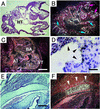
FIG. 1.
FIG. 1.
Expression of ADAM15 mRNA in mouse embryos. All sections were hybridized with an antisense ADAM15 probe (as described in Materials and Methods). Digital images were obtained by bright-field (A, D, and E) and dark-field (B, C, and F) microscopy. (A and B) Section of E9.5 embryo, showing a prominent expression in mesenchymal tissue in the developing head (red arrows), developing heart (HT), arteries (blue arrows). Scale bar, 100 μm. (C) Section of the heart of an E11.5 embryo. Strong expression was observed in the surface lining the ventricle (VT), atrium (AT), and bulbus arteriosus (BA). Scale bar, 200 μm. (D) Bright-field view (magnification, ×400) of the ventricle of an E13.5 embryo shows high-level expression in the endocardium (arrowheads). Scale bar, 50 μm. (E and F) In the cartilage primordium of occipital bone of an E16.5 embryo, positive signals can be seen in hypertrophic cells (arrowheads). Scale bar, 200 μm.
FIG. 2.
FIG. 2.
Expression of ADAM15 (A, C, and E) and PECAM-1 (B, D, and F) in the endocardium and in the developing vasculature at E13.5. (A and B) Consecutive serial sections of the endocardial cushion of the heart of an E13.5 embryo stained with anti-ADAM15 antibody (A) or anti-PECAM-1 antibody (B). Strong expression of ADAM15 can be seen in endocardial cells, which here line the surface of the endocardial cushion. These cells are also positive for PECAM-1 staining. Scale bar, 100 μm. (C to F) Consecutive sections of the lower trunk and abdomen of an E13.5 embryo reveal an almost identical staining pattern for ADAM15 (C) as for PECAM-1 (D). Scale bar, 200 μm. At high magnification (the boxed areas in panels C and D), positive-staining reveals similar capillary structures in the sections stained with ADAM15 (E) and PECAM-1 (F). Scale bar, 100 μm. The staining of endothelial cells is specific for ADAM15, since it was not observed in mice lacking ADAM15 (see below, Fig. 4). However, the staining of blood cells by the ADAM15 antibody is nonspecific, since this staining is not abolished in adam15−/− mice (see Fig. 4D). (G) Western blot analysis confirmed that ADAM15 is expressed in HUVECs.
FIG. 3.
FIG. 3.
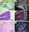
Expression of ADAM15 mRNA in adult mouse brain. Brain sections were hybridized with antisense ADAM15 mRNA probe and analyzed by using bright-field (A, C, and E) and corresponding dark-field (B, D, and F) images. Coronal sections through the hippocampus (A and B) show strong expression in the pyramidal layer and in the granular layer (arrowheads). HP, hippocampus proper; DG, dentate gyrus. Strong expression can be seen in the Purkinje cell layer (C and D, arrowheads) and in a subset of cells in the central part of the hypothalamus (E and F, arrowheads). Scale bars: A, 500 μm; C and E, 200 μm.
FIG. 4.
FIG. 4.
Targeted mutation of ADAM15. (A) The targeting vector is shown at the top. The targeted exon was disrupted by insertion of a pMC1neoPolyA cassette, which introduces a BssHII site that is not present in the wild-type allele. A diphtheria toxin gene cassette was added at the 3′ end of the targeting construct to select against nonhomologous recombination events. A schematic of the wild-type adam15 allele is shown in the middle. The position of the 5′ probe used for Southern blot analysis is indicated, as well as the exon that codes for the initial methionine of ADAM15, and KpnI sites. The bottom panel shows key features of the targeted allele. The BssHII site introduced into the targeted allele reduces the length of the KpnI/BssHII genomic fragment recognized by the 5′ probe from 6.5 to 4.9 kb. (B) Southern blot analysis of KpnI/BssHII-digested genomic DNA from wild-type, heterozygous adam15+/−, and homozygous adam15−/− mice. (C) Western blot analysis of heart extracts from wild-type and adam15−/− mice confirmed that ADAM15 protein expression is abolished in adam15−/− mice. (D) Sections of the descending aorta (arrows) and subcardinal vein (arrowheads) of wild-type (WT) and adam15−/− (−/−) E13.5 embryos. An immunohistochemical analysis shows staining of vascular cells in wild-type mice but not in adam15−/− mice. Scale bar, 100 μm. This finding confirms that the vascular staining pattern of anti-ADAM15 polyclonal antibodies is specific. In contrast, blood cells within the vessels are stained in both wild-type and adam15−/− mice, and therefore this staining is not specific for ADAM15. The polyclonal anti-ADAM15 antibodies used in panels C and D and in Fig. 2 were from the same bleed of a New Zealand White rabbit that was immunized with an Fc fusion protein with the ectodomain of mouse ADAM15.
FIG. 5.
FIG. 5.
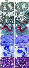
Histological sections of the heart, bone, brain and renal glomerulus of wild-type (WT) and adam15−/− (−/−) mice. (A to D) Sections of the heart are from E13.5 wild-type (A and C) and adam15−/− (B and D) embryos stained with PECAM-1, a marker for endothelial and endocardial cells (magnification, ×50; scale bar, 500 μm [A and B]; magnification, ×200; scale bar, 100 μm [C and D]). (E and F) Section of the distal femur of 4-week-old wild-type (E) or adam15−/− (F) mice stained with safranin-o (which stains cartilage in red), fast green, and hematoxylin. The overall thickness of the growth plate (GP) and the relative size of the zone of hypertrophic cells (HC) were comparable in wild-type and adam15−/− mice. Scale bar, 500 μm. (G to J) Morphological comparison of the hippocampus (G and H) and cerebellum (I and J) of adult wild-type (G and I) and adam15−/− (H and J) mice stained with cresyl violet. No histological aberrations were apparent in the brains of adam15−/− mice. Scale bar, 500 μm. (K and L) Kidney glomerulus of a wild-type (K) and adam15−/− (L) mouse are indistinguishable from each other at the light-microscopic level at a magnification of ×400. Scale bar, 50 μm.
FIG. 6.
FIG. 6.
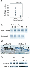
Decreased retinal angiogenesis in adam15−/− mice in a mouse ROP model. (A) Wild-type (WT) and adam15−/− (−/−) mice were subjected to the ROP model, and the ensuing neovascularization in the retina was evaluated as described in Materials and Methods. A significantly decreased angiogenic response was observed in adam15−/− mice compared to wild-type animals. The bars represent the average value. (B) Western blot analysis of ADAM15 expression in the retinas of mice subjected to the ROP model, analyzed at different time points after the return to room air (top panel). As a control, a Western blot of retinas from age-matched untreated mice is shown in the lower panel. Each lane contains the lysate of one retina. ADAM15 expression is significantly upregulated 1 day after the return to room air (P13). The upregulation persists for at least 4 days and ADAM15 expression in the retina returns to the basal levels by P19. (C) Serial sections of the retinas of wild-type mice subjected to the ROP model stained with ADAM15 or PECAM-1. Prominent expression of ADAM15 is found in the endothelial cells migrating into the vitreous body. The staining pattern of PECAM-1 is very similar to that of ADAM15. Scale bar, 50 μm. (D) Confluent HUVECs were incubated without additional supplement (lanes −), with VEGF (20 ng/ml) (lanes V), or bFGF (20 ng/ml) (lanes F) after 12 h of starvation. Neither addition of VEGF nor addition of bFGF detectably affected the expression level of ADAM15 protein in HUVECs.
FIG. 7.
FIG. 7.
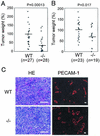
Tumor growth is retarded in adam15−/− mice. Wild-type (WT) and adam15−/− (−/−) mice were injected subcutaneously with 106 B16F0 cells. (A) Comparison of the final weight of tumors grown in mixed-genetic-background (129/SvJ and C57BL/6) wild-type and adam15−/− mice. The results represent the combination of five independent experiments. (B) Comparison of the final weight of the tumors grown in wild-type and adam15−/− mice in a predominantly C57BL/6 background (six backcrosses). The data represent the combination of three independent experiments. To account for possible differences in the number of injected cells and their growth potential between individual experiments, as well as different durations of the experiments, the average tumor weight in wild-type animals in each experiment was used as a reference to calculate the weight of each tumor in percentages of this average. The bars in panels A and B represent the average value for wild-type and adam15−/− mice. (C) Sections of the tumors grown in wild-type and adam15−/− mice stained with hematoxylin and eosin or immunostained with anti-PECAM-1 antibody. No apparent difference in histological morphology or vascular density was observed between the two genotypes. Scale bar, 100 μm.

Similar articles

Cited by

References

    1. Abe, M., and Y. Sato. 2001. cDNA microarray analysis of the gene expression profile of VEGF-activated human umbilical vein endothelial cells. Angiogenesis 4:289-298. - PubMed
    1. Alfandari, D., H. Cousin, A. Gaultier, K. Smith, J. M. White, T. Darribere, and D. W. DeSimone. 2001. Xenopus ADAM 13 is a metalloprotease required for cranial neural crest-cell migration. Curr. Biol. 11:918-930. - PubMed
    1. Asakura, M., M. Kitakaze, S. Takashima, Y. Liao, F. Ishikura, T. Yoshinaka, H. Ohmoto, K. Node, K. Yoshino, H. Ishiguro, H. Asanuma, S. Sanada, Y. Matsumura, H. Takeda, S. Beppu, M. Tada, M. Hori, and S. Higashiyama. 2002. Cardiac hypertrophy is inhibited by antagonism of ADAM12 processing of HB-EGF: metalloproteinase inhibitors as a new therapy. Nat. Med. 8:35-40. - PubMed
    1. Black, R. A., C. T. Rauch, C. J. Kozlosky, J. J. Peschon, J. L. Slack, M. F. Wolfson, B. J. Castner, K. L. Stocking, P. Reddy, S. Srinivasan, N. Nelson, N. Boiani, K. A. Schooley, M. Gerhart, R. Davis, J. N. Fitzner, R. S. Johnson, R. J. Paxton, C. J. March, and D. P. Cerretti. 1997. A metalloproteinase disintegrin that releases tumour-necrosis factor-α from cells. Nature 385:729-733. - PubMed
    1. Black, R. A., and J. M. White. 1998. ADAMs: focus on the protease domain. Curr. Opin. Cell Biol. 10:654-659. - PubMed

Publication types

MeSH terms