. 2003 Aug;77(16):8857-71.
doi: 10.1128/jvi.77.16.8857-8871.2003.

Investigation of structural and functional motifs within the vaccinia virus A14 phosphoprotein, an essential component of the virion membrane

Affiliations

Investigation of structural and functional motifs within the vaccinia virus A14 phosphoprotein, an essential component of the virion membrane

Jason Mercer et al. J Virol. 2003 Aug.

Abstract

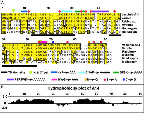
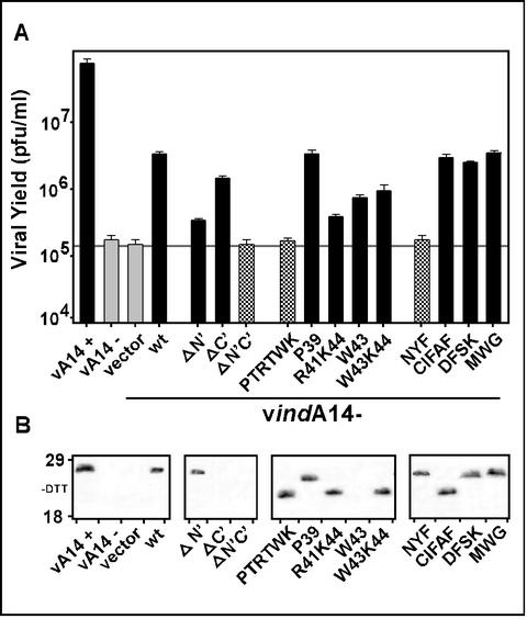
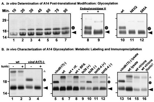
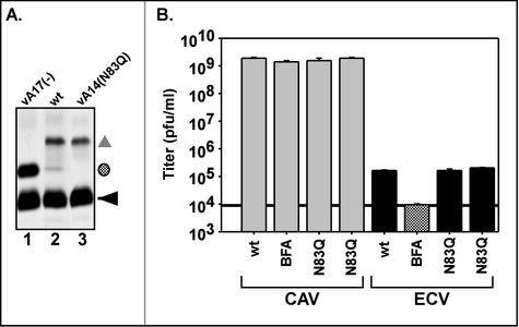
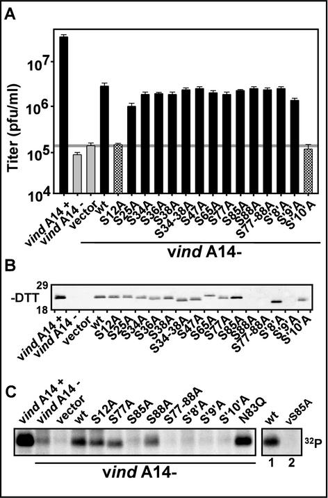
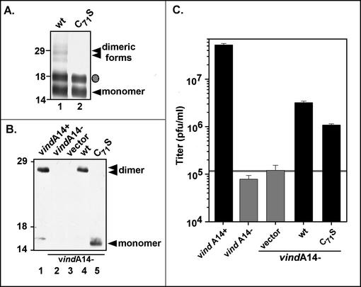
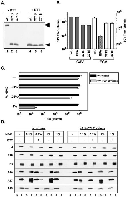
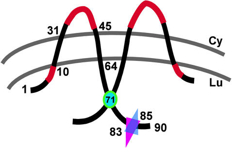
We have previously reported the construction and characterization of an inducible recombinant virus in which expression of the vaccinia virus membrane protein A14 is experimentally regulated using the tetracycline operator-repressor system. Repression of A14, which results in a 1,000-fold reduction in viral yield, leads to an early block in viral morphogenesis characterized by the accumulation of large virosomes, empty "crescents" that fail to contact these virosomes, and, most strikingly, large numbers of aberrant 25-nm vesicles. Here we report the establishment of a transient-complementation system for the structure-function analysis of A14. We have constructed numerous mutant alleles of A14 designed to identify and test the importance of key structural and sequence motifs within A14, including sites of posttranslational modification, such as glycosylation, phosphorylation, and dimerization. From these studies we have determined that robust complementation ability requires an intact N terminus and two regions flanking the first membrane-spanning domain of A14. We show that A14 is modified by N-linked glycosylation both in vitro and in vivo. However, only a minority of A14 molecules are glycosylated in vivo and these are not encapsidated. In this report we also identify the sole phosphorylated serine residue of A14 as lying within the NHS(85) motif that undergoes glycosylation. Additionally, we show that the Cys(71) residue is required for intermolecular disulfide bond formation and describe the properties of a virus expressing an allele of A14 that cannot form disulfide-linked dimers.


Figures

FIG. 1.
FIG. 1.
VV A14 is highly conserved among the Poxviridae family. (A) Alignment of VV A14 with several of its orthopox homologs. Putative membrane-spanning regions are underlined in black. The A14 mutants constructed for this study are depicted as follows: green triangles indicate the new initiation and termination sites engineered for alleles containing N′ and/or C′ truncations. Several sequences chosen for their high level of conservation among the Poxviridae homologs were changed to an equivalent number of Ala residues: N9YF (dark blue overline), C26IFAF (cyan), D32FSK (dark green), P37TRTWK (purple), and M66WG (magenta). Asn83 (orange square), part of a highly conserved N-linked glycosylation motif (NXS) at the C terminus of A14, was changed to Gln. Cys71 (blue circle), likely to direct the covalent dimerization of A14 dimer, was changed to Ser. Red circles highlight Ser residues at positions 12, 25, 34, 36, 38, 47, 65, 77, 85, and 88 that were mutated to Ala, individually and in various combinations (see text). (B) Hydrophobicity plot of VV A14 as determined by the Kyte-Doolittle method. The highly hydrophobic A14 protein is thought to span the membrane twice; the two transmembrane domains comprise residues 13 to 31 and 45 to 64, which are predicted to adopt inside-to-outside and outside-to-inside orientations, respectively (http://www.ch.embnet.org/software/TMPRED_form.html). The hydrophobic domains are separated by a 13-aa hydrophilic loop region.
FIG. 2.
FIG. 2.
Transient rescue of vindA14 performed with structural mutants of A14. (A) Determination of complementation competency by titration of viral yield from infections and transfections. BSC40 cells were infected at an MOI of 5 with vindA14 virus. Infections were performed in the presence (+) or absence (−) of TET, followed by transfection of 10 μg of empty vector or vector encoding the indicated alleles of A14. Extracts were harvested at 24 hpi and were titrated in order to assess the ability of the various A14 alleles to substitute for endogenous virus A14 in a transient-rescue assay. Cells infected in the presence (+) and absence (−) of TET served as positive and negative controls, respectively; cells infected in the absence of inducer and transfected with empty vector or with the plasmid encoding wt A14 served as the benchmarks for transient rescue. The horizontal grey line shows the titer obtained upon transfection of empty vector. The data shown represent the average of three independent experiments. (B) Immunoblot analysis of A14 expression. Aliquots of the extracts described above were resolved by nonreducing SDS-17% PAGE and were subjected to immunoblot analysis with anti-A14 serum to monitor A14 expression from the endogenous and transfected alleles. The positions of the 18,000- and 29,000-Mr standards are shown at the left.
FIG. 3.
FIG. 3.
A highly conserved N-linked glycosylation motif found at the carboxy terminus of A14 is functional both in vitro and in vivo. (A) In vitro characterization of posttranslational modification of A14 by N-linked glycosylation. Lanes 1 to 7, IVTT time course of A14. A 50-μl IVTT reaction was programmed with pTM1-A14 and was performed in the presence of microsomal membranes according to the manufacturer's instructions. Five-microliter aliquots were removed at the times indicated (10 to 90 min) and were analyzed by SDS-PAGE (under reducing conditions) and autoradiography (lanes 1 to 7). The black arrow indicates the primary translation product; the shaded circle indicates a species of lower mobility that is predicted to represent a glycosylated form of A14. Lanes 8 and 9, removal of N-linked carbohydrate moiety from IVTT-synthesized A14 by EndoH. wt A14 synthesized in an IVTT reaction in the presence of microsomal membranes was subsequently incubated at 37°C for 24 h in the absence (lane 8) or presence (lane 9) of EndoH. EndoH treatment led to the disappearance of the slowly migrating form of A14 (shaded circle). Lanes 10 to 12, IVTT reactions programmed with alleles of A14 containing a disrupted N-linked glycosylation motif. Parallel IVTT reactions performed in the presence of microsomal membranes were programmed with plasmids encoding wt A14 (lane 10) or mutant alleles of A14 containing Asn83-to-Gln (lane 11, N83Q) or Ser85-to-Ala (lane 12, S85A) substitutions. Both of these amino acid substitutions prevent the appearance of the slowly migrating form of A14 (shaded circle). For all three panels, reactions were analyzed by SDS-PAGE and autoradiography; the positions of the 14,000-, 18,000-, and 29,000-Mr standards are shown at the left. (B) Determination of N-linked glycosylation status of A14 expressed in vivo. Lanes 1 to 4, low levels of glycosylated A14 are seen during wt virus infections. BSC40 monolayers were infected with either wt (lanes 1 and 2) or vindA17(−) virus (lanes 3 and 4) at an MOI of 10 in the presence (+) or absence (−) of tunicamycin (tunic). Cultures were metabolically labeled with [35S]methionine from 6 to 9 hpi; cell lysates were prepared and were subjected to immunoprecipitation with anti-A14 serum. Immunoprecipitates were resolved by SDS-PAGE and were visualized by autoradiography. ◂ indicates unmodified A14; the shaded circle indicates the putative glycosylated form of A14; ▴ indicates coprecipitated A17 protein. Lanes 5 to 12, early blocks to virion morphogenesis increase the levels of glycosylated A14. Cells were infected with either vindA17(−) (lane 5), wt virus (lane 6), wt virus plus rifampin (rif, lane 7), wt virus plus BFA (lane 8), ts28 at permissive (+) and nonpermissive (−) temperatures (lanes 9 and 10), or tsH5-4 at permissive (+) and nonpermissive (−) temperatures (lanes 11 and 12). Cells were metabolically labeled with [35S]methionine from 6 to 9 hpi; cell lysates were subjected to immunoprecipitation with anti-A14 serum. Immunoprecipitates were resolved by SDS-PAGE and were visualized by autoradiography. The symbols indicate the unmodified (◂) and glycosylated (shaded circle) forms of A14 as well as A14's binding partner, A17 (▴). Lanes 13 to 16, the glycosylated form of A14 is not present within virions. BSC40 cells were infected at an MOI of 2 with wt virus in the absence (lane 15) or presence (lane 16) of tunicamycin. Virions were harvested 48 hpi and were purified by ultracentrifugation through a 36% sucrose cushion followed by banding on a 25 to 40% sucrose gradient. Virions were disrupted and subjected to immunoprecipitation with anti-A14 serum. Immunoprecipitates were resolved by SDS-PAGE and were visualized by fluorography. Immunoprecipitates from vindA17(−)- and wt virus-infected cell lysates (lanes 13 and 14, respectively) served as controls for high and low levels of glycosylated A14 (shaded circle) as well as the coprecipitation of the A17 protein (▴). For all three panels, the positions of the 14,000-, 18,000-, and 29,000-Mr standards are shown at the left.
FIG. 4.
FIG. 4.
Characterization of vA14(N83Q). (A) The A14 protein encoded by vA14(N83Q) is not glycosylated in vivo. Cells were infected with vindA17 (without IPTG) (lane 1), wt virus (lane 2), or vA14(N83Q) (lane 3) and were metabolically labeled with [35S]methionine from 6 to 9 hpi. Cell lysates were subjected to immunoprecipitation with anti-A14 serum; immunoprecipitates were resolved by SDS-PAGE and were visualized by fluorography. As above, the symbols indicate unmodified A14 (◂), glycosylated A14 (shaded circle), and coprecipitated A17 protein (▴). (B) Determination of CAV and ECV yields. Cells were infected at an MOI of 2 with wt virus in the absence or presence of BFA or with two isolates of vA14(N83Q). At 24 hpi cells and culture media were harvested and viral yield was determined by plaque assay. CAV is largely IMV but also scores low levels of IEV and cell-associated enveloped virus (see introduction). The black horizontal line, set at the titer of virus found in the supernatant fluid of cultures infected in the presence of BFA, represents the background level of IMV that had leaked into the supernatant fluid. Titers above this threshold are interpreted as representing bona fide EEV. No significant differences are seen in the levels of CAV or ECV produced by vA14(N83Q) versus those produced by wt virus.
FIG. 5.
FIG. 5.
Transient rescue of vindA14 performed with Ser-to-Ala mutants of A14. (A) Determination of complementation competence by titration of viral yield from infections and transfections. Infection-transfection assays were performed as described above. Cells infected with vindA14 in the absence of inducer were transfected with empty vector, plasmid encoding wt A14, or plasmids encoding the various Ser-to-Ala mutants in order to assess their ability to substitute for endogenous A14. All experiments were performed in triplicate, and the titers of viral yield were averaged. The horizontal grey line illustrates the titer of virus obtained upon transfection of empty vector. (B) Immunoblot analysis of A14 expression. Aliquots of the extracts described above were resolved by nonreducing SDS-17% PAGE and were subjected to immunoblot analysis to monitor A14 expression from the endogenous and transfected alleles. The positions of the 18,000- and 29,000-Mr markers are shown at the left. (C) 32PPi labeling of A14 Ser-to-Ala mutants. Infections and transfections were performed as described earlier. Cells were metabolically labeled with 32PPi from 6 to 24 hpi; cell lysates were subjected to immunoprecipitation analysis with anti-A14 serum in order to determine the phosphorylation status of the various A14 proteins. In the case of wt and vA14(S85A) virus infections (rightmost lanes 1 and 2), cells were labeled with [35S]methionine from 5 to 6 hpi and with 32PPi from 6 to 9 hpi prior to being harvested at 9 hpi and subjected to immunoprecipitation with anti-A14 serum as described above. In all cases, immunoprecipitates were resolved by SDS-PAGE and were visualized by autoradiography.
FIG. 6.
FIG. 6.
Cys71 is necessary and sufficient for intermolecular disulfide bond formation by A14, both in vitro and in vivo. (A) Synthesis of wt A14 (lane 1) and the C71S A14 mutant (lane 2) by using IVTT. [35S]methionine-labeled IVTT products were subjected to SDS-PAGE in the absence of reducing agents in order to assess the ability of the A14 proteins to undergo covalent dimerization; proteins were visualized by autoradiography. The unmodified (◂) and glycosylated (shaded circle) monomers of A14 were seen, as were several more slowly migrating forms that represent dimeric forms of wt A14 (stacked triangles). The positions of the 14,000-, 18,000-, and 29,000-Mr standards are shown at the left. (B) Immunoblot analysis of the wt and C71S A14 proteins expressed in vivo. Transient-complementation analysis was performed by using the infection-transfection protocol described above. Lysates were resolved by nonreducing SDS-PAGE; A14 species were visualized by immunoblot analysis. Infections with vindA14 were performed in the presence (lane 1) or absence (lanes 2 to 5) of TET. The latter were transfected with vector alone (lane 3) or plasmids encoding wt A14 (lane 4) or the C71S allele of A14 (lane 5). The A14 monomer ([◂]) and dimer (stacked triangles) are indicated; no dimer is seen in cells expressing the C71S allele of A14 (lane 5). The positions of the 14,000-, 18,000-, and 29,000-Mr standards are shown at the left. (C) Transient-complementation assay using A14 C71S. BSC40 monolayers were infected with vindA14 in the presence (+) or absence (−) of TET and were transfected with empty vector or plasmids expressing wt A14 or the C71S allele. Twenty-four-hour viral yield was determined by plaque assay; the horizontal grey line represents the titer obtained upon transfection of empty vector.
FIG. 7.
FIG. 7.
Characterization of vA14(C71S). (A) Immunoblot analysis of A14 encapsidated within purified wt or vA14(C71S) virions. One microgram of virions purified from cells infected with wt virus (lanes 1 and 4) or with two isolates of vA14(C71S) (lanes 2 and 5 and 3 and 6) was resolved by SDS-PAGE under nonreducing (lanes 1 to 3) or reducing (lanes 4 to 6) conditions and was subjected to immunoblot analysis with anti-A14 serum. The monomeric and dimeric forms of A14 are indicated (◂ and stacked triangles, respectively). (B) Determination of CAV and ECV yields. Cells were infected at an MOI of 2 with wt virus in the absence or presence of BFA or with vA14(C71S). Harvesting and titration of virus were performed as described for Fig. 4B and in Materials and Methods. Note the different scales used to plot the yields of CAV (left axis) and ECV (right axis). (C) Biological sensitivity of wt and vA14(C71S) virions to detergent treatment. A total of 2.0 × 10⁸ wt or vA14(C71S) virions were incubated at 37°C for 30 min in various concentrations of NP-40 (0, 0.01, 0.05, or 0.1%) and were then titrated to determine the impact on viral titer (number of PFU/milliliter). (D) Permeabilization of wt and vA14(C71S) virions and fractionation of membrane and core components. wt and vA14(C71S) virions were subjected to detergent permeabilization and centrifugation to separate membrane and core components. Virions were incubated at 37°C for 30 min in 100 mM Tris (pH 9.0) containing NP-40 (0.1 or 1%) in the absence or presence of 50 mM DTT (− and +, respectively). After permeabilization, solubilized (S) and particulate (P) fractions were separated by sedimentation. Samples were resolved by SDS-PAGE, and the partitioning of various viral proteins was catalogued by immunoblot analysis by using sera directed against core (L4, F18, and H5) and membrane (A14, A17, and A13) proteins.
FIG. 8.
FIG. 8.
Predicted topology and functional motifs of VV A14. A model of membrane-spanning, disulfide-linked dimers of A14 is depicted. The N′ and C′ termini are predicted to be luminal (Lu) upon synthesis in the ER; the hydrophilic loop region is predicted to be cytosolic (Cy). Regions highlighted in red represent essential sequence elements within A14 (N9YFS and P39TRTWK). The position of Cys71, the residue required for disulfide formation, is indicated by a cyan circle. The overlapping triangles indicate the sites of N-linked glycosylation (Asn83) and phosphorylation (Ser85).

Similar articles

Cited by

References

    1. Betakova, T., E. J. Wolffe, and B. Moss. 1999. Regulation of vaccinia virus morphogenesis: phosphorylation of the A14L and A17L membrane proteins and C-terminal truncation of the A17L protein are dependent on the F10L kinase. J. Virol. 73:3534-3543. - PMC - PubMed
    1. Bogan, A. A., and K. S. Thorn. 1998. Anatomy of hot spots in protein interfaces. J. Mol. Biol. 280:1-9. - PubMed
    1. Byrd, C. M., T. C. Bolken, and D. E. Hruby. 2002. The vaccinia virus I7L gene product is the core protein proteinase. J. Virol. 76:8973-8976. - PMC - PubMed
    1. Cassetti, M. C., M. Merchlinsky, E. J. Wolffe, A. S. Weisberg, and B. Moss. 1998. DNA packaging mutant: repression of the vaccinia virus A32 gene results in noninfectious, DNA-deficient, spherical, enveloped particles. J. Virol. 72:5769-5780. - PMC - PubMed
    1. da Fonseca, F. G., E. J. Wolffe, A. Weisberg, and B. Moss. 2000. Characterization of the vaccinia virus H3L envelope protein: topology and posttranslational membrane insertion via the C-terminal hydrophobic tail. J. Virol. 74:7508-7517. - PMC - PubMed

Publication types

LinkOut - more resources