. 2003 Jul 21;162(2):305-15.
doi: 10.1083/jcb.200302033.

Regulation of phospholipase D1 subcellular cycling through coordination of multiple membrane association motifs

Affiliations

Regulation of phospholipase D1 subcellular cycling through coordination of multiple membrane association motifs

Guangwei Du et al. J Cell Biol. .

Abstract

The signaling enzyme phospholipase D1 (PLD1) facilitates membrane vesicle trafficking. Here, we explore how PLD1 subcellular localization is regulated via Phox homology (PX) and pleckstrin homology (PH) domains and a PI4,5P2-binding site critical for its activation. PLD1 localized to perinuclear endosomes and Golgi in COS-7 cells, but on cellular stimulation, translocated to the plasma membrane in an activity-facilitated manner and then returned to the endosomes. The PI4,5P2-interacting site sufficed to mediate outward translocation and association with the plasma membrane. However, in the absence of PX and PH domains, PLD1 was unable to return efficiently to the endosomes. The PX and PH domains appear to facilitate internalization at different steps. The PH domain drives PLD1 entry into lipid rafts, which we show to be a step critical for internalization. In contrast, the PX domain appears to mediate binding to PI5P, a lipid newly recognized to accumulate in endocytosing vesicles. Finally, we show that the PH domain-dependent translocation step, but not the PX domain, is required for PLD1 to function in regulated exocytosis in PC12 cells. We propose that PLD1 localization and function involves regulated and continual cycling through a succession of subcellular sites, mediated by successive combinations of membrane association interactions.


Figures
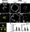
Figure 1.
Figure 1.
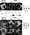
PLD1 recycles between PM and intracellular vesicles in an activity-facilitated manner on PMA stimulation. COS-7 cells were transiently transfected with an HA-tagged PLD1 expression plasmid. 36 h later, the cells were stimulated with PMA for varying periods of time (A, C, and D) and then fixed and immunostained using an anti-HA mAb. Images were captured using a confocal microscope (TCS SP2; Leica) using a green fluorophore-labeled secondary antibody. Colocalization with EEA1, GM130, and TfR (B) was performed using far-red as the second fluorophore. On stimulation by PMA, PLD1 translocates to the PM; shown is a 2-h time point (C). C′ and D′ show additional images of cells in which translocation or return was not complete at the respective time points. E depicts a tabulation of percent localization for 300 cells at each time point, including SDs that were determined by conducting the experiment three times. Vesicles (V), plasma membrane (PM), and intermediate cells (PM/V) were scored as indicated by the labels in the bottom right corner of each image.
Figure 2.
Figure 2.
Summary of the PLD1 constructs used in this paper. The constructs were NH2-terminally HA- or EGFP-tagged as indicated. See Results for details.
Figure 3.
Figure 3.
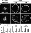
The NH2 terminus contains targeting signals for the endosomes and Golgi and is required for internalization, whereas the PI4,5P2-interacting motif mediates recruitment to the PM. (A and C) COS-7 cells were transiently transfected with deletion or mutated PLD1 constructs as shown in Fig. 2, and were processed as described in Fig. 1. In C, two examples are shown for each time point. (B) Characterization of the PLD1 PI4,5P2-binding motif. Wild-type (WT) and R691G,R695G (RG) mutant PLD1 proteins purified from baculovirus-infected sf9 cells were mixed with different amounts of liposomes containing PC, PE, and 5% PIP or 5% PI4,5P2. The vesicles were sedimented by centrifugation and the pellets were analyzed by Western blotting for cosedimentation of PLD1.
Figure 4.
Figure 4.
The PX domain is required for PLD1 internalization potentially through phosphoinositide interactions, but is not required for localization to at least some types of endosomes. COS-7 cells were transiently transfected with deletion or mutated PLD1 constructs as shown in Fig. 2, and were processed as described in Fig. 1. Two examples are shown for each time point to indicate how cells were scored.
Figure 5.
Figure 5.
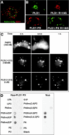
The PX domain mediates endosomal association independent of PI3 kinase products or lipid interactions, but may facilitate reentry through interaction with PI5P. (A and B) COS-7 cells were transiently transfected with wild-type or mutated full-length PLD1 or isolated PX domain constructs as shown in Fig. 2, and were processed as described in Fig. 1. (A) The isolated PLD1 PX domain localizes to cytosolic vesicles, but does not colocalize with EEA1 endosomes (or GM130, not depicted). Not depicted are similar results that were observed for the isolated domain and for the full NH2 terminus (1–212, which contains an extra 70 amino acids, as shown in Fig. 2). (C) COS-7 cells transiently transfected with p40 or PLD1 EGFP-PX domain constructs were followed over time after the addition of varying concentrations of wortmannin. (D) Purified bacterial Nus protein or Nus–PLD1 PX domain fusion protein was used to probe a set of phospholipids spotted on nitrocellulose strips, after which the proteins were detected using an anti-Nus mAb.
Figure 6.
Figure 6.
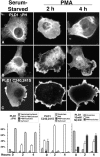
The PLD1 PH domain targets endosomes in quiescent cells and mediates internalization from the PM through controlling entry into lipid rafts. COS-7 cells were transiently transfected with deletion or mutated PLD1 constructs as shown in Fig. 2, and were processed as described in Fig. 1. (A and B) Two examples are shown for the deletion construct at each time point. Where indicated (rightmost quantification panel), methyl-β-cyclodextrin was added to the culture medium for 45 min and then thoroughly washed away before PMA stimulation.
Figure 7.
Figure 7.
The isolated PLD1 PH domain associates only weakly with internal and plasma membranes, both in resting cells and stimulated cells. COS-7 cells were transfected with plasmids expressing the PH domain flanked by leader and trailing sequences.
Figure 8.
Figure 8.
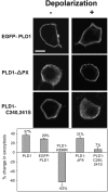
PLD1 localization affects regulated exocytosis in PC12 cells. PC12 cells were cotransfected with plasmids encoding hGH and tagged PLD1 alleles. 48 h after transfection, cells were incubated for 10 min in calcium-free Locke's solution (nondepolarized) or in 59 mM K+ solution (depolarized). (Top) Cells were immunostained to visualize the PLD1 protein by confocal microscopy. Bar, 5 μm. (Bottom) Assay for regulated exocytosis. The total GH content in control and PLD1-expressing cells was in the range of 5 ng/well. The net K+-evoked secretory response was obtained by subtracting the basal hGH release from the hGH release evoked by 59 mM K+. In these experiments, basal release ranged from 5.0 to 5.7%. The net K+-evoked GH release was 25.7% ± 0.4 from empty plasmid-transfected control cells, and, depending on the allele, ranged from 12.8 to 32.3% in PLD1-expressing cells. Finally, the net secretion under experimental conditions was divided by the net secretion in control cultures to estimate the percent change in regulated exocytosis. The experiments were performed twice in triplicate, and SEM is presented.
Figure 9.
Figure 9.
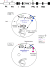
A model for regulated cycling of PLD1. See Discussion for details. In brief, in quiescent COS-7 cells, PLD1 localizes to a complex set of perinuclear and cytosolic vesicles (top schema, heavy black circle). This is mediated by a non-PI interaction by the PX domain, the palmitoylated PH domain, and weak interactions with PI4,5P2 by the central basic amino acid–rich motif, although the different domains most likely preferentially target distinct subpopulations of vesicles. In PC12 cells, a combination of PX domain interactions and interactions with PI4,5P2 suffice to recruit PLD1 to the PM (bottom schema, heavy black circle). With higher levels of stimulation, such as exposure to PMA for COS-7 cells, the PI4,5P2 interaction alone suffices to promote PM association. However, to reenter the cell with normal kinetics, PLD1 needs to enter into lipid rafts, and for this, the PH domain to be palmitoylated. Once in rafts, the PX domain association with the PM ceases; but a new PI-dependent interaction takes place, potentially through the binding of PX to PI5P, which facilitates translocation of PLD1 to vesicles internalizing through endocytosis. In PC12 cells, secretagogue-evoked stimulation (depolarization) recruits PLD1 into sites of active exocytosis, which are found in rafts and are marked by increased levels of PI4,5P2. Palmitoylation of the PH domain is similarly required for this recruitment, but once it happens, the PX domain interaction is no longer required for membrane association or for PLD1 functional promotion of exocytosis. How PLD1 returns to its original location remains to be determined.

Similar articles

Cited by

References

    1. Brown, F.D., N. Thompson, K.M. Saqib, J.M. Clark, D. Powner, N.T. Thompson, R. Solari, and M.J. Wakelam. 1998. Phospholipase D1 localises to secretory granules and lysosomes and is plasma-membrane translocated on cellular stimulation. Curr. Biol. 8:835–838. - PubMed
    1. Ceresa, B.P., A.W. Kao, S.R. Santeler, and J.E. Pessin. 1998. Inhibition of clathrin-mediated endocytosis selectively attenuates specific insulin receptor signal transduction pathways. Mol. Cell. Biol. 18:3862–3870. - PMC - PubMed
    1. Chamberlain, L.H., R.D. Burgoyne, and G.W. Gould. 2001. SNARE proteins are highly enriched in lipid rafts in PC12 cells: implications for the spatial control of exocytosis. Proc. Natl. Acad. Sci. USA. 98:5619–5624. - PMC - PubMed
    1. Chen, Y.G., A. Siddhanta, C.D. Austin, S.M. Hammond, T.C. Sung, M.A. Frohman, A.J. Morris, and D. Shields. 1997. Phospholipase D stimulates release of nascent secretory vesicles from the trans-Golgi network. J. Cell Biol. 138:495–504. - PMC - PubMed
    1. Choi, W.S., Y.M. Kim, C. Combs, M.A. Frohman, and M.A. Beaven. 2002. Phospholipase D1 and 2 regulate different phases of exocytosis in mast cells. J. Immunol. 168:5682–5689. - PubMed

Publication types

MeSH terms