. 2003 Jul;112(2):209-21.
doi: 10.1172/JCI18242.

Neph1 and nephrin interaction in the slit diaphragm is an important determinant of glomerular permeability

Affiliations

Neph1 and nephrin interaction in the slit diaphragm is an important determinant of glomerular permeability

Gang Liu et al. J Clin Invest. 2003 Jul.

Abstract

Neph1-deficient mice develop nephrotic syndrome at birth, indicating the importance of this protein in the development of a normal glomerular filtration barrier. While the precise subcellular localization of Neph1 remains unknown, its relationship with other components of the glomerular filtration barrier is of great interest in this field. In this paper, we localize the expression of Neph1 to the glomerular slit diaphragm by immunogold electron microscopy in rodents and describe its direct interaction with two other components of the slit diaphragm, nephrin and ZO-1. Both native and recombinant Neph1 associate with each other as dimers and multimers and interact with nephrin via their extracellular segments. Disruption of the Neph1-nephrin interaction in vivo by injecting combinations of individual subnephritogenic doses of anti-Neph1 and anti-nephrin results in complement- and leukocyte-independent proteinuria with preserved foot processes. This disruption modestly reduces Neph1 and nephrin protein expression in podocytes and dramatically reduces ZO-1 protein expression via the interaction of ZO-1 PDZ domains with the cytoplasmic tail of Neph1, independent of changes in mRNA expression of all three genes. The interaction between nephrin and Neph1 is specific and not shared by either protein with P-cadherin, another integral slit diaphragm protein. The interaction between nephrin and Neph1 therefore appears to be an important determinant of glomerular permeability.


Figures
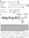
Figure 1
Figure 1
Cloning and characterization of mouse Neph1. (a) The missing base pair after 1,204 in the original published sequence, and the assembly of full-length mouse cDNA by 3′ RACE. (b) A representation of mouse Neph1 protein structure, including five Ig-like domains in the extracellular segment. (c) The full-length Neph1 protein prior to signal peptide (underlined) cleavage, along with potential phosphorylation sites.
Figure 2
Figure 2
Characterization of native and recombinant mouse Neph1 by Western blot. (a and b) A distinct band is seen at approximately 110 kDa in rat and mouse glomerular extracts, respectively, under reducing conditions. (c) There is a band at approximately 220 kDa (arrow) and a higher band in mouse glomerular extracts under nonreducing conditions, suggesting dimer and multimer formation. (d) Migration of anti-Neph1 immunoprecipitates (Immunoppt.) from mouse glomerular extracts at approximately 90 kDa following deglycosylation with PNGase F. (e and f) Composite Western blots of the extracellular segment (clone 3,261) and intracellular segment (clone 3,285) expressed in E. coli by IPTG induction, run under nonreducing (e) and reducing (f) conditions, also demonstrating dimer formation under nonreducing conditions. IPTG, isopropyl-β-D-thiogalactoside.
Figure 3
Figure 3
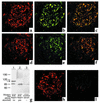
Localization of Neph1 in adult and developing rodent glomeruli. (ac) Immunofluorescence images of adult rat glomeruli (3-μm sections; magnification, ×400) stained for Neph1 (a), CD2 AP (b), and merged images (c), localizing Neph1 to podocytes. (df) Immunofluorescence images of day 15 developing mouse glomeruli stained for Neph1 (d), CD2 AP (e), and merged images (f), localizing Neph1 to developing podocytes. (g) Western blot of rat glomerular extracts under reducing conditions with anti-Neph1 antibody prior to (lane 1) and following (lane 2) affinity absorption with a Neph1 peptide column, showing near-complete loss of immunoreactivity. (h and i) Immunofluorescence staining of 3-μm rat kidney sections prior to (h) and following (i) affinity absorption of anti-Neph1 with the Neph1 peptide affinity column, showing loss of immunoreactivity.
Figure 4
Figure 4
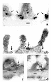
Immunogold localization of Neph1 to the slit diaphragm in adult rat glomeruli. (a) A control electron micrograph (magnification, ×50,000) shows staining with goat anti–CD2 AP and rabbit anti-goat IgG coupled with 15-nm gold particles localizing CD2 AP predominantly to the cytoplasmic aspect of the slit diaphragm (arrows). (b) Staining with rabbit anti-Neph1 (magnification, ×27,000) and goat anti-rabbit IgG coupled with 10-nm gold particles, localizing Neph1 mostly to the slit diaphragm (arrows), though some labeling of the foot process is also present (see text). (c and d) Staining of kidney sections with goat anti-rabbit IgG coupled with 10-nm gold particles (magnification, ×30,000) from rats injected with either rabbit anti-Neph1 (c) or rabbit anti-nephrin (d). Both anti-Neph1 (c) and anti-nephrin (d) bind specifically to the slit diaphragm (arrows), as shown by the cluster of gold particles. FP, foot process; GBM, glomerular basement membrane; E, endothelial cell; PB, podocyte body.
Figure 5
Figure 5
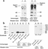
Composite Western blots showing the association of native (a) and recombinant (b) mouse Neph1 and nephrin. (a) Native Neph1 from mouse glomerular extracts coimmunoprecipitates with anti-nephrin (lane 2), and nephrin with anti-Neph1 (lane 3) under nonreducing conditions. All lanes, including control immunoprecipitations (lane 1 and lane 4), show the prominent band of nonreduced rabbit IgG at 150–160 kDa and traces of heavy chain (50 kDa) and light chain (25 kDa). (b) Lane 1 shows the absence of FLAG-reactive proteins in native mouse glomerular extracts. In lanes 2–11, native glomerular extracts were combined with FLAG-tagged recombinant extracellular Neph1 and also supplemented with an excess of recombinant extracellular Neph1 digested with enterokinase to remove the FLAG tag (labeled “unFLAG”) in lane 5 (unFLAG/FLAG ratio, 5:1) and lane 7 (unFLAG/FLAG ratio, 10:1). Coimmunoprecipitation of recombinant Neph1 by anti-Neph1 (lane 4) and anti-nephrin (lane 6); coimmunoprecipitation of nephrin (lane 9) and native Neph1 (lane 11) by anti-FLAG; and competition between FLAG-tagged and non–FLAG-tagged protein (lane 5 and lane 7) are demonstrated (see text for details). GE, glomerular extract.
Figure 6
Figure 6
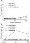
The induction of heterologous-phase proteinuria in rats by combinations of anti-Neph1 and anti-nephrin in individual subnephritogenic doses. (a) A dose-response plot of each individual Ab or preimmune serum to assess the optimum subnephritogenic dose. Some increase in permeability is noted at 500 μl for both study Ab’s compared with preimmune serum (P < 0.05). (b) Induction of heterologous-phase proteinuria in rats by a combination of 150 μl each of anti-Neph1 and anti-nephrin, whereas 300 μl of each individual antibody and preimmune serum do not cause proteinuria.
Figure 7
Figure 7
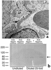
(a) Electron micrograph of rat kidney taken 24 hours after injection of the anti-Neph1 and anti-nephrin combination, showing that heterologous-phase proteinuria occurs in the absence of foot process effacement (arrow). Magnification, ×5,000. (b) GelCode Blue–stained reducing SDS PAGE for urinary protein from rats injected with either anti-Neph1 or anti-nephrin (undiluted), or a combination of both (diluted 25-fold). Urine from only two of four rats injected with each individual antibody is shown. The rats in the combination group showed a prominent band at approximately 70 kDa (arrow) that was confirmed to be rat albumin by Western blot (see text).
Figure 8
Figure 8
Composite Western blot analysis of (a) the expression of recombinant FLAG-tagged PDZ domains of mouse ZO-1 and (b) the association of Neph1 with FLAG-tagged ZO-1 PDZ domains by coimmunoprecipitation. (b) Lane 1 shows the absence of anti-FLAG reactivity in native mouse glomerular protein extracts. All other experiments shown were conducted with a combination of native mouse glomerular proteins and recombinant ZO-1 PDZ domains. Lane 4 shows the coimmunoprecipitation of ZO-1 PDZ domains by anti-Neph1; lane 8 shows the coimmunoprecipitation of Neph1 by anti-FLAG.
Figure 9
Figure 9
Composite image showing protein (a and b) and mRNA (cg) expression in rat glomeruli of Neph1, nephrin, and ZO-1 24 hours after the injection into rats of anti-Neph1 or anti-nephrin alone, or a combination of both. (a) Composite Western blot of glomerular extracts for each protein in the three experimental subgroups or controls. (b) Densitometric analysis of bands shown in a (mean ± SE). Modest reduction of Neph1 and nephrin and dramatic reduction of ZO-1 protein expression were noted in the combination group. Modest reduction in ZO-1 expression was also noted in the anti-Neph1 group. (cf) Real-time PCR tracings (n = 9 per group) for the three genes or 18S controls in the anti-nephrin (red), anti-Neph1 (green), and combination (blue) groups. (g) ΔCt (mean ± SE) values of each gene in the three groups. No significant difference in mRNA expression was noted for the individual genes between the combination and single-Ab groups (P > 0.05).

Similar articles

Cited by

References

    1. Quaggin SE, et al. The basic-helix-loop-helix protein pod1 is critically important for kidney and lung organogenesis. Development. 1999;126:5771–5783. - PubMed
    1. Rohr C, et al. The LIM-homeodomain transcription factor Lmx1b plays a crucial role in podocytes. J. Clin. Invest. 2002;109:1073–1082. doi:10.1172/JCI200213961. - PMC - PubMed
    1. Miner JH, et al. Transcriptional induction of slit diaphragm genes by Lmx1b is required in podocyte differentiation. J. Clin. Invest. 2002;109:1065–1072. doi:10.1172/JCI200213954. - PMC - PubMed
    1. Schnabel E, Anderson JM, Farquhar MG. The tight junction protein ZO-1 is concentrated along slit diaphragms of the glomerular epithelium. J. Cell. Biol. 1990;111:1255–1263. - PMC - PubMed
    1. Orikasa M, Matsui K, Oite T, Shimizu F. Massive proteinuria induced in rats by a single intravenous injection of a monoclonal antibody. J. Immunol. 1988;141:807–814. - PubMed

Publication types

MeSH terms