. 2003 Jul 15;17(14):1755-67.
doi: 10.1101/gad.1098303.

Mrc1 is a replication fork component whose phosphorylation in response to DNA replication stress activates Rad53

Affiliations

Mrc1 is a replication fork component whose phosphorylation in response to DNA replication stress activates Rad53

Alexander J Osborn et al. Genes Dev. .

Abstract

When DNA replication is stalled, a signal transduction pathway is activated that promotes the stability of stalled forks and resumption of DNA synthesis. In budding yeast, this pathway includes the kinases Mec1 and Rad53. Here we report that the Mediator protein Mrc1, which is required for normal DNA replication and for activation of Rad53, is present at replication forks. Mrc1 initially binds early-replicating sequences and moves along chromatin with the replication fork. Blocking initiation of DNA replication blocks Mrc1 loading onto origins, providing an explanation for why so many mutants in DNA replication show checkpoint defects. In the presence of replication blocks, we find that Mec1 is recruited to regions of stalled replication, where it encounters and presumably phosphorylates Mrc1. Mutation of the canonical Mec1 phosphorylation sites on Mrc1 prevents Mrc1 phosphorylation and blocks Rad53 activation, but does not alter Mrc1's role in DNA replication. Our results suggest a model whereby in response to DNA replication interference, the Mec1 kinase is recruited to sites of replication blocks and phosphorylates a component of the DNA replication complex, Mrc1, thereby setting up a solid-state Rad53 activation platform to initiate the checkpoint response.


Figures

Figure 1.
Figure 1.
Compromised viability of mrc1AQ mutants in the face of replication stress. (A) mrc1Δ rad9Δ cells carrying wild-type MRC1 (Y1133; •), mrc1AQ (Y2296; ▴), or vector alone (Y1131; ○) were arrested in G1 with α-factor, released into YPD containing 200 mM HU at 30°C, and assessed for viability over time. (B) mrc1Δ rad9Δ cells carrying either MRC1 (Y1133), mrc1AQ (Y2296), or vector alone (Y1131) were struck onto YPD plates containing the indicated amounts of HU and grown at 30°C for 3 d. (C) mrc1Δ rad9Δ cells carrying RAD9 (Y1132; ▪), MRC1 (Y1133; •), mrc1AQ (Y2296; ▴), or vector alone (Y1131; ○) were exposed to increasing doses of UV radiation and assessed for viability.
Figure 2.
Figure 2.
mrc1AQ fails to activate Rad53 in response to replication stress. (A) mrc1Δ rad9Δ cells carrying MRC1 (Y1133; •), mrc1AQ (Y2296; ▴), or vector alone (Y1131; ○) were arrested in G1 with α-factor and released into YPD containing 200 mM HU at 30°C. Samples were removed at the indicated times and processed for tubulin staining to assess spindle elongation. (B) Representative fluorescence microscopy images from the experiment described in A. Tubulin staining (green) and DAPI staining (blue) represent the mitotic spindle and chromatin masses, respectively. (C) mrc1Δ rad9Δ cells carrying either MRC1 (Y1133) or mrc1AQ (Y2296) were arrested in G1 with α-factor and released into YPD containing 200 mM HU at 30°C. Samples were removed at the indicated times, and both Rad53 and Mrc1 phosphorylation were assessed by Western blot. (D) mrc1Δ rad9Δ cells carrying either MRC1 (Y1133) or mrc1AQ (Y2296) were arrested in G1 with α-factor and released into YPD containing 0.1% MMS at 30°C. Samples were removed at the indicated times, and both Rad53 and Mrc1 phosphorylation were assessed by Western blot.
Figure 3.
Figure 3.
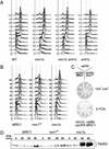
mrc1AQ is competent for DNA replication. (A) The defect of Mrc1 during S phase is not suppressed by deleting SML1. Wild-type (WT; Y2305), mrc1Δ (Y2306), mrc1Δ sml1Δ (Y2307), and sml1Δ (Y2308) cells were arrested in G1 with α-factor and released into YPD at 30°C. Samples were removed at the indicated times and subjected to FACS analysis. The first and second broken vertical lines in each profile represent the 1n and 2n peaks of DNA content, respectively. (B) mrc1AQ mutants undergo a normal S phase. mrc1Δ (Y1127) cells carrying MRC1 (pMRC1), mrc1AQ (pAO138), or vector alone (pRS416) were arrested in G1 with α-factor and released into normal YPD at 30°C. Samples were removed at the indicated times and subjected to FACS analysis. The broken lines represent the peaks of DNA content as in A. (C) mrc1AQ mutants can suppress the lethality of mrc1Δ rad9Δ mutants. mrc1Δ rad9Δ cells carrying MRC1 on a URA3 vector (Y2297) were transformed with MRC1 (pAO122), mrc1AQ (pAO139), or empty LEU2 (pRS416) vectors. The strains containing both URA3 and LEU2 vectors were struck onto 5-FOA plates to select for cells that lost the MRC1-containing URA3 vector. Growth of the strains on plates selecting for both plasmids is shown as a control for the effect of the LEU2 plasmid on viability. (D) mrc1AQ mutants do not induce DNA damage during S phase. The strains used in B were arrested in G1 with α-factor and released into YPD at 30°C. Samples were removed at the indicated times, and Rad53 activation was assessed by mobility shift on a Western blot.
Figure 4.
Figure 4.
Mrc1 associates with replication forks. (A) Wild-type cells were arrested in G1 with α-factor and released into YPD at 30°C. Samples were removed at the indicated times, and total cellular protein was separated into soluble (s) and insoluble (c) fractions. The presence of Mrc1 in the two fractions was assessed by Western blot. (Right) Cells were also processed for FACS analysis. (B) Cells containing a MYC13-tagged genomic copy of MRC1 (Y1134) were arrested in G1 with α-factor and released into YPD at 19°C. Samples were taken at the indicated times and subjected to formaldehyde cross-linking. The DNA that coimmunoprecipitated with Mrc1–MYC13 was analyzed for the presence of the ARS305 locus, DNA 8 kb and 17 kb centromeric to this origin (ARS 305 + 8 kb and ARS 305 + 17 kb, respectively), and the ARS603 locus by PCR. (C) FACS profiles for cultures at 30°C and 19°C, which were used in A and B, respectively. (D) The PCR products in A were quantified and compared with those obtained from whole-cell DNA to determine the fraction of total DNA brought down in the IP. The percentage bound is represented graphically for ARS305 (•), ARS305 + 8 kb (□), ARS305 + 17 kb (▵), and ARS603 (⋄).
Figure 5.
Figure 5.
mrc1AQ moves with replication forks. (A) Cells containing a MYC13-tagged genomic copy of mrc1AQ (Y2298) were arrested in G1 with α-factor and released into YPD at 19°C. Samples were taken at the indicated times and subjected to formaldehyde cross-linking. DNA that coimmunoprecipitated with mrc1AQ–MYC13 was analyzed for the presence of the ARS305 locus, DNA 8 kb and 17 kb centromeric to this origin (ARS 305 + 8 kb and ARS 305 + 17 kb, respectively), and the ARS603 locus by PCR. (B) FACS profile for the culture. (C) The PCR products in A were quantified and compared with those obtained from whole-cell DNA to determine the fraction of total DNA brought down in the IP. The percentage bound is represented graphically for ARS305 (•), ARS305 + 8 kb (□), ARS305 + 17 kb (▵), and ARS603 (⋄).
Figure 6.
Figure 6.
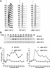
Replication initiation is required for Mrc1's chromatin association. dbf4-1 cells containing a genomic MRC1–MYC13 construct (Y2299) were arrested in G1 with α-factor and released into YPD at 20°C. Wild-type and dbf4-1 cells with a genomic MRC1–MYC13 construct (Y1134 and Y2299, respectively) were arrested in G1 with α-factor and released into YPD with 100 mM HU at 34°C. Samples were taken at the indicated times and subjected to FACS analysis (A) and to formaldehyde cross-linking (B). DNA coimmunoprecipitating with Mrc1–MYC13 was analyzed for the presence of ARS305 DNA by PCR. (C) The PCR products in B were quantified and compared with those obtained from input DNA to determine the fraction of total DNA brought down in the IP. The fold increase in signal over time from the 0-min and 24-min average baseline is represented for dbf4-1 cells at 34°C(•), wild-type (WT) cells at 34°C(•), and dbf4-1 cells at 20°C(○).
Figure 7.
Figure 7.
Mec1 is recruited to sites of DNA replication interface. Wild-type cells with a genomic MEC1–MYC18 construct (YLL447.32/1A) were arrested in G1 and released at 19°C into YPD or YPD containing 200 mM HU. Aliquots were removed at the indicated times and subjected to formaldehyde cross-linking (A) and to FACS analysis (B). DNA that associated with Mec1–MYC18 was analyzed for the presence of origin-associated (ARS 305) and nonorigin (ARS 305 + 8 kb, URA3) sequences by PCR. (C) A model for the role of Mec1 and Mrc1 in the activation of Rad53 in response to DNA replication stress. DNA replication begins at origins of replication, which are constitutively bound by the Orc complex. Dbf4 activity promotes the replication complex and Mrc1 to assemble at the origin and begin the task of replication. Once a replication blocking lesion (×) is encountered or deoxyribonucleotides are depleted, the replication complex stalls and abnormal structures are generated. These abnormal structures contain stretches of Rpa-coated single-stranded DNA, which recruits Mec1/Ddc2 kinase to the site of stalled replication (Zou and Elledge 2003). Mec1/Ddc2 then phosphorylates Mrc1, which is uniquely located to transduce the stress signal. This phosphorylation on Mrc1 allows it to mediate the activation of Rad53, which may subsequently further phosphorylate Mrc1.

Similar articles

Cited by

References

    1. Alcasabas A.A., Osborn, A.J., Bachant, J., Hu, F., Werler, P.J., Bousset, K., Furuya, K., Diffley, J.F., Carr, A.M., and Elledge, S.J. 2001. Mrc1 transduces signals of DNA replication stress to activate Rad53. Nat. Cell Biol. 3: 958–965. - PubMed
    1. Allen J.B., Zhou, Z., Siede, W., Friedberg, E.C., and Elledge, S.J. 1994. The SAD1/RAD53 protein kinase controls multiple checkpoints and DNA damage-induced transcription in yeast. Genes & Dev. 8: 2401–2415. - PubMed
    1. Aparicio O.M., Weinstein, D.M., and Bell, S.P. 1997. Components and dynamics of DNA replication complexes in S. cerevisiae: Redistribution of MCM proteins and Cdc45p during S phase. Cell 91: 59–69. - PubMed
    1. Araki H., Leem, S.H., Phongdara, A., and Sugino, A. 1995. Dpb11, which interacts with DNA polymerase II(ε) in Saccharomyces cerevisiae, has a dual role in S-phase progression and at a cell cycle checkpoint. Proc. Natl. Acad. Sci. 92: 11791–11795. - PMC - PubMed
    1. de la Torre-Ruiz M.A., Green, C.M., and Lowndes, N.F. 1998. RAD9 and RAD24 define two additive, interacting branches of the DNA damage checkpoint pathway in budding yeast normally required for Rad53 modification and activation. EMBO J. 17: 2687–2698. - PMC - PubMed

Publication types

MeSH terms

Substances

LinkOut - more resources