Canonical Wnt signals are essential for homeostasis of the intestinal epithelium
- PMID: 12865297
- PMCID: PMC196179
- DOI: 10.1101/gad.267103
Canonical Wnt signals are essential for homeostasis of the intestinal epithelium
Abstract
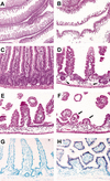
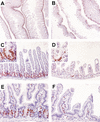
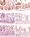
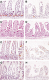
To assess the critical role of Wnt signals in intestinal crypts, we generated transgenic mice ectopically expressing Dickkopf1 (Dkk1), a secreted Wnt inhibitor. We find that epithelial proliferation is greatly reduced coincidentally with the loss of crypts. Although enterocyte differentiation appears unaffected, secretory cell lineages are largely absent. Disrupted intestinal homeostasis is reflected by an absence of nuclear beta-catenin, inhibition of c-myc expression, and subsequent up-regulation of p21CIP1/WAF1. Thus, our data are the first to establish a direct requirement for Wnt ligands in driving proliferation in the intestinal epithelium, and also define an unexpected role for Wnts in controlling secretory cell differentiation.
Figures


Similar articles
-
Kremen proteins are Dickkopf receptors that regulate Wnt/beta-catenin signalling.Nature. 2002 Jun 6;417(6889):664-7. doi: 10.1038/nature756. Epub 2002 May 26. Nature. 2002. PMID: 12050670
-
When Wnts antagonize Wnts.J Cell Biol. 2003 Sep 1;162(5):753-5. doi: 10.1083/jcb.200307181. J Cell Biol. 2003. PMID: 12952929 Free PMC article.
-
Phases of canonical Wnt signaling during the development of mouse intestinal epithelium.Gastroenterology. 2007 Aug;133(2):529-38. doi: 10.1053/j.gastro.2007.04.072. Epub 2007 May 3. Gastroenterology. 2007. PMID: 17681174
-
Wnt signaling in the intestinal epithelium: from endoderm to cancer.Genes Dev. 2005 Apr 15;19(8):877-90. doi: 10.1101/gad.1295405. Genes Dev. 2005. PMID: 15833914 Review.
-
Regulation of beta-catenin signaling in the Wnt pathway.Biochem Biophys Res Commun. 2000 Feb 16;268(2):243-8. doi: 10.1006/bbrc.1999.1860. Biochem Biophys Res Commun. 2000. PMID: 10679188 Review.
Cited by
-
Cooperation of Wnt/β-catenin and Dll1-mediated Notch pathway in Lgr5-positive intestinal stem cells regulates the mucosal injury and repair in DSS-induced colitis mice model.Gastroenterol Rep (Oxf). 2024 Oct 23;12:goae090. doi: 10.1093/gastro/goae090. eCollection 2024. Gastroenterol Rep (Oxf). 2024. PMID: 39444950 Free PMC article.
-
BMP suppresses Wnt signaling via the Bcl11b-regulated NuRD complex to maintain intestinal stem cells.EMBO J. 2024 Oct 21. doi: 10.1038/s44318-024-00276-1. Online ahead of print. EMBO J. 2024. PMID: 39433900
-
Pathways regulating intestinal stem cells and potential therapeutic targets for radiation enteropathy.Mol Biomed. 2024 Oct 10;5(1):46. doi: 10.1186/s43556-024-00211-0. Mol Biomed. 2024. PMID: 39388072 Free PMC article. Review.
-
A living organoid biobank of patients with Crohn's disease reveals molecular subtypes for personalized therapeutics.Cell Rep Med. 2024 Oct 15;5(10):101748. doi: 10.1016/j.xcrm.2024.101748. Epub 2024 Sep 26. Cell Rep Med. 2024. PMID: 39332415 Free PMC article.
-
A Ctnnb1 enhancer transcriptionally regulates Wnt signaling dosage to balance homeostasis and tumorigenesis of intestinal epithelia.Elife. 2024 Sep 25;13:RP98238. doi: 10.7554/eLife.98238. Elife. 2024. PMID: 39320349 Free PMC article.
References
-
- Andl T., Reddy, S.T., Gaddapara, T., and Millar, S.E. 2002. WNT signals are required for the initiation of hair follicle development. Dev. Cell 2: 643–653. - PubMed
-
- Bafico A., Liu, G., Yaniv, A., Gazit, A., and Aaronson, S.A. 2001. Novel mechanism of Wnt signalling inhibition mediated by Dickkopf-1 interaction with LRP6/Arrow. Nat. Cell Biol. 3: 683–686. - PubMed
-
- Batlle E., Henderson, J.T., Beghtel, H., van den Born, M.M., Sancho, E., Huls, G., Meeldijk, J., Robertson, J., van de Wetering, M., Pawson, T., et al. 2002. β-Catenin and TCF mediate cell positioning in the intestinal epithelium by controlling the expression of EphB/ephrinB. Cell 111: 251–263. - PubMed
-
- Bienz M. and Clevers, H. 2000. Linking colorectal cancer to Wnt signaling. Cell 103: 311–320. - PubMed
-
- Booth C., Brady, G., and Potten, C.S. 2002. Crowd control in the crypt. Nat. Med. 8: 1360–1361. - PubMed
MeSH terms
Substances
LinkOut - more resources
Full Text Sources
Other Literature Sources
Molecular Biology Databases
