. 2003 Jul 1;22(13):3411-20.
doi: 10.1093/emboj/cdg315.

The histone deacetylase inhibitor valproic acid selectively induces proteasomal degradation of HDAC2

Affiliations

The histone deacetylase inhibitor valproic acid selectively induces proteasomal degradation of HDAC2

Oliver H Krämer et al. EMBO J. .

Abstract

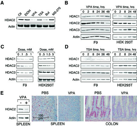
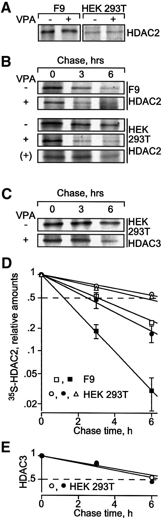
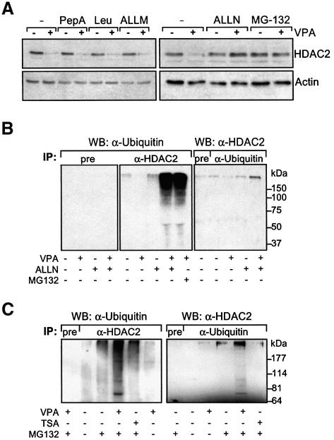
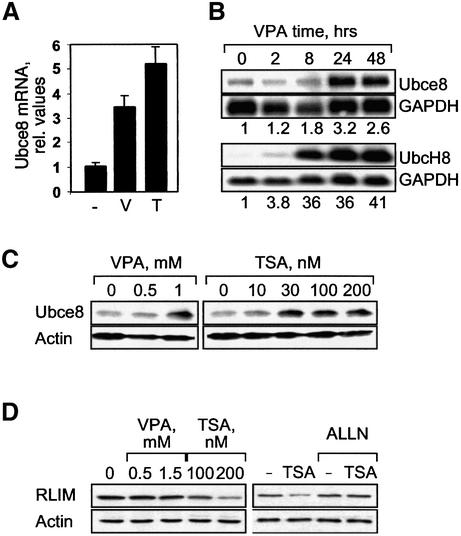
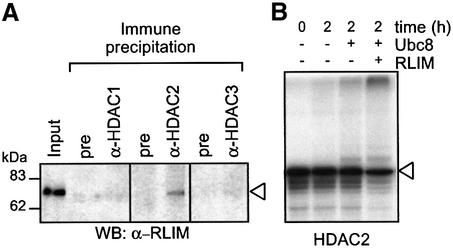
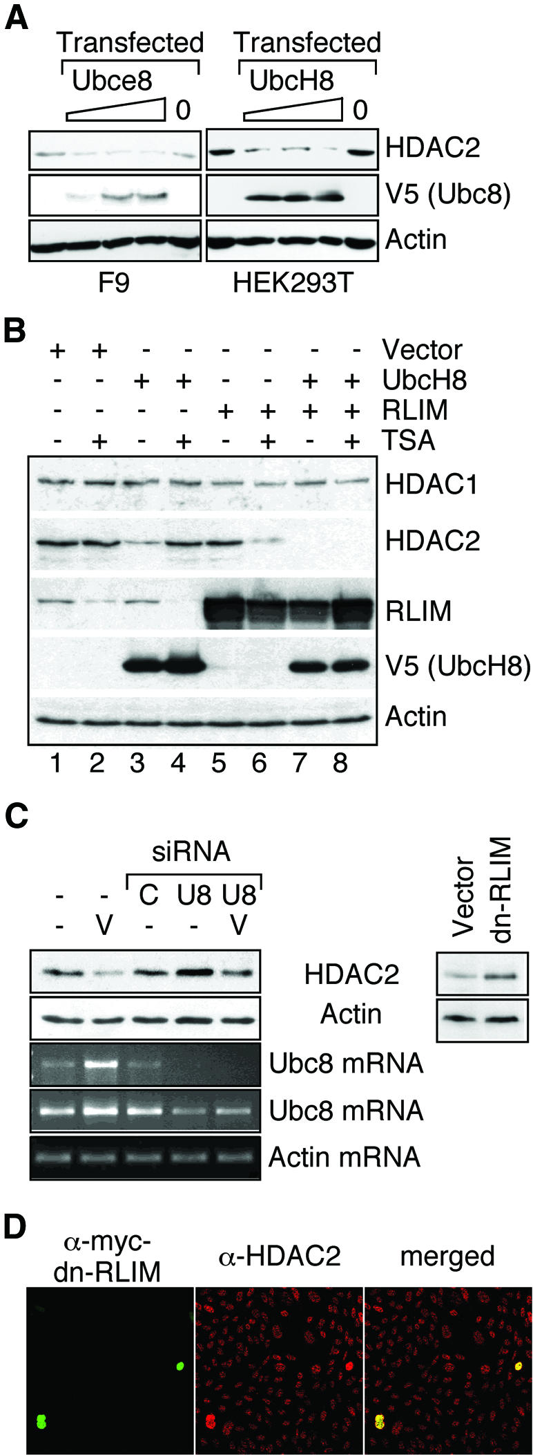
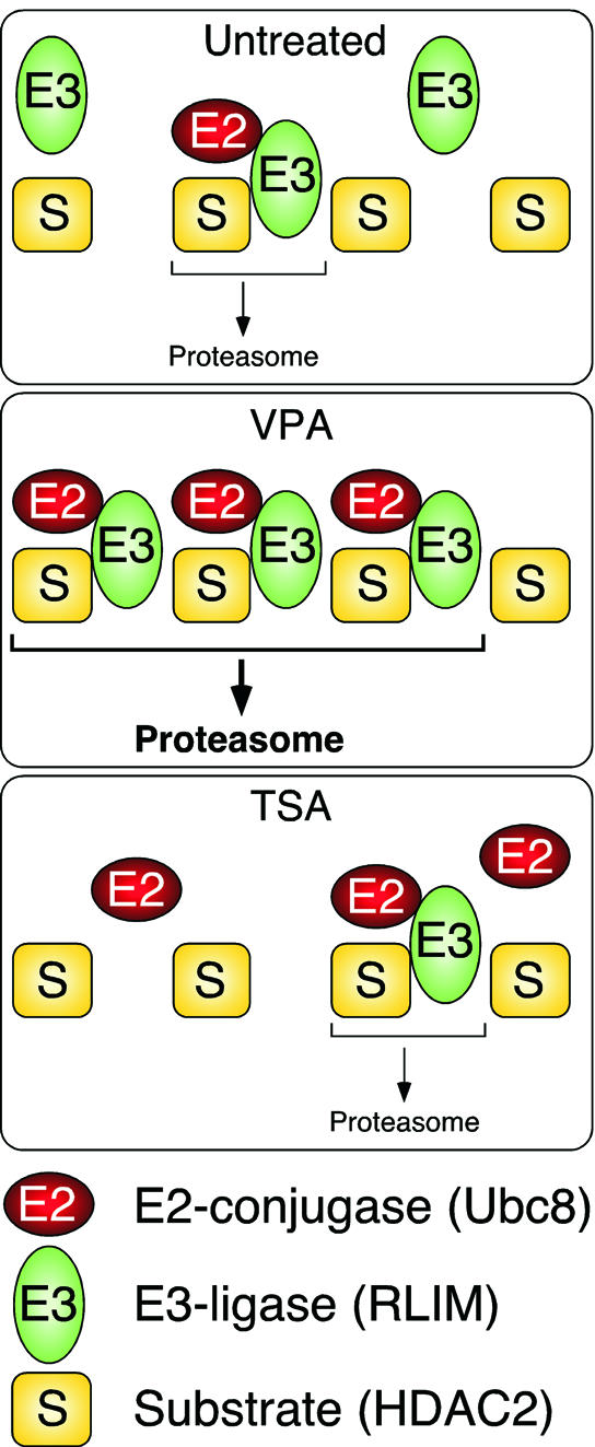
Histone-modifying enzymes play essential roles in physiological and aberrant gene regulation. Since histone deacetylases (HDACs) are promising targets of cancer therapy, it is important to understand the mechanisms of HDAC regulation. Selective modulators of HDAC isoenzymes could serve as efficient and well-tolerated drugs. We show that HDAC2 undergoes basal turnover by the ubiquitin-proteasome pathway. Valproic acid (VPA), in addition to selectively inhibiting the catalytic activity of class I HDACs, induces proteasomal degradation of HDAC2, in contrast to other inhibitors such as trichostatin A (TSA). Basal and VPA-induced HDAC2 turnover critically depend on the E2 ubiquitin conjugase Ubc8 and the E3 ubiquitin ligase RLIM. Ubc8 gene expression is induced by both VPA and TSA, whereas only TSA simultaneously reduces RLIM protein levels and therefore fails to induce HDAC2 degradation. Thus, poly-ubiquitination and proteasomal degradation provide an isoenzyme-selective mechanism for downregulation of HDAC2.


Figures

None
Fig. 1. VPA but not TSA leads to reduction of HDAC2 protein levels. (A) K562 human erythroleukemia cells were treated for 24 h as indicated with the HDAC inhibitors VPA (1.5 mM), TSA (100 nM), butyrate (1.5 mM) or MS-27–275 (5 µM). Amounts of HDAC2 protein were determined by western blot analysis of whole-cell extracts. Actin protein levels were determined to verify equal loading of samples. (B) F9 mouse teratocarcinoma or HEK293T human embryonic kidney carcinoma cells were exposed to 1 mM VPA for the indicated periods of time. Protein levels of HDAC2 as well as HDAC1, HDAC3 and actin were determined by western blot analysis. (C) The dose-dependent reduction of HDAC2 protein levels was determined in F9 or HEK293T cells after exposure to VPA for 30 or 24 h, respectively. (D) Time course analyses in F9 and HEK293T cells confirmed that TSA (100 nM) does not affect the amount of HDAC2 protein. (E) Reduction of HDAC2 protein levels after treatment of mice with VPA was tested by western blot analysis of tissue extracts and immunohistochemistry. Similar results were obtained in at least two sets of independent experiments.
None
Fig. 2. VPA induces degradation of HDAC2 protein. Synthesis and degradation rates of HDAC2 were determined by [35S]methionine labeling followed by chase analyses. [35S]HDAC2 was detected by HDAC2-specific immunoprecipitation, SDS–PAGE and autoradiography. (A) Pulse labeling for 1 h was performed in F9 and HEK293T cells, which were precultured for 24 h in the absence or presence of 1 mM VPA (F9) or 1.5 mM VPA (HEK293T). (B) For pulse–chase analysis, cells were cultured for 24 h either in the absence or presence of 1 mM VPA (F9) or 1.5 mM VPA (HEK293T) and labeled with [35S]methionine for an additional hour without or with VPA. After removal of [35S]methionine and addition of non-labeled methionine, the elimination of radiolabeled HDAC2 was followed over a period of 6 h. A pulse–chase analysis was also performed in non-pretreated HEK293T cells to which VPA was added only at the time when the chase was started [row marked (+)]; VPA at time of chase, triangles. Efficiency of immunoprecipitations was confirmed by proving depletion of HDAC2 from the precipitation supernatants. Comparable loading between lanes was controlled by Coomassie staining of gels for the amounts of antibodies. Similar results were obtained in at least a second independent experiment. (C) HDAC3 was also precipitated from HEK293T cell extracts. (D and E) Phosphoimager analyses of the experiments in (B) and (C) were quantitatively evaluated by subtraction of local background. The graphs show averages of two or three experiments (control, open symbols; VPA, filled symbols; VPA at time of chase, triangles) and were used for half-life determination. Standard deviations are shown if bars exceed symbol sizes.
None
Fig. 3. VPA induces polyubiquitination and proteasome-dependent degradation of HDAC2. (A) HEK293T cells were treated for 24 h with VPA or left untreated. The protease inhibitors pepstatin A (20 µM), leupeptin (10 µM), or ALLM (20 µM), or the proteasome inhibitors ALLN (5 µM), or MG 132 (10 µM), were added at the same time as VPA. HDAC2 protein levels were determined by western blot analysis. (B) Precipitation of poly-ubiquitinated proteins with anti-HDAC2 antibody as well as high molecular weight anti-HDAC2-reactive proteins with anti-ubiquitin antibody was tested by immunoprecipitation analysis from extracts of HEK293T cells that had been left untreated or were treated with 1.5 mM VPA for 36 h. The proteasome inhibitors ALLN (25 µM) or MG 132 (20 µM) were added 4 h before cell harvest. The presence of ubiquitinated proteins in anti-HDAC2 immunoprecipitates is shown in the middle panel. The left panel shows the corresponding control experiment with preimmune serum instead of the anti-HDAC2 antibody. The right panel shows precipitation with an anti-ubiquitin or non-immune (pre) serum and detection of poly-ubiquitinated HDAC2 protein by western blot analysis. (C) Ubiquitinated HDAC2 was also precipitated from F9 cells that had been treated with 1 mM VPA in experiments comparable to those in (B). One representative example of two independent experiments is shown.
None
Fig. 4. The HDAC inhibitors VPA and TSA induce expression of the E2 ubiquitin-conjugating enzyme Ubc8, whereas the E3 ligase RLIM is differentially regulated by TSA and VPA. (A) F9 cells were treated for 17 h with 1 mM VPA (V) or 100 nM TSA (T) and expression levels of Ubce8 mRNA were determined by real-time RT–PCR assuming a 1.5-fold amplification per cycle. The amplicon was part of the coding region of the Ubce8 mRNA. Results were normalized for GAPDH expression. Average values ± range of two independent experiments, each with triplicate determinations. (B) Northern blot analysis of 5 µg of poly(A)+ mRNA from F9 or HEK293T cells treated for the indicated times with 1 mM VPA (F9 cells) or 1.5 mM VPA (HEK293T cells) was performed to confirm RT–PCR results. Probes were derived from the 3′-UTR of murine Ubce8 or the 5′ coding region of human UbcH8. Phosphoimager analysis was performed for quantitative evaluation and relative values for Ubc8 mRNA abundance normalized for GAPDH are presented below the panels. One of two experiments with similar results is shown. (C) Ubce8 protein levels were determined by western blot analysis in total extracts of F9 cells that had been treated for 24 h as indicated. (D) HEK293T cells were treated for 24 h with VPA, TSA or ALLN (2.5 µM) as indicated. Abundance of the E3 ubiquitin ligase RLIM was determined by western blot analysis. Actin protein levels were determined to verify equal loading.
None
Fig. 5. RLIM interacts with HDAC2 and induces poly-ubiquitination in vitro. (A) Interaction of HDACs 1, 2 and 3 with RLIM was tested by co-immunoprecipitation from whole-cell extracts of HEK293T cells that had been pretreated with the proteasome inhibitor ALLN (25 µM) for 4 h. Immunoprecipitations efficiently depleted HDACs from the cell extracts (not shown) and coprecipitated RLIM was detected by western blot analysis. Control immunoprecipitations were performed with non-immune serum. The left lane shows 5% of the extract used for immunoprecipitation. (BIn vitro translated [35S]methionine-labeled HDAC2 was incubated for 2 h in buffer with E1 ubiquitin ligase and ubiquitin only, or in reactions containing the recombinantly expressed E2 ubiquitin ligase Ubce8 without or with the E3 ligase RLIM. For control (left lane), the reaction was stopped immediately after addition of ubiquitin and E1 ligase. Loss of input HDAC2 and appearance of high molecular weight radiolabeled proteins were determined by SDS–PAGE followed by autoradiography.
None
Fig. 6. Identification of HDAC2-ubiquitinating E2 and E3 enzymes as limiting factors for HDAC2 degradation. (A) F9 or HEK293T cells were either left untransfected (left lanes) or transfected with 0.5, 1 or 1.5 µg of Ubce8, UbcH8 or empty expression vector (0). Twenty-four hours after transfection, HDAC2 levels were determined in whole-cell lysates by western blot analysis. The amount of transfected murine Ubce8 or human UbcH8 was analyzed by western blotting against the V5 epitope tag. Similar results were obtained in at least three independent experiments. (B) HEK293T cells were transfected as indicated with expression vectors for UbcH8 or RLIM and empty expression vector. If indicated, cells were treated with 300 nM TSA 24 h after transfection and whole-cell extracts for western blot analysis were prepared an additional 24 h later. One representative of two similar experiments is shown. (C) Endogenous Ubc8 levels were lowered in HEK293T cells (left panel) by transfection of two siRNAs (AACCTCCCTACC ACCTGAAAG and AACTGGAAGCCTTGCACCAAG) directed against the Ubc8 mRNA (U8) or a non-related mRNA (C). HDAC2 levels were determined by western blot analysis in cells without further treatment or after treatment with 1.5 mM VPA (V). Semi-quantitative analysis of Ubc8 mRNA was performed by 30 (upper) or 35 (lower) cycle RT–PCR reactions in comparison to actin (25 cyles). Right panel: HEK293T cells were transfected at high efficiency with an expression vector for a myc-tagged dominant-negative mutant form of RLIM or the empty expression vector. HDAC2 levels were determined 48 h later by western blot analysis. The panels show one representative of at least three independent experiments. Quantitative values for HDAC2 levels were normalized for actin signals. (D) CHO cells were cultured on glass slides and transfected at low efficiency with the myc-tagged dominant-negative mutant form of RLIM. Expression of the transgene (left panel) and endogenous HDAC2 (middle panel) was detected by immunostaining. Representative frames and a merged picture (right panel) are shown.
None
Fig. 7. Schematic representation of the mechanism of HDAC2 degradation. Proteasomal degradation of HDAC2 involves the E3 ubiquitin ligase RLIM and in untreated cells is limited by the abundance of an E2 ubiquitin conjugase, presumably Ubc8. We cannot exclude the involvement of additional E2 and E3 enzymes. In VPA-treated cells, Ubc8 is induced and the degradation of HDAC2 is enhanced. In TSA-treated cells, the amount of RLIM is reduced in addition to induction of Ubc8. Under these conditions, the abundance of the ubiquitin ligase RLIM limits the rate of HDAC2 degradation to a level similar to that in untreated cells.

Similar articles

Cited by

References

    1. Alland L., Muhle,R., Hou,H.,Jr, Potes,J., Chin,L., Schreiber-Agus,N. and DePinho,R.A. (1997) Role for N-CoR and histone deacetylase in Sin3-mediated transcriptional repression. Nature, 387, 49–55. - PubMed
    1. Bach I. et al. (1999) RLIM inhibits functional activity of LIM homeodomain transcription factors via recruitment of the histone deacetylase complex. Nat. Genet., 22, 394–399. - PubMed
    1. Carrano A.C., Eytan,E., Hershko,A. and Pagano,M. (1999) SKP2 is required for ubiquitin-mediated degradation of the CDK inhibitor p27. Nat. Cell Biol., 1, 193–199. - PubMed
    1. David G., Neptune,M.A. and DePinho,R.A. (2002) SUMO-1 modification of histone deacetylase 1 (HDAC1) modulates its biological activities. J. Biol. Chem., 277, 23658–23663. - PubMed
    1. Driever P.H., Knüpfer,M.M., Cinatl,J. and Wolff,J.E. (1999) Valproic acid for the treatment of pediatric malignant glioma. Klin. Pädiatr., 211, 323–328. - PubMed

Publication types