

Scansite 2.0: Proteome-wide prediction of cell signaling interactions using short sequence motifs
- PMID: 12824383
- PMCID: PMC168990
- DOI: 10.1093/nar/gkg584
Scansite 2.0: Proteome-wide prediction of cell signaling interactions using short sequence motifs
Abstract
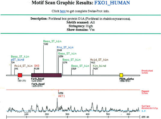
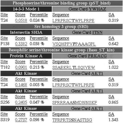
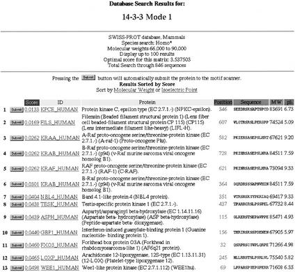
Scansite identifies short protein sequence motifs that are recognized by modular signaling domains, phosphorylated by protein Ser/Thr- or Tyr-kinases or mediate specific interactions with protein or phospholipid ligands. Each sequence motif is represented as a position-specific scoring matrix (PSSM) based on results from oriented peptide library and phage display experiments. Predicted domain-motif interactions from Scansite can be sequentially combined, allowing segments of biological pathways to be constructed in silico. The current release of Scansite, version 2.0, includes 62 motifs characterizing the binding and/or substrate specificities of many families of Ser/Thr- or Tyr-kinases, SH2, SH3, PDZ, 14-3-3 and PTB domains, together with signature motifs for PtdIns(3,4,5)P(3)-specific PH domains. Scansite 2.0 contains significant improvements to its original interface, including a number of new generalized user features and significantly enhanced performance. Searches of all SWISS-PROT, TrEMBL, Genpept and Ensembl protein database entries are now possible with run times reduced by approximately 60% when compared with Scansite version 1.0. Scansite 2.0 allows restricted searching of species-specific proteins, as well as isoelectric point and molecular weight sorting to facilitate comparison of predictions with results from two-dimensional gel electrophoresis experiments. Support for user-defined motifs has been increased, allowing easier input of user-defined matrices and permitting user-defined motifs to be combined with pre-compiled Scansite motifs for dual motif searching. In addition, a new series of Sequence Match programs for non-quantitative user-defined motifs has been implemented. Scansite is available via the World Wide Web at http://scansite.mit.edu.
Figures
Similar articles
-
Computational prediction of protein-protein interactions.Methods Mol Biol. 2004;261:445-68. doi: 10.1385/1-59259-762-9:445. Methods Mol Biol. 2004. PMID: 15064475 Review.
-
[A motif-based scanning approach for prediction of protein phosphorylation].Sheng Wu Gong Cheng Xue Bao. 2004 Jul;20(4):623-6. Sheng Wu Gong Cheng Xue Bao. 2004. PMID: 15969001 Chinese.
-
ADAN: a database for prediction of protein-protein interaction of modular domains mediated by linear motifs.Bioinformatics. 2009 Sep 15;25(18):2418-24. doi: 10.1093/bioinformatics/btp424. Epub 2009 Jul 14. Bioinformatics. 2009. PMID: 19602529
-
GlobPlot: Exploring protein sequences for globularity and disorder.Nucleic Acids Res. 2003 Jul 1;31(13):3701-8. doi: 10.1093/nar/gkg519. Nucleic Acids Res. 2003. PMID: 12824398 Free PMC article.
-
Identification of motifs in protein sequences.Curr Protoc Cell Biol. 2001 May;Appendix 1:Appendix 1C. doi: 10.1002/0471143030.cba01cs00. Curr Protoc Cell Biol. 2001. PMID: 18228275 Review.
Cited by
-
An IκB Kinase-Regulated Feedforward Circuit Prolongs Inflammation.Cell Rep. 2015 Jul 28;12(4):537-44. doi: 10.1016/j.celrep.2015.06.050. Epub 2015 Jul 16. Cell Rep. 2015. PMID: 26190110 Free PMC article.
-
Autoinhibition of Mint1 adaptor protein regulates amyloid precursor protein binding and processing.Proc Natl Acad Sci U S A. 2012 Mar 6;109(10):3802-7. doi: 10.1073/pnas.1119075109. Epub 2012 Feb 21. Proc Natl Acad Sci U S A. 2012. PMID: 22355143 Free PMC article.
-
Generation of high-performance binding proteins for peptide motifs by affinity clamping.Methods Enzymol. 2013;523:285-302. doi: 10.1016/B978-0-12-394292-0.00013-8. Methods Enzymol. 2013. PMID: 23422435 Free PMC article.
-
The alternatively-included 11a sequence modifies the effects of Mena on actin cytoskeletal organization and cell behavior.Sci Rep. 2016 Oct 17;6:35298. doi: 10.1038/srep35298. Sci Rep. 2016. PMID: 27748415 Free PMC article.
-
Computational analysis of interactomes: current and future perspectives for bioinformatics approaches to model the host-pathogen interaction space.Methods. 2012 Aug;57(4):508-18. doi: 10.1016/j.ymeth.2012.06.011. Epub 2012 Jun 28. Methods. 2012. PMID: 22750305 Free PMC article. Review.
References
Publication types
MeSH terms
Substances
Grants and funding
LinkOut - more resources
Full Text Sources
Other Literature Sources
