

Mesenchymal stem cell engraftment in lung is enhanced in response to bleomycin exposure and ameliorates its fibrotic effects
- PMID: 12815096
- PMCID: PMC166242
- DOI: 10.1073/pnas.1432929100
Mesenchymal stem cell engraftment in lung is enhanced in response to bleomycin exposure and ameliorates its fibrotic effects
Abstract
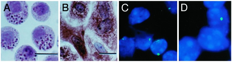
Previously we described a reliable method based on immunodepletion for isolating mesenchymal stem cells (MSCs) from murine bone marrow and showed that, after intracranial transplantation, the cells migrated throughout forebrain and cerebellum and adopted neural cell fates. Here we systemically administered MSCs purified by immunodepletion from male bleomycin (BLM)-resistant BALB/c mice into female BLM-sensitive C57BL/6 recipients and quantified engraftment levels in lung by real-time PCR. Male DNA accounted for 2.21 x 10(-5)% of the total lung DNA in control-treated mice but was increased 23-fold (P = 0.05) in animals exposed to BLM before MSC transplantation. Fluorescence in situ hybridization revealed that engrafted male cells were localized to areas of BLM-induced injury and exhibited an epithelium-like morphology. Moreover, purification of type II epithelial cells from the lungs of transplant recipients resulted in a 3-fold enrichment of male, donor-derived cells as compared with whole lung tissue. MSC administration immediately after exposure to BLM also significantly reduced the degree of BLM-induced inflammation and collagen deposition within lung tissue. Collectively, these studies demonstrate that murine MSCs home to lung in response to injury, adopt an epithelium-like phenotype, and reduce inflammation and collagen deposition in lung tissue of mice challenged with BLM.
Figures


Similar articles
-
Expression of TNF and the necessity of TNF receptors in bleomycin-induced lung injury in mice.Exp Lung Res. 1998 Nov-Dec;24(6):721-43. doi: 10.3109/01902149809099592. Exp Lung Res. 1998. PMID: 9839161
-
Therapeutic benefits of young, but not old, adipose-derived mesenchymal stem cells in a chronic mouse model of bleomycin-induced pulmonary fibrosis.Transl Res. 2015 Dec;166(6):554-67. doi: 10.1016/j.trsl.2015.09.004. Epub 2015 Sep 18. Transl Res. 2015. PMID: 26432923 Free PMC article.
-
Human bone marrow-derived mesenchymal stromal cells cultured in serum-free media demonstrate enhanced antifibrotic abilities via prolonged survival and robust regulatory T cell induction in murine bleomycin-induced pulmonary fibrosis.Stem Cell Res Ther. 2021 Sep 16;12(1):506. doi: 10.1186/s13287-021-02574-5. Stem Cell Res Ther. 2021. PMID: 34530920 Free PMC article.
-
Induced pluripotent stem cell-derived mesenchymal stem cells reverse bleomycin-induced pulmonary fibrosis and related lung stiffness.Biomed Pharmacother. 2024 Sep;178:117259. doi: 10.1016/j.biopha.2024.117259. Epub 2024 Aug 7. Biomed Pharmacother. 2024. PMID: 39116786
-
Human placental mesenchymal stem cells of fetal origins-alleviated inflammation and fibrosis by attenuating MyD88 signaling in bleomycin-induced pulmonary fibrosis mice.Mol Immunol. 2017 Oct;90:11-21. doi: 10.1016/j.molimm.2017.06.032. Mol Immunol. 2017. PMID: 28662409
Cited by
-
Treatment of Cystic Fibrosis: From Gene- to Cell-Based Therapies.Front Pharmacol. 2021 Mar 16;12:639475. doi: 10.3389/fphar.2021.639475. eCollection 2021. Front Pharmacol. 2021. PMID: 33796025 Free PMC article. Review.
-
Shh Signaling from the Injured Lung Microenvironment Drives BMSCs Differentiation into Alveolar Type II Cells for Acute Lung Injury Treatment in Mice.Stem Cells Int. 2024 Sep 28;2024:1823163. doi: 10.1155/2024/1823163. eCollection 2024. Stem Cells Int. 2024. PMID: 39372681 Free PMC article.
-
The role of immunosuppression of mesenchymal stem cells in tissue repair and tumor growth.Cell Biosci. 2012 Mar 5;2(1):8. doi: 10.1186/2045-3701-2-8. Cell Biosci. 2012. PMID: 22390479 Free PMC article.
-
Hepatocyte Growth Factor Is Required for Mesenchymal Stromal Cell Protection Against Bleomycin-Induced Pulmonary Fibrosis.Stem Cells Transl Med. 2016 Oct;5(10):1307-1318. doi: 10.5966/sctm.2015-0337. Epub 2016 Jul 7. Stem Cells Transl Med. 2016. PMID: 27388243 Free PMC article.
-
Stem cell-based therapy for pulmonary fibrosis.Stem Cell Res Ther. 2022 Oct 4;13(1):492. doi: 10.1186/s13287-022-03181-8. Stem Cell Res Ther. 2022. PMID: 36195893 Free PMC article. Review.
References
-
- American Thoracic Society/European Respiratory Society (2002) Am. J. Respir. Crit. Care Med. 165 277-304. - PubMed
-
- Jiang, Y., Balkrishna, N., Reinhardt, R. L., Schwartz, R. E., Keene, C. D., Ortiz-Gonzalez, X. R., Reyes, M., Lenvik, T., Lund, T., Blackstad, M., et al. (2002) Nature 418 41-49. - PubMed
-
- Krause, D. S., Theise, N. D., Collector, M. I., Henegariu, O., Hwang, S., Gardner, R., Neutzel, S. & Sharkis, S. J. (2001) Cell 105 369-377. - PubMed
-
- Kotton, D. N., Yang-Ma, B., Cardoso, W. B., Sanderson, E. A., Summer, R. S., Williams, M. C. & Fine, A. (2001) Development (Cambridge, U.K.) 128 5181-5188. - PubMed
-
- Bowden, D. H. (1984) Lab. Invest. 50 487-488. - PubMed
Publication types
MeSH terms
Substances
Grants and funding
LinkOut - more resources
Full Text Sources
Other Literature Sources
Medical
