. 2003 Jul 1;17(13):1630-45.
doi: 10.1101/gad.260003. Epub 2003 Jun 18.

A novel regulation mechanism of DNA repair by damage-induced and RAD23-dependent stabilization of xeroderma pigmentosum group C protein

Affiliations

A novel regulation mechanism of DNA repair by damage-induced and RAD23-dependent stabilization of xeroderma pigmentosum group C protein

Jessica M Y Ng et al. Genes Dev. .

Abstract

Primary DNA damage sensing in mammalian global genome nucleotide excision repair (GG-NER) is performed by the xeroderma pigmentosum group C (XPC)/HR23B protein complex. HR23B and HR23A are human homologs of the yeast ubiquitin-domain repair factor RAD23, the function of which is unknown. Knockout mice revealed that mHR23A and mHR23B have a fully redundant role in NER, and a partially redundant function in embryonic development. Inactivation of both genes causes embryonic lethality, but appeared still compatible with cellular viability. Analysis of mHR23A/B double-mutant cells showed that HR23 proteins function in NER by governing XPC stability via partial protection against proteasomal degradation. Interestingly, NER-type DNA damage further stabilizes XPC and thereby enhances repair. These findings resolve the primary function of RAD23 in repair and reveal a novel DNA-damage-dependent regulation mechanism of DNA repair in eukaryotes, which may be part of a more global damage-response circuitry.


Figures
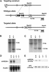
Figure 1.
Figure 1.
Targeted disruption of the mHR23A gene by homologous recombination. (A) Genomic organization and disruption strategy for mHR23A depicting the gene, the targeting construct, and the targeted mHR23A allele. Exons III to VI (and part of exons II and VII) were replaced by the dominant selectable neomycin-resistance marker transcribed in an antisense orientation. (B) Southern blot analysis of BamHI-digested DNA from ES cells showing the 5.0-kb and 3.5-kb fragments representing the wild-type and targeted allele of mHR23A, respectively. (C) Southern blot analysis of BamHI-digested tail DNA from mHR23A+/+, mHR23A+/–, and mHR23A–/– mice. (D, top) RNA blot analysis of mHR23A mRNA from mHR23A+/+, mHR23A+/–, and mHR23A–/– MEFs using full-length mHR23A cDNA as a probe. (Bottom) As a loading control for the amount of RNA, the blot was reprobed with β-actin cDNA. (E) Immunoblot analysis of mHR23A protein in cellular extracts from mHR23A+/+, mHR23A+/–, and mHR23A–/– MEFs loaded in equal amounts. Polyclonal antibodies against the human HR23A protein (top panel) and the human XPC protein (bottom panel) were used. The asterisk indicates an aspecific cross-reacting band.
Figure 2.
Figure 2.
Repair characteristics of mHR23A–/– E13.5 and DKO E8.5 MEFs. (A) UV survival curves of primary mHR23A+/+, mHR23A+/–, and mHR23A–/– E13.5 MEFs. XPC–/– fibroblasts were included as a negative control. Cells were exposed to different doses of UV (254 nm). After 4–5 d, the number of proliferating cells was estimated from the amount of radioactivity incorporated during a 3-h pulse with [3H]thymidine. For each genotype, identical results were obtained with three other cell lines (data not shown). (B) Global genome repair (UDS) in primary mHR23A+/+, mHR23A+/–, and mHR23A–/– E13.5 MEFs. Cells were irradiated with 16 J/m2 of UV (254 nm) and labeled with [3H]thymidine. Incorporation of radioactivity was measured by autoradiography and grain counting (average of 50 nuclei per cell line; the standard error of the mean is indicated). XPA–/– fibroblasts were measured as a negative control. For each genotype, consistent results were obtained with three other independent cell lines (data not shown). (C) RNA synthesis recovery (RRS) after UV exposure of primary mHR23A+/+, mHR23A+/–, and mHR23A–/– E13.5 MEFs. Cells were UV-irradiated (10 J/m2, 254 nm) and allowed to recover for 16 h. After a 1-h pulse labeling with [3H]uridine, cells were processed for autoradiography. The relative rate of RNA synthesis was expressed as the quotient of the number of autoradiographic grains over the UV-exposed nuclei and the number of grains over the nuclei of unirradiated cells (average of 50 nuclei per cell line; the standard error of the mean is indicated). CSB–/– cells were used as a negative control. For each genotype, three other independent lines were assayed with similar outcomes (data not shown). (D) UV survival of E8.5 MEF lines of wild-type, XPC–/–, mHR23A–/–/B+/–, mHR23A+/–/B–/–, and mHR23A–/–/B–/– (DKO). (E) UV-induced UDS in wild-type, XPC–/–, and DKO E8.5 MEFs. (F) RNA synthesis recovery after UV irradiation of wild-type, XPC–/–, and DKO E8.5 MEFs. For details for panels D–F, see legends to panels A–C, respectively, and Materials and Methods. Two independent experiments using two other DKO cell lines (before the cultures extinguished; data not shown) showed a similar effect on UDS and RNA synthesis recovery.
Figure 3.
Figure 3.
XPC expression in DKO E8.5 MEFs. (A) Phase contrast (left panels) and epifluorescence (middle and right panels) images of fixed wild-type (WT, labeled with latex beads), XPC–/– (XPC), and mHR23A–/–/B–/– (DKO) MEFs. Cells were fixed by paraformaldehyde, permeabilized by 0.1% Triton X-100, and subsequently immunolabeled with affinity-purified polyclonal antibodies against the human XPC protein (middle panels; stained green with goat anti-rabbit Alexa 488-labeled secondary antibody). Monoclonal antibodies recognizing the p62 subunit of TFIIH (right panels; stained red with goat antimouse Cy3-labeled secondary antibody) were used as an internal control. All images were taken at the same magnification. (B,C) Immunoblot analysis of XPC protein in cellular extracts from wild-type, XPC–/–, and DKO E8.5 MEFs using polyclonal anti-human XPC antibodies (B). (C) Monoclonal anti-p62 antibodies were used as an internal reference for the amount of protein in each lane.
Figure 4.
Figure 4.
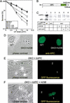
Characterization of DKO cells expressing hHR23B and XPC-GFP. (A) UV survival of wild-type, XPC–/–, DKO, and DKO MEFs cotransfected with hHR23B (h23B), human XPC-GFP (hXPC), and h23B and hXPC-GFP cDNAs. Cells were exposed to different doses of UV (254 nm). After 4–5 d, the number of proliferating cells was estimated from the amount of radioactivity incorporated during a 3-h pulse with [3H]thymidine. For details, see Materials and Methods. For each cDNA construct, similar results were obtained with at least two other independent stably transfected cell lines (data not shown). (B) Schematic representation of XPC-EGFP–His6HA-N3 fusion protein (1208 amino acids). Indicated are the human XPC protein (940 amino acids), the enhanced green fluorescent protein tag (EGFP; 238 amino acids), and the hexameric histidine-hemagglutinin double-epitope tag (His6HA; 17 amino acids). (C, top) Immunoblot analysis of XPC expression in cellular extracts of wild-type (WT; lane 1), XPC (lane 2), DKO (lane 3), and DKO MEFs cotransfected with h23B (lane 4), hXPC-GFP (lane 5), and h23B and hXPC-GFP (lane 6) cDNAs, using a polyclonal antibody against the C terminus of human XPC. (Bottom) Monoclonal anti-p62 antibodies were used as a loading control. (D) Phase contrast (left) and epifluorescence (right) images of fixed WT (labeled with latex beads) and DKO cells cotransfected with hHR23B cDNA. Cells were fixed by paraformaldehyde, followed by 0.1% Triton X-100 permeabilization, and subsequently immunolabeled with affinity-purified polyclonal anti-human XPC (right; stained green with goat anti-rabbit Alexa 488-labeled secondary antibody). Monoclonal anti-p62 antiserum was used as an internal control (stained red with goat anti-mouse Cy3-labeled secondary antibody; data not shown). Images were taken at the same magnification. Similar results were obtained with DKO cells cotransfected with hXPC-GFP, and hHR23B and hXPC-GFP cDNAs (data not shown). (E,F) Phase contrast (left) and epifluorescence (right) images of living DKO cells cotransfected with hXPC-GFP (E) or hHR23B and hXPC-GFP (F) cDNAs. All images were taken at the same magnification.
Figure 5.
Figure 5.
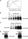
Analyses of XPC-GFP expression after UV irradiation and treatment with proteasome inhibitor CBZ-LLL. (A) Kinetic analysis of living DKO cells expressing XPC-GFP/hHR23B after exposure to 10 J/m2 of UV-C in time over a period of 30 h. Percentage XPC-GFP, the percentage of GFP-expressing fluorescent cells of the total number of cells. (B) Immunoprecipitation study of UV-irradiated DKO cells expressing XPC-GFP/hHR23B in time. XPC-GFP was immunoprecipitated (IP) from WCEs with monoclonal anti-GFP antibodies. The precipitates were analyzed by immunoblotting using polyclonal anti-XPC antiserum. Untreated WCE, and extracts from cells isolated at 45 min, 90 min, 3 h, 6 h, and 9 h after exposure to 10 J/m2 of UV-C. The amount of expressed XPC-GFP was also visualized by immunoblot analysis of the total cell lysate with anti-XPC or anti-GFP antibodies, and with a monoclonal antibody against the p62 subunit of TFIIH as a loading control (data not shown). XPC-GFP expression was also dose-dependent (4, 8, 12, and 16 J/m2 of UV-C; data not shown). (C) Immunoprecipitation analysis of DKO cells expressing XPC-GFP/hHR23B after CBZ-LLL treatment. IPs of WCEs from cells treated with 10 μM CBZ-LLL for 45 min, 90 min, 3 h, 6 h, and 9 h, were performed as in B. XPC-GFP expression was confirmed by immunoblot analysis of the total cell lysate using anti-XPC or anti-GFP antibodies, and anti-p62 as a loading control (data not shown). (D) Large magnification of the higher migrating XPC species (arrows) detected in the 3-h samples after UV (left, see panel B) and CBZ-LLL treatment (right, see panel C).
Figure 6.
Figure 6.
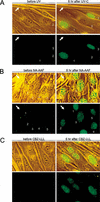
Effect of UV, NA-AAF, and CBZ-LLL on hHR23B-dependent XPC-GFP levels in living DKO cells. (A) Combined phase contrast (transmission light) and fluorescence (green) images (top panels), and epifluorescence images (bottom panels) of the same living DKO cells expressing XPC-GFP/hHR23B before UV (left panels) and 6 h after 10 J/m2 of UV-C (right panels). White arrows indicate the scratch mark on glass coverslips for proper orientation. Numbers represent the same living cells before and after UV exposure. Identical results were obtained with two other independent DKO cell lines expressing XPC-GFP/hHR23B (data not shown). All images were taken at the same magnification. (B) Combined phase contrast (transmission light) and fluorescence (green) images (top panels), and epifluorescence images (bottom panels) of living DKO cells expressing XPC-GFP/hHR23B before NA-AAF (left panels) and 8 h after 50 μM NA-AAF (right panels). White arrows indicate the scratch on glass coverslips for proper comparison. The numbers represent the corresponding living cells on coverslips before and after NA-AAF treatment. Identical results were obtained with two other independent DKO cell lines expressing XPC-GFP/hHR23B (data not shown). All images were taken at the same magnification. (C) Combined phase contrast (transmission light) and fluorescence (green) images (top panels), and only epifluorescence images (bottom panels) of living DKO cells expressing XPC-GFP/hHR23B before treatment with proteasome inhibitor CBZ-LLL (left panels) and 6 h after 10 μM CBZ-LLL (right panels). All images were taken at the same magnification.
Figure 7.
Figure 7.
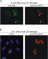
Local UV damage induces overall XPC stabilization in nuclei of DKO cells expressing XPC-GFP/hHR23B. (A,B) DKO cells expressing XPC-GFP/hHR23B were exposed to 64 J/m2 of UV-C through 5.0-μm pore filters and fixed 5 min (A) and 2 h (B) later with paraformaldehyde. Double immunofluorescent labeling using antibodies against XPA (A, left panel; stained green with goat anti-rabbit Alexa 488-labeled secondary antibody) and HA epitope (A, right panel; stained red with goat anti-rat Alexa 594-labeled secondary antibody); DAPI stained (B, left panel) and HA antibody labeling (B, right panel). Arrowheads indicate the site of UV-induced local damage in the nuclei of DKO cells expressing hXPC-GFP/hHR23B. Note: Compare the increased fluorescence signal over the entire nucleus of damaged cells to the signal of undamaged nuclei for overall stabilization of XPC (B).
Figure 8.
Figure 8.
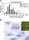
Enhanced DNA repair correlates with high levels of XPC in UV-induced UDS in DKO cells expressing XPC-GFP/hHR23B. (A) Histogram of UV-induced UDS in DKO cells expressing XPC-GFP/hHR23B. Five hours after exposure to 10 J/m2 of UV-C, cells were subsequently irradiated with 16 J/m2 of UV-C and labeled with [3H]thymidine for 1 h (white columns, mean of UDS level is 25 ± S.E.M. 1). In parallel, nonprechallenged cells only exposed to 16 J/m2 of UV-C were used as controls (black columns, mean of UDS level is 16 ± S.E.M. 0.6). Asterisks indicate the mean values of the UDS levels. Incorporation of radioactivity was measured by autoradiography and grain counting (130 fixed squares counted per cell line; each square represented ∼50% of the nucleus surface). UV-induced UDS of wild-type (mean 17 ± S.E.M. 0.8) fibroblasts were measured as a control (data not shown). (B) Effect of microinjection of XPC-GFP cDNA on UV-induced UDS in human wild-type (C5RO) fibroblasts. Shown is a micrograph of a wild-type homodikaryon (numbered 1) microinjected with XPC-GFP in one of the nuclei and subjected to UV-induced UDS. Prior to UDS, fluorescence images were captured (inset in B). The injected cell has a considerably larger number of grains above its nuclei than the noninjected, surrounding mononuclear cells (numbered 2).
Figure 9.
Figure 9.
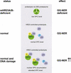
Model for the DNA damage and HR23-dependent regulation of XPC and GG-NER. In the total absence of the HR23 proteins (mHR23A/B-deficient), XPC is intrinsically unstable and targeted for ubiquitin-dependent proteolysis via the 26S proteasome. As a consequence, the steady-state level of XPC is decreased, resulting in reduced GG-NER capacity (top panel). Under normal conditions, HR23 proteins (indicated as 23) control XPC degradation, leading to partial stabilization of XPC [in a complex with HR23 and CEN2 (C)]. Higher steady-state levels of XPC result in proficient GG-NER (middle panel). NER-type DNA damage (e.g., UV irradiation) induces a transient further increase in XPC/HR23/CEN2 protein levels through nuclear accumulation of XPC bound to lesions, and accordingly enhances GG-NER capacity (bottom panel). A comparable HR23-mediated stabilization mechanism may hold for other factors (here indicated by “?”) and cellular pathways in which HR23 proteins are implicated (see Discussion for further explanation).

Similar articles

Cited by

References

    1. Adimoolam S. and Ford, J.M. 2002. p53 and DNA damage-inducible expression of the xeroderma pigmentosum group C gene. Proc. Natl. Acad. Sci. 99: 12985–12990. - PMC - PubMed
    1. Amundson S.A., Patterson, A., Do, K.T., and Fornace Jr., A.J. 2002. A nucleotide excision repair master-switch: p53 regulated coordinate induction of global genomic repair genes. Cancer Biol. Ther. 1: 145–149. - PubMed
    1. Araki M., Masutani, C., Takemura, M., Uchida, A., Sugasawa, K., Kondoh, J., Ohkuma, Y., and Hanaoka, F. 2001. Centrosome protein centrin 2/caltractin 1 is part of the xeroderma pigmentosum group C complex that initiates global genome nucleotide excision repair. J. Biol. Chem. 276: 18665–18672. - PubMed
    1. Bertolaet B.L., Clarke, D.J., Wolff, M., Watson, M.H., Henze, M., Divita, G., and Reed, S.I. 2001. UBA domains of DNA damage-inducible proteins interact with ubiquitin. Nat. Struct. Biol. 8: 417–422. - PubMed
    1. Biggins S., Ivanovska, I., and Rose, M.D. 1996. Yeast ubiquitin-like genes are involved in duplication of the microtubule organizing center. J. Cell. Biol. 133: 1331–1346. - PMC - PubMed

Publication types

MeSH terms