. 2003 Jul;23(13):4559-72.
doi: 10.1128/MCB.23.13.4559-4572.2003.

H1 linker histones are essential for mouse development and affect nucleosome spacing in vivo

Affiliations

H1 linker histones are essential for mouse development and affect nucleosome spacing in vivo

Yuhong Fan et al. Mol Cell Biol. 2003 Jul.

Abstract

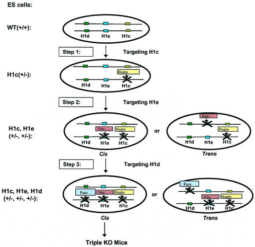
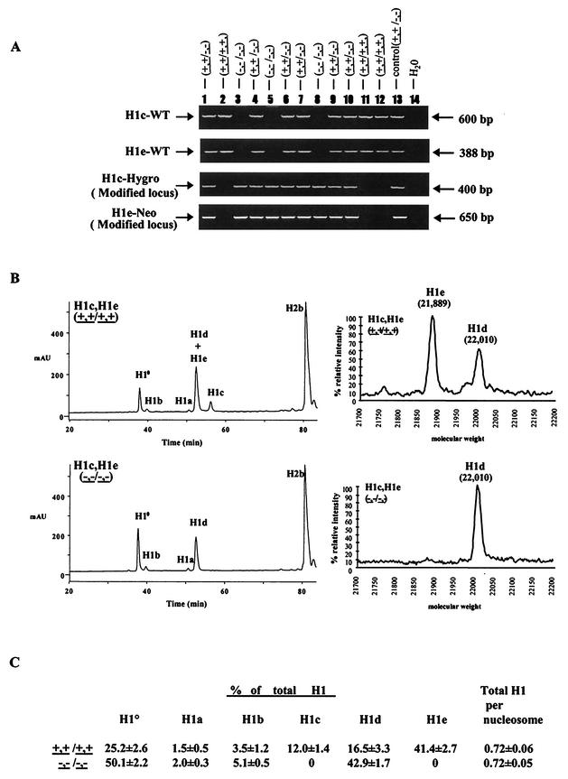
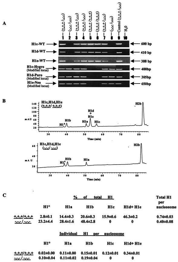
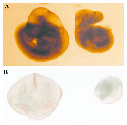
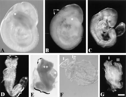
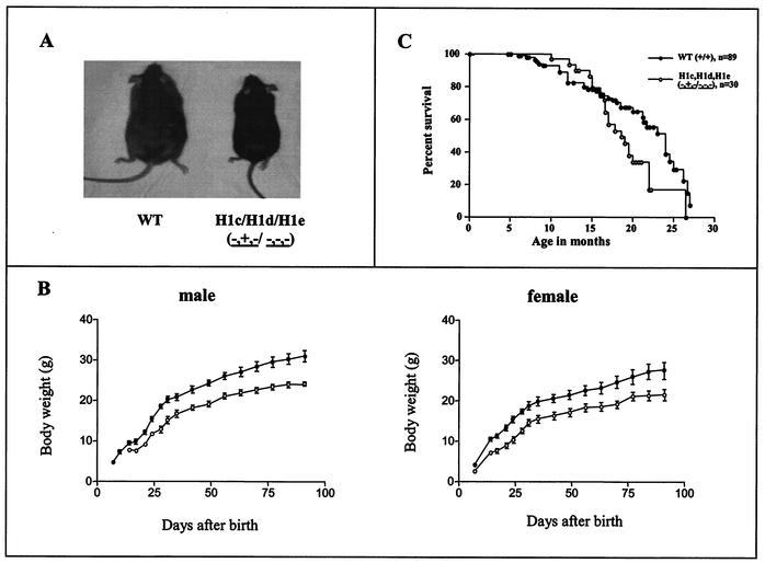
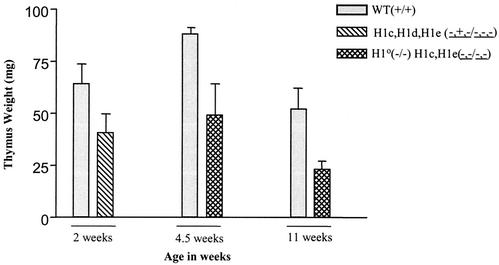
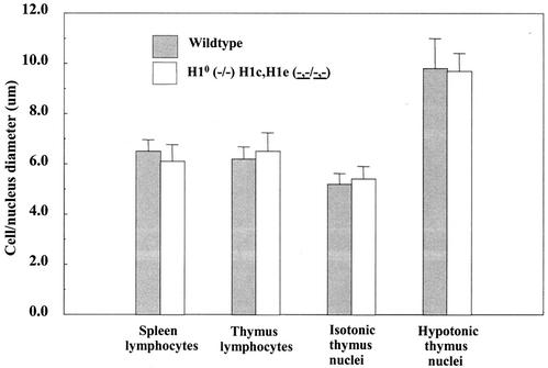
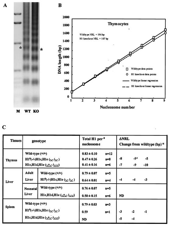
Most eukaryotic cells contain nearly equimolar amounts of nucleosomes and H1 linker histones. Despite their abundance and the potential functional specialization of H1 subtypes in multicellular organisms, gene inactivation studies have failed to reveal essential functions for linker histones in vivo. Moreover, in vitro studies suggest that H1 subtypes may not be absolutely required for assembly of chromosomes or nuclei. By sequentially inactivating the genes for three mouse H1 subtypes (H1c, H1d, and H1e), we showed that linker histones are essential for mammalian development. Embryos lacking the three H1 subtypes die by mid-gestation with a broad range of defects. Triple-H1-null embryos have about 50% of the normal ratio of H1 to nucleosomes. Mice null for five of these six H1 alleles are viable but are underrepresented in litters and are much smaller than their littermates. Marked reductions in H1 content were found in certain tissues of these mice and in another compound H1 mutant. These results demonstrate that the total amount of H1 is crucial for proper embryonic development. Extensive reduction of H1 in certain tissues did not lead to changes in nuclear size, but it did result in global shortening of the spacing between nucleosomes.


Figures

FIG. 1.
FIG. 1.
Strategy for sequential inactivation of three H1 histone genes in mouse ES cells. Depicted are the chromosome homologues containing the three linked H1 genes (those encoding H1c, H1d, and H1e) to be targeted and the three steps used to target the genes, each with a different selectable marker gene. Also depicted are the cis and trans configurations of gene targetings that can occur in steps 2 and 3. WT, wild type; KO, knockout.
FIG.2.
FIG.2.
Analysis of H1c H1e double-knockout (KO) mice. (A) Genotype analysis of F2 mice from intercrosses of H1cH1e++/−− mice. Mouse tail DNA was analyzed by PCR (as described in Materials and Methods) for the wild-type H1c allele (H1c-WT), the wild-type H1e allele (H1e-WT), the modified H1c allele (H1c-Hygro), and the modified H1e allele (H1e-Neo). The deduced genotype of each embryo is indicated above each lane. The positions of the PCR products from the wild-type and modified alleles are indicated. Control reaction mixtures contained tail DNA from an H1c H1e double-heterozygous mutant mouse. (B) Analysis of histones extracted from livers of wild-type and H1c H1e double-homozygous mutant mice. The graphs on the left show results of reverse-phase HPLC analyses of approximately 100 μg of total liver histone extracts from a 20-week-old wild-type mouse (top) and an H1c H1e double-homozygous mutant (bottom). The abscissa represents elution time, and the ordinate represents absorbency at 214 Å. The identity of the histone subtype(s) in each peak is indicated. mAU, milli-absorbency units. The graphs on the right show results of time-of-flight mass spectrometry analysis of a fraction eluting between 52 and 54 min (corresponding to the peak marked H1d + H1e). The identities of the H1d and H1e subtypes detected in this analysis were shown previously (51). (C) H1 subtype composition of liver chromatin from wild-type and H1c H1e double-mutant mice. Data were calculated from HPLC analyses of wild-type and H1c H1e double-mutant strains like that shown in panel B. Values are means ± standard deviations of individual determinations made on three 5-month-old mice of each of the indicated genotypes. The percentage of total H1 was determined by the ratio of the A214 of the indicated H1 peak to the total A214 of all of the H1 peaks. Total H1 per nucleosome was determined by the ratio of the total A214 of all of the H1 peaks to half of the A214 of the H2b peak. The A214 values of the individual H1 peaks and the H2b peak were adjusted to account for the differences in the number of peptide bonds in each H1 subtype and H2b.
FIG.3.
FIG.3.
Analysis of chromatin from H1c H1d H1e triple-null mouse embryos. (A) PCR genotype analysis of E7.5 embryos from intercrosses of H1c+/− H1d+/− H1e+/− mice. Embryo DNA was prepared and analyzed by PCR assays for H1c, H1d, and H1e wild-type (WT) and modified loci as described in Materials and Methods. The deduced genotype of each embryo is indicated above each lane. The positions of the PCR products from the wild-type and modified alleles are indicated. Control reaction mixtures contained tail DNA from an H1c H1d H1e triple-heterozygous mutant mouse. (B) Reverse-phase HPLC analysis of histones in extracts from E10.5 wild-type and homozygous H1c H1d H1e mutant embryos. Approximately 20 μg of total histone extract of chromatin from wild-type (top) and homozygous triple H1c H1d H1e mutant (bottom) E10.5 embryos were fractionated by reverse-phase HPLC. Other details are as in the legend to Fig. 2B. mAU, milli-absorbency units. (C) H1 subtype composition of chromatin from wild-type and H1c−/− H1d−/− H1e−/− E10.5 embryos. Data were calculated from HPLC analyses of wild-type and H1c H1d H1e triple-mutant embryos like that shown in panel B. Other details are as described in the legend to Fig. 2C.
FIG. 4.
FIG. 4.
Gross morphology of wild-type and H1c H1d H1e triple-mutant embryos at E10.5. Approximately one-fourth of the expected number of homozygous mutant embryos are recovered at E10.5. (A) Morphology of an E10.5 wild-type embryo (left) and an H1c H1dH1e−−−/−− littermate (right). (B) Whole view of E10.5 conceptuses with yolk sacs, i.e., the wild type (left) and an H1cH1d H1e−−−/−−− littermate (right).
FIG. 5.
FIG. 5.
Phenotype of H1cH1dH1e−−−/−−− triple-homozygous mutant embryos recovered on E9.5. Approximately 60% of the expected number of homozygous mutant embryos are recovered at E9.5. The broad spectrum of abnormalities observed in recovered homozygous mutant embryos fall into three general classes. In the most predominant category (∼50%), representatives of which are shown in panels B and C, are embryos that were slightly developmentally delayed relative to their littermates (a representative wild-type embryo is shown in panel A) and presented a variety of incompletely penetrant abnormalities. Abnormalities observed in this class of mutants included occlusion of the rhombencephalic ventricle (arrows in panel B), a short tail (compare asterisks at the tail tips in panels A and B), and failure of the allantois to fuse with the chorion (asterisk in panel C). Mutant embryos falling into a second class, representatives of which are shown in panels D and E, are very developmentally delayed compared to their littermates and again variably presented developmental perturbations, including splayed anterior neural tubes (arrows in panels D and E), regions of excess tissue (++ in panel E), pericardial expansion (asterisk in panel E), failure of chorioallantoic fusion (asterisk in panel D), and caudal dysgenesis (braces in panels D and E). Mutant embryos in a third class are severely abnormal (F and G). They had not progressed past an early somite stage and in each case showed morphological evidence of partial (I and I+ in panel F) or complete (I and II in panel G) axis duplication. Embryos are viewed from the right side in panels A to C and E, from nearly dorsal in panel D, from ventral in panel F, and from a midline between the axes in panel G. Scale bar = 300 μm.
FIG. 6.
FIG. 6.
H1cH1dH1e−+−/−−− mice are growth retarded. (A) Photograph of a representative H1cH1dH1e−+−/−−− mouse and a representative wild-type (WT) littermate at 4 months of age. (B) Growth curves of H1cH1dH1e−+−/−−− mice and littermate control mice. The left graph shows the mean body weight in grams ± the standard error of the mean for male mice (n = 7 for H1cH1e++/−− littermate controls, and n = 6 for H1cH1dH1e−+−/−−− mutant mice). The right graph shows the body weights of female mice (n = 7 for H1cH1e++/−− littermate controls, and n = 9 for H1cH1d−+−/−−− mutant mice). Symbols: •, controls; ○, mutants. (C) Kaplan-Meier survival curve of H1cH1dH1e−+−/−−− mutant mice and age-matched wild-type mice (P = 0.03).
FIG. 7.
FIG. 7.
Thymus weight is reduced in compound H1 knockout mice. Thymi from mice with the indicated genotypes were dissected and weighed at 2 weeks (wild type [WT] or littermate control, n = 9; H1cH1dH1e−+−/−−−, n = 10), 4.5 weeks (wild type, n = 2; H1o−/−; H1cH1e−−/−−−, n = 4), and 11 weeks (wild type, n = 7; H1o−/−; H1cH1e−−/−−, n = 2). Weights are presented as averages ± standard deviations.
FIG. 8.
FIG. 8.
Nuclear size is not changed in lymphocytes depleted of H1. Spleen and thymus cells and nuclei of wild-type and H10 H1c H1e triple-null mice were equilibrated and measured in isotonic and chelating hypotonic buffers. Bars show mean diameters and standard deviations (n = 70 and 62, respectively, for wild-type and mutant spleen lymphocytes; n = 123 and 75, respectively, for wild-type and mutant thymus lymphocytes; n = 76 and 117, respectively, for wild-type and mutant isotonic thymus nuclei; n = 152 and 38, respectively, for wild-type and mutant hypotonic thymus nuclei).
FIG. 9.
FIG. 9.
Nucleosome spacing is reduced in mutants with extensive reductions in total H1. (A and B) Thymus nuclei from wild-type (WT) and H10 H1c H1e triple-null (KO) mice. (A) Nucleosome repeat ladder obtained by micrococcal nuclease digestion of thymus nuclei as described in Materials and Methods. Lane 1 contains marker (M) DNA. Chromatin from the H1-deficient mutant nuclei migrates faster than that from wild-type nuclei, as illustrated by the asterisks at the pentanucleosome bands. (B) Plot of nucleosome number versus DNA length for thymus chromatin shown in panel A. Symbols show the data points, and lines are the linear regressions of the data points, which indicate a ΔNRL of 9 bp for these samples (see Materials and Methods for details). (C) Relationship between changes in H1 content and NRL. Superscripts: α, results of separate experiments (each comparison was done with different wild-type and knockout mice); #, from data in Table 2; *, data shown in panels A and B. ND, not determined.

Similar articles

Cited by

References

    1. Adenot, P. G., E. Campion, E. Legouy, C. D. Allis, S. Dimitrov, J. Renard, and E. M. Thompson. 2000. Somatic linker histone H1 is present throughout mouse embryogenesis and is not replaced by variant H1 degrees. J. Cell Sci.113:2897-2907. - PubMed
    1. Annunziato, A. T., and R. L. Seale. 1983. Chromatin replication, reconstitution and assembly. Mol. Cell. Biochem. 55:99-112. - PubMed
    1. Barra, J. L., L. Rhounim, J.-L. Rossignol, and G. Faugeron. 2000. Histone H1 is dispensable for methylation-associated gene silencing in Ascobolus immersus and essential for long life span. Mol. Cell. Biol. 20:61-69. - PMC - PubMed
    1. Bates, D. L., and J. O. Thomas. 1981. Histones H1 and H5: one or two molecules per nucleosome? Nucleic Acids Res. 9:5883-5894. - PMC - PubMed
    1. Blank, T. A., and P. B. Becker. 1995. Electrostatic mechanism of nucleosome spacing. J. Mol. Biol. 252:305-313. - PubMed

Publication types

MeSH terms