. 2003 Jun;14(6):2303-13.
doi: 10.1091/mbc.e02-12-0831. Epub 2003 Feb 6.

Mdm30 is an F-box protein required for maintenance of fusion-competent mitochondria in yeast

Affiliations

Mdm30 is an F-box protein required for maintenance of fusion-competent mitochondria in yeast

Stefan Fritz et al. Mol Biol Cell. 2003 Jun.

Abstract

Mitochondrial fusion and fission play important roles for mitochondrial morphology and function. We identified Mdm30 as a novel component required for maintenance of fusion-competent mitochondria in yeast. The Mdm30 sequence contains an F-box motif that is commonly found in subunits of Skp1-Cdc53-F-box protein ubiquitin ligases. A fraction of Mdm30 is associated with mitochondria. Cells lacking Mdm30 contain highly aggregated or fragmented mitochondria instead of the branched tubular network seen in wild-type cells. Deltamdm30 cells lose mitochondrial DNA at elevated temperature and fail to fuse mitochondria in zygotes at all temperatures. These defects are rescued by deletion of DNM1, a gene encoding a component of the mitochondrial division machinery. The protein level of Fzo1, a key component of the mitochondrial fusion machinery, is regulated by Mdm30. Elevated Fzo1 levels in cells lacking Mdm30 or in cells overexpressing Fzo1 from a heterologous promoter induce mitochondrial aggregation in a similar manner. Our results suggest that Mdm30 controls mitochondrial shape by regulating the steady-state level of Fzo1 and point to a connection of the ubiquitin/26S proteasome system and mitochondria.


Figures
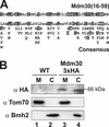
Figure 1.
Figure 1.
Mdm30 contains an F-box and is partially associated with mitochondria. (A) The F-box motif of Mdm30 is aligned with an F-box consensus sequence according to Patton et al. (1998). Residues that match the consensus sequence are highlighted by black boxes. (B) Wild-type yeast cells (WT; lanes 1 and 2) and yeast cells expressing Mdm30 with a triple HA epitope (Mdm30-3xHA; lanes 3 and 4) were fractionated into cytosol and mitochondria. Mitochondria were further purified on a sucrose step gradient. Then 50 μg of each fraction was analyzed by immunoblotting. Markers were Tom70 for mitochondria and Bmh2 for cytosol. M, mitochondria (lanes 1 and 3); C, cytosol (lanes 2 and 4).
Figure 2.
Figure 2.
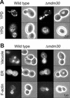
Δmdm30 cells exhibit grossly altered mitochondrial morphology. (A) Mitochondrial morphology. Wild-type and Δmdm30 cells expressing mtGFP were grown to log phase in glucose-containing medium (YPD) or glycerol-containing medium (YPG) and examined by fluorescence (left) and phase contrast (right) microscopy. (B) Morphology of other cellular structures in wild-type and Δmdm30 cells. For staining of vacuoles, cells were grown to log phase in YPD medium and stained with 5-(and-6)-carboxy-2′,7′-dichlorofluorescein diacetate. For visualization of the ER, cells expressing ER-targeted GFP were grown to log phase in glucose-containing synthetic minimal medium lacking methionine. For staining of F-actin, cells were grown to log phase in YPD medium, fixed, and stained with rhodamine-phalloidin. Samples were analyzed by fluorescence and phase contrast microscopy.
Figure 3.
Figure 3.
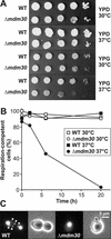
Deletion of the MDM30 gene leads to respiratory deficiency at elevated temperature and loss of mtDNA. (A) Growth phenotype of the Δmdm30 mutant. Wild-type and Δmdm30 cells were grown overnight in glucose-containing medium at 30°C. Then, 10-fold serial dilutions were spotted onto plates containing glucose (YPD) or glycerol (YPG) as carbon source. YPD plates were incubated for 2 d, and YPG plates were incubated for 3 d at the indicated temperatures. (B) Loss of respiratory competence in Δmdm30 cells grown at elevated temperature. Wild-type and Δmdm30 cells were precultured on YPG plates at 30°C to select for cells containing mtDNA. These colonies were used to inoculate liquid cultures in YPD medium to allow for loss of mtDNA. YPD cultures growing at 30 or 37°C were maintained at log phase. At the indicated time points, aliquots of these cultures were plated on glucose-containing medium. The next day, colonies were replica-plated to glycerol-containing medium, and the percentage of respiratory-competent colonies was determined. (C) Δmdm30 cells lose mtDNA. Wild-type and Δmdm30 cells harvested from YPD cultures growing for 20 h at 37°C were stained with DAPI and viewed by fluorescence (left) and phase contrast (right) microscopy. Arrowheads point to mtDNA chondrolites. Big fluorescent spots represent nuclei.
Figure 4.
Figure 4.
Δfzo1 and Δdnm1 mutations are epistatic to Δmdm30. (A) Growth phenotypes of Δmdm30/Δfzo1 and Δmdm30/ Δdnm1 mutants. Growth of strains was assessed as described in Figure 3A. (B) Mitochondrial phenotypes of Δmdm30/Δfzo1 and Δmdm30/Δdnm1 mutants. Mitochondrial morphology of mtGFP-expressing cells grown in glucose-containing medium was analyzed as described in Figure 2A.
Figure 5.
Figure 5.
Mitochondrial fusion is blocked in Δmdm30 cells and can be restored by deletion of the DNM1 gene. (A) Wild-type (WT) cells of opposite mating types containing mitochondria preloaded with mtGFP or mtRFP were mated and analyzed by fluorescence microscopy. The distribution of mtGFP and mtRFP in a representative zygote containing a medial bud (asterisk) is shown. (B) Δmdm30 zygotes and (C) Δmdm30/Δdnm1 zygotes were analyzed as described above.
Figure 6.
Figure 6.
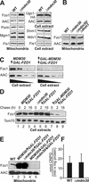
The protein level of Fzo1 depends on Mdm30. (A) Steady-state levels of mitochondrial proteins in wild-type (WT) and Δmdm30 cells. Equal amounts of mitochondria or total cell extracts isolated from wild-type and Δmdm30 cells were analyzed by immunoblotting by using specific antisera directed against the indicated proteins. For Ugo1, cells expressing an epitope-tagged protein, Ugo1-HA (Sesaki and Jensen, 2001), were analyzed. Note that Mgm1 is present in mitochondria in two forms of different size (Shepard and Yaffe, 1999). (B) Steady-state levels of Fzo1 in Δmdm30/Δdnm1 cells. Mitochondria of wild-type, Δmdm30, and Δmdm30/Δdnm1 cells were analyzed as described above. (C) Level of overexpressed Fzo1 in the absence or presence of Mdm30 overexpression. Cells carrying a chromosomal insertion of the GAL1 promoter upstream of the FZO1 coding region (left; MDM30/↑GAL-FZO1) and cells that in addition harbored a plasmid with an MDM30 allele under control of the GAL1 promoter (right; ↑GAL-MDM30/↑GAL-FZO1) were grown overnight in galactose-containing minimal medium under selection for the plasmid. Total cell extracts were prepared from log phase cultures, and twofold serial dilutions were analyzed by immunoblotting by using antisera against Fzo1 and, as a loading control, AAC. (D) Mdm30-dependent turnover of Fzo1. Cells carrying a chromosomal insertion of the GAL1 promoter in front of the FZO1 coding region in MDM30 wild-type (lanes 1–3), Δmdm30 (lanes 4–6), or GAL1-controlled MDM30 overexpression (lanes 7–9) backgrounds were grown overnight in galactose-containing medium. The next day, cultures were diluted with fresh medium and allowed to grow logarithmically for two generation times. Then, 7.5 mg/ml cycloheximide was added to stop cytosolic protein synthesis. After 0, 2, and 16 h, aliquots were harvested and cell extracts were analyzed as described above. Tom70 served as a loading control. Note that for lanes 7–9, a longer exposure of the Western blot was used to visualize the Fzo1 signal. (E) Fzo1 levels in mitochondria of GAL1-regulated strains. Wild-type cells (lane 1) and cells carrying a chromosomal insertion of the GAL1 promoter upstream of the FZO1 coding region in MDM30 (lanes 2 and 4) and Δmdm30 (lanes 2 and 5) backgrounds were grown in galactose-containing medium (lanes 2 and 3; MDM30/↑GAL-FZO1 and Δmdm30/↑GAL-FZO1) or in glucose-containing medium (lanes 4 and 5; MDM30/↓GAL-FZO1 and Δmdm30/↓GAL-FZO1). Mitochondria were isolated and equal amounts of mitochondrial protein were analyzed by immunoblotting by using antisera against Fzo1 and, as a loading control, AAC. (F) FZO1 promoter activity in wild-type and Δmdm30 cells. Cell extracts were prepared from logarithmically growing cultures of wild-type and Δmdm30 cells carrying a β-galactosidase reporter gene under control of the FZO1 promoter. β-Galactosidase activity was determined as nanomoles of o-nitrophenyl-β-d-galactopyranoside (ONPG) hydrolyzed/(min × mg protein). Error bars indicate standard deviations of 10 measurements obtained with five independently isolated clones. Strains lacking the reporter construct did not exhibit any significant activity.
Figure 7.
Figure 7.
Mitochondrial morphology depends on FZO1 and MDM30. Cells carrying a chromosomal insertion of the GAL1 promoter upstream of the FZO1 coding region in MDM30 (A and C) and Δmdm30 (B and D) backgrounds were grown in galactose-containing medium (YPGal; A and B) or in glucose-containing medium (YPD; C and D). Mitochondrial morphology of mtGFP-expressing cells was analyzed in log phase cultures as described in Figure 2A.

Similar articles

Cited by

References

    1. Amberg, D.C. (1998). Three-dimensional imaging of the yeast actin cytoskeleton through the budding cell cycle. Mol. Biol. Cell 9, 3259-3262. - PMC - PubMed
    1. Bähler, J., Wu, J.Q., Longtine, M.S., Shah, N.G., McKenzie, A., 3rd, Steever, A.B., Wach, A., Philippsen, P., and Pringle, J.R. (1998). Heterologous modules for efficient and versatile PCR-based gene targeting in Schizosaccharomyces pombe. Yeast 14, 943-951. - PubMed
    1. Bai, C., Sen, P., Hofmann, K., Ma, L., Goebl, M., Harper, J.W., and Elledge, S.J. (1996). SKP1 connects cell cycle regulators to the ubiquitin proteolysis machinery through a novel motif, the F-box. Cell 86, 263-274. - PubMed
    1. Bereiter-Hahn, J., and Vöth, M. (1994). Dynamics of mitochondria in living cells: shape changes, dislocations, fusion, and fission of mitochondria. Microsc. Res. Tech. 27, 198-219. - PubMed
    1. Berger, K.H., and Yaffe, M.P. (2000). Mitochondrial DNA inheritance in Saccharomyces cerevisiae. Trends Microbiol. 8, 508-513. - PubMed

Publication types

MeSH terms