. 2003 Jun 9;161(5):911-21.
doi: 10.1083/jcb.200211021.

BMPs signal alternately through a SMAD or FRAP-STAT pathway to regulate fate choice in CNS stem cells

Affiliations

BMPs signal alternately through a SMAD or FRAP-STAT pathway to regulate fate choice in CNS stem cells

Prithi Rajan et al. J Cell Biol. .

Abstract

The ability of stem cells to generate distinct fates is critical for the generation of cellular diversity during development. Central nervous system (CNS) stem cells respond to bone morphogenetic protein (BMP) 4 by differentiating into a wide variety of dorsal CNS and neural crest cell types. We show that distinct mechanisms are responsible for the generation of two of these cell types, smooth muscle and glia. Smooth muscle differentiation requires BMP-mediated Smad1/5/8 activation and predominates where local cell density is low. In contrast, glial differentiation predominates at high local densities in response to BMP4 and is specifically blocked by a dominant-negative mutant Stat3. Upon BMP4 treatment, the serine-threonine kinase FKBP12/rapamycin-associated protein (FRAP), mammalian target of rapamycin (mTOR), associates with Stat3 and facilitates STAT activation. Inhibition of FRAP prevents STAT activation and glial differentiation. Thus, glial differentiation by BMP4 occurs by a novel pathway mediated by FRAP and STAT proteins. These results suggest that a single ligand can regulate cell fate by activating distinct cytoplasmic signals.


Figures

Figure 1.
Figure 1.
BMP4-treated cortical stem cells will differentiate into smooth muscle or glia depending on local density. (A) Low-density treatment schedule. E14.5 cortical stem cells were plated at clonal density (35 cell/cm2) and instructed immediately with BMP4 along with bFGF (10 ng/ml each) for 2 d, followed by differentiation with BMP4 alone for 4 d. (B) This results in small clones with a low local cell density and differentiation into almost exclusively SMA+ smooth muscle. (C) High-density treatment schedule. E14.5 cortical stem cells were plated at clonal density, expanded in bFGF to increase local cell density, and then instructed with BMP4 along with bFGF for 2 d, followed by differentiation with BMP4 alone for 4 d. (D) This results in clones with a high central density (GFAP+ glia) and a low density around the periphery (SMA+ smooth muscle). These regions meet at a boundary of interspersed smooth muscle, glia, and cells with a transitional morphology that have weak or absent staining for these markers. Rectangles in cartoon clones indicate area shown in fluorescent images. Bars, 100 μm.
Figure 2.
Figure 2.
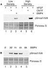
BMP4 causes SMAD activation in both low- and high-density stem cell cultures. Cells were treated with bFGF ± CNTF or BMP4 (10 ng/ml each) for 1 h. Normalized whole cell extracts were used for Western blot analysis with an anti-phosphoSMAD antibody recognizing activated Smad1, 5, and 8 (47 kD for Smad1 and 5; 40 kD for Smad8). (A) BMP4 treatment causes SMAD activation in both low- (lane 3) and high-density (lane 6) cultures. Control (lanes 1 and 4) or CNTF-treated (lanes 2 and 5) stem cells have no SMAD activation. (B) Time course of SMAD phosphorylation during BMP4 (10 ng/ ml) ± bFGF (10 ng/ml) treatment of high-density cultures. BMP4 causes SMAD activation within 30 min (lane 2) that decreases at 8 h (lane 6). The lower panels of both figures show a Ponceau S staining of the nitrocellulose filter to demonstrate normalized loading of proteins across all lanes.
Figure 3.
Figure 3.
BMP4 causes STAT activation only in high-density stem cell cultures. (A) Cells were treated with bFGF ± CNTF for 1 h or BMP4 for 8 h (10 ng/ml each). Nuclear extracts were subjected to EMSA analysis using a 32P-radiolabeled STAT binding site (double-stranded oligonucleotide) to measure STAT activation. Basal (bFGF treated only) levels of STAT binding are minimal in low-density cultures (lane 1) but are increased in high-density cultures (lane 4). CNTF treatment causes a strong increase in STAT binding in both low- (lane 2) and high-density (lane 5) cultures. BMP4 treatment of low-density cultures (lane 3) has no effect on STAT binding, whereas BMP4 treatment of high-density cultures (lane 6) causes increased STAT binding. The arrow marks complexes of Stat3 homodimers with STAT binding site oligonucleotides. The fainter bands below are complexes of Stat1 and Stat3 heterodimers with STAT binding site oligonucleotides. (B, top) Cortical stem cells were treated with BMP4 for 20, 40, or 60 min at 10 ng/ml, denaturing extracts prepared and analyzed on SDS-PAGE gels. The immunoblot was probed sequentially with antibodies specific for Stat3 phosphorylated on Ser727 and panStat3 for total levels of Stat3. BMP causes phosphorylation of Stat3 on Ser727, as seen in the top band of the figure. (B, bottom) Mouse cortical stem cells were cultured as described in the Materials and methods section and treated with BMP4 for 20 min before fixation. Cells were then stained with an antibody specific to the phosphor-ser727 Stat3 protein and viewed by indirect immunofluorescence. The DAPI staining is blue, and pser727Stat3 staining is red.
Figure 4.
Figure 4.
BMP4-mediated activation of Stat3 does not require transcription. Cortical stem cells were expanded in mass culture to high local density using bFGF (10 ng/ml) and then treated with 10 ng/ml BMP4 for the time points indicated in the absence (lanes 1–5) or presence (lanes 6–10) of 5 μg/ml α-amanitin. Control samples were treated with α-amanitin alone (lanes 11–14). Nuclear extracts were subjected to EMSA analysis using a 32P-radiolabeled STAT binding site (double-stranded oligonucleotide) to measure STAT activation. Inhibition of transcription with α-amanitin does not prevent Stat3 binding in response to BMP4.
Figure 5.
Figure 5.
BMP4-mediated activation of Stat3, and glial differentiation, requires FRAP. (A) BMP4 causes dissociation of BMPR-IA and FKBP12. Cells were treated with BMP4 (10 ng/ml) for the duration of 30 min to 8 h, control sample was treated with bFGF for 8 h. Normalized, nondenatured whole cell lysates were immunoprecipitated with anti-FKBP12 before Western blot analysis using anti–BMPR-IA. BMP4 treatment causes a dissociation of BMPR-IA–FKBP12 complexes (arrow indicates 64-kD band). The lower panel shows the levels of FKBP12 in the immunoprecipitated complexes across all lanes. (B) BMP- mediated STAT activation is blocked by rapamycin. High-density stem cell cultures with enhanced STAT binding (lane 1) showed a further increase in STAT binding after BMP4 (10 ng/ml) treatment for 9 h (lane 2). Pretreatment with rapamycin for 12 h before BMP4 addition blocked further Stat3 binding (lane 3). (C) BMP4 treatment causes increased complex formation between FRAP and Stat3. Cells were treated with bFGF alone or with CNTF for 15 min or BMP4 for 4 h (10 ng/ml each). Note that 15 min represents the time of maximal Stat3 activation by CNTF. Normalized, nondenatured whole cell lysates were immunoprecipitated with anti-FRAP before Western blot analysis using anti-Stat3. Arrow indicates 91-kD band. Estimation of the levels of FRAP in the immunoprecipitated complexes could not be performed under the conditions optimal for Stat3 detection because FRAP is a substantially larger protein, 287 kD. (D) Activated FRAP is required for BMP-mediated glial differentiation. Cortical stem cells were transfected with NH2-terminal FLAG-tagged dominant-negative FRAP mutants, Asp2357Glu, which is kinase dead, and Trp2027Phe, which is FRB defective. Cells were transfected for 3 h, treated with BMP4 in the presence of bFGF (both 10 ng/ml) for 2 d and BMP4 alone for 4 d, and then fixed and stained for GFAP and FLAG. Cells were counted by indirect immunofluorescence, and those expressing GFAP were calculated as a percentage of cells expressing FLAG protein. Control cultures were transfected with a GFP-expressing plasmid, which had no effect on BMP-mediated glial differentiation. (E) 80% of GFP-positive cells coexpress GFAP, whereas 25% of mutant FRAP–expressing cells coexpress GFAP, indicating that dominant-negative FRAP inhibits BMP-mediated GFAP expression. Mean GFAP+ cells as a percentage of total transfected cells, **P < 0.001. Bar, 20 μm.
Figure 6.
Figure 6.
BMP4-mediated glial differentiation is rapamycin sensitive, whereas smooth muscle differentiation is rapamycin insensitive. CNS stem cells were grown to medium or high local density before treatment. (A–E) Dense core of E14.5 cortical stem cell colonies after treatment and immunostaining for GFAP. (A) Simple withdrawal of bFGF (control) yields glia with immature morphology. (B) Treatment with CNTF before mitogen withdrawal yields CNS astrocytes with mature morphology. (C) Treatment with BMP4 before mitogen withdrawal yields glia with more flattened morphology. (D) Cotreatment with rapamycin blocks BMP4-mediated glial differentiation. (E) Cotreatment with FK506 has no effect on BMP4-mediated glial differentiation. (F and G) Quantitation of GFAP and SMA staining at the transitional zones in (F) medium-size/density E14.5 colonies, (G) large, high-density E14.5 colonies, and (H) medium-size/density adult colonies. Rectangles in cartoon colonies mark transitional zones chosen for quantitation; note that these zones have sizeable numbers of GFAPSMA cells. Mean GFAP+ or SMA+ cells as percentage of total cells ± SEM (n = 6–9). Bar, 100 μm.
Figure 7.
Figure 7.
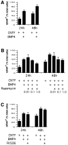
BMP4 augments CNTF-mediated glial differentiation of CNS stem cells. (A) Cells were treated with bFGF + CNTF (10 ng/ml) for 24 or 48 h and stained with anti-GFAP to measure glial differentiation. Where indicated, cells were treated with BMP4 for 4 h before CNTF treatment. BMP4 augments the effect of CNTF in generating glia at both 24 and 48 h of treatment. (B) Rapamycin blocks this augmentation in a dose-dependent manner (doses in μM). (C) Using FK506 (10 nM) instead of rapamycin does not affect the BMP-mediated augmentation of glial differentiation. Mean GFAP+ cells as percentage of total cells ± SEM, n = 4.
Figure 8.
Figure 8.
Model for recruitment of BMP signals in fate choice decisions. BMP4 causes the activation of at least two signaling pathways in neural stem cells. Upon ligand–receptor binding, the BMPR receptor complex releases FKBP12 and activates SMAD proteins, resulting in smooth muscle differentiation. The released FKBP12 may bind with rapamycin to inhibit FRAP or may act in some modified form to activate FRAP. FRAP then catalyzes serine phosphorylation (S*) of STAT to augment its prior activation by tyrosine phosphorylation (Y*) by another signal. High cell density (described in text) acts to promote basal STAT activation and DNA binding by an unknown signaling mechanism. This enhanced activation of STAT (Y*S*) causes efficient glial differentiation. Levels of activated STAT proteins in the cell dictate whether the STAT or SMAD signal acquires precedence in the fate choice between smooth muscle and glia.

Similar articles

Cited by

References

    1. Abraham, R.T., and G.J. Wiederrecht. 1996. Immunopharmacology of rapamycin. Annu. Rev. Immunol. 14:483–510. - PubMed
    1. Bjornson, C.R., R.L. Rietze, B.A. Reynolds, M.C. Magli, and A.L. Vescovi. 1999. Turning brain into blood: a hematopoietic fate adopted by adult neural stem cells in vivo. Science. 283:534–537. - PubMed
    1. Boeuf, H., K. Merienne, S. Jacquot, D. Duval, M. Zeniou, C. Hauss, B. Reinhardt, Y. Huss-Garcia, A. Dierich, D.A. Frank, et al. 2001. The ribosomal S6 kinases, cAMP-responsive element-binding, and STAT3 proteins are regulated by different leukemia inhibitory factor signaling pathways in mouse embryonic stem cells. J. Biol. Chem. 276:46204–46211. - PubMed
    1. Bonni, A., Y. Sun, M. Nadal-Vicens, A. Bhatt, D.A. Frank, I. Rozovsky, N. Stahl, G.D. Yancopoulos, and M.E. Greenberg. 1997. Regulation of gliogenesis in the central nervous system by the JAK-STAT signaling pathway. Science. 278:477–483. - PubMed
    1. Chen, Y.G., F. Liu, and J. Massague. 1997. Mechanism of TGFβ receptor inhibition by FKBP12. EMBO J. 16:3866–3876. - PMC - PubMed

Publication types

MeSH terms

Substances