. 2003 Jun;23(12):4356-70.
doi: 10.1128/MCB.23.12.4356-4370.2003.

A novel jmjC domain protein modulates heterochromatization in fission yeast

Affiliations

A novel jmjC domain protein modulates heterochromatization in fission yeast

Nabieh Ayoub et al. Mol Cell Biol. 2003 Jun.

Abstract

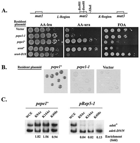
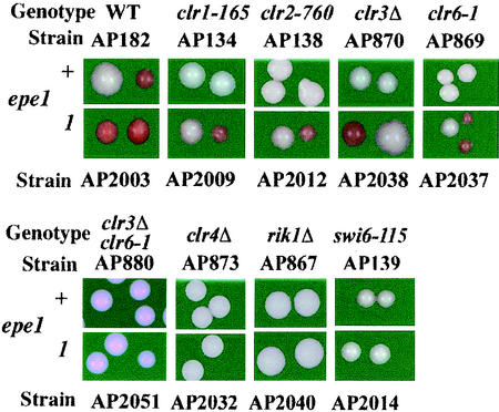
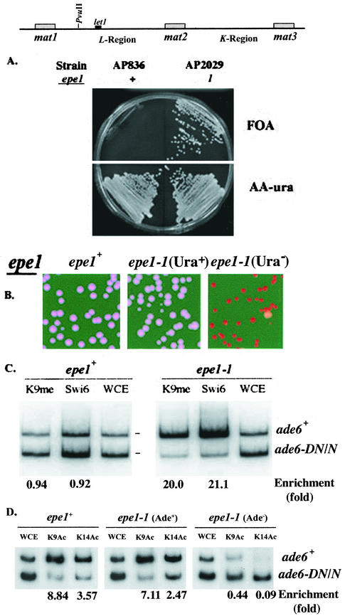
The heterochromatin domain at the mat locus of Schizosaccharomyces pombe is bounded by the IR-L and IR-R barriers. A genetic screen for mutations that promote silencing beyond IR-L revealed a novel gene named epe1, encoding a conserved nuclear protein with a jmjC domain. Disruption of epe1 promotes continuous spreading of heterochromatin-associated histone modifications and Swi6 binding to chromatin across heterochromatic barriers. It also enhances position effect variegation at heterochromatic domains, suppresses mutations in silencing genes, and stabilizes the repressed epigenetic state at the mat locus. However, it does not enhance silencing establishment. Our analysis suggests that the jmjC domain is essential for Epe1 activity and that Epe1 counteracts transcriptional silencing by negatively affecting heterochromatin stability. Consistent with this proposition, the meiotic stability of established heterochromatin beyond IR-L is diminished by Epe1 activity, and overexpression of Epe1 disrupts heterochromatin through acetylation of H3-K9 and H3-K14 and methylation of H3-K4. Furthermore, overexpression of Epe1 elevates the rate of chromosome loss. We propose that Epe1 helps control chromatin organization by down-regulating the stability of epigenetic marks that govern heterochromatization.


Figures

FIG. 1.
FIG. 1.
The epe1-1 mutation promotes silencing beyond the heterochromatin barriers at IR-L and IR-R. (A) The mat locus of S. pombe. The mat2-P and mat3-M mating type donor cassettes are located within a heterochromatin domain that is flanked by the IR-L and IR-R barriers. mat2-P and mat3-M are separated from each other by a repressed region named K. mat2-P is separated from the transcriptionally active mat1 by the L region. The locations of the mat cassettes, the essential let1 gene, the cen2 homology (cenH), IR-L, IR-R, and relevant restriction sites are indicated. (B) Cells of strains with a ura4+ insertion at the HpaI site in the L region or at the SpeI site on the centromere-distal side of IR-R were inoculated from colonies on the indicated media into a nonselective liquid medium (YEA) and grown to a density of 2 × 10⁷ cells per ml. The indicated media were spotted with 10-fold serial dilutions of the cultures (N.S., nonselective medium), and plates were incubated at 33°C for 4 days. The epe1+ and epe1-1 strains with a ura4+ insertion at the HpaI site are AP219 and AP2007, respectively. The epe1+ and epe1-1 strains with a ura4+ insertion at the SpeI site are AP876 and AP2050, respectively. The control strains are AP114 (ura4+) and SP1172 (ura4-D18).
FIG. 2.
FIG. 2.
epe1-1 enhances PEV at the centromeres and cenH-dependent silencing at an ectopic site. (A) Cells of strains with ura4+ insertions within centromere 1 outer repeats (otr1) or inner core (cnt1) were grown on nonselective (N/S) medium (YEA), subjected to 10-fold serial dilutions, and applied as spots to plates with the indicated media. Because lower temperature enhances repression (3), the effect of epe1 genotype on centromere silencing was measured at 36°C. Vertical lines on the cen1 map indicate ura4+ insertion sites. The strains with a ura4+ insertion within otr1 are FY988 (epe1+) and AP2026 (epe1-1). The strains with a ura4+ insertion within cnt1 are FY312 (epe1+) and AP2027 (epe1-1). (B) Cells of strains with a cenH-ura4+ insertion within ade6 and the indicated epe1 genotype were grown on AA −ura or FOA plates and then transferred to nonselective medium. The indicated media were spotted with 10-fold serial dilutions of the cultures and incubated at 33°C for 4 days. The epe1+ strain is AP274. The epe1-1 strain is AP2023.
FIG. 3.
FIG. 3.
epe1-1 is a recessive nonsense mutation in a gene encoding a jmjC domain nuclear protein. (A) Cells of haploid and diploid strains with an ade6+ insertions at the SacI site in the L region (Fig. 1A) were patched onto a YE plate and photographed after 4 days at 30°C. The haploid strains are AP161 (epe1+) and AP2002 (epe1-1). The diploid strains were constructed and maintained as described in Materials and Methods. (B) Genetic analysis and complementation testing identified epe1 as the C622.16C open reading frame on chromosome III. The location of epe1 on a 70-kb fragment of chromosome III and the locations of the jmjC domain and the putative nuclear localization signal (NLS) on Epe1 are indicated. (C) A complementation test. Cells of an epe1-1 mutant with an ade6+ insertion at the L(SacI) site (AP2005) were transformed with pRep-1 (30) or with its derivatives, containing the C622.16C sequence of an epe1+ strain (pepe1+), an epe1-1 mutant (pepe1-1), or an epe1 sequence with a Y307A mutation (pepe1-Y307A). Transformants were plated on leucine-depleted low-adenine medium and incubated at 30°C. (D) Comparative sequence analysis reveals that epe1-1 is a G-to-A transition mutation within the jmjC domain of the gene. This mutation replaced a TGG tryptophan codon with a TGA stop codon (underlined) at position 996 of the gene. Numbers indicate nucleotide locations with respect to the beginning of the jmjC domain (positions 251 to 290), and the beginning of the open reading frame (positions 980 to 1019). (E) Subcellular localization of Epe1. Cells of an epe1-1 mutant (AP2005) harboring a plasmid encoding an Epe1-GFP fusion protein (pepe1-GFP) or the vector plasmid (pRep1-GFP) were examined by phase microscopy (Phase), fluorescence microscopy to localize the fusion protein or GFP (GFP), and DAPI staining of DNA (DAPI).
FIG. 4.
FIG. 4.
Epe1 is a conserved protein. Alignment of Epe1 with the six most-related protein sequences. The sequences and accession numbers are as follows: S. pombe NP_588188 (aa 138 to 473), A. gambiae EAA01048 (aa 29 to 371), D. melanogaster AAF54335 (aa 73 to 417), H. sapiens BAA76848 (aa 41 to 368); M. musculus AAD21792 (aa 89 to 431), Caenorhabditis elegans AAL02526 (aa 1 to 313), S. cerevisiae AAB64586 (aa 130 to 474), and the jmjC domain consensus sequence (pfam02373). Comparison and alignment were performed with the Pileup procedure of the GCG Wisconsin Package, version 10.2, with default parameters. Solid boxes represent identical amino acids, dark gray boxes indicate a high degree of similarity, and light gray boxes indicate weak similarity. Dotted lines indicate gaps introduced to maximize alignment.
FIG. 5.
FIG. 5.
The mutated epe1 allele is required for meiotic stability of the repressed state beyond the IR-L barrier. Segregation of the mat1-M and mat1-P alleles is followed in all three crosses by the linkage of mat1-P to LEU2. (A and B) Both parent strains in crosses A and B are epe1-1. The mat1-M parent displayed an Ade+ epitype (white) in cross A and an Ade epitype (red) in cross B. Cosegregation of the expressed (A) or repressed (B) epialleles of L(HpaI)::ade6+ with the genetically linked mat1-M allele indicates that the respective epialleles are inherited in cis. (C) The meiotic stability of the repressed epiallele of L(HpaI)::ade6+ depends on the maintenance of the epe1-1 allele. The mat1-P parent in this cross is epe1+, and the mat1-M parent is epe1-1, displaying an Ade epitype. Examples of different segregation patterns of crosses in which mat1-M cosegregated with the epe1-1 allele (rows 1 and 3) or with the epe1+ allele (rows 2 and 4) are presented. Segregation of the different alleles and epialleles in all three crosses is presented in Table 3.
FIG. 6.
FIG. 6.
Overexpression of Epe1 disrupts heterochromatin structure at the mat silent domain. (A) Cultures of AP312 cells [K(XbaI)::ura4+] harboring the indicated plasmids were subjected to 10-fold serial dilutions and applied as spots to leucine-depleted minimal medium (AA −leu) and on the same medium with uracil also depleted (AA −ura) or supplemented with FOA. Plates were incubated at 33°C for 5 days before photography. (B) Colonies of AP312 cells harboring the indicated plasmids were replica plated from leucine-depleted minimal medium onto leucine-depleted sporulation medium and stained with iodine vapor following 3 days of incubation at 30°C. (C) ChIP analysis of extracts from cells with an ade6+ insertion within mat2 (AP284), harboring the indicated plasmids, with antibodies against acetylated H3-K9 (K9Ac), acetylated H3-K14 (K14Ac), and dimethylated H3-K4 (K4Me). The locations of the electrophoretically separated ade6+ and ade6-DN/N PCR products are indicated. Enrichment values (ade6+/ade6-DN-N) were normalized for the ratio in whole-cell extract (WCE).
FIG. 7.
FIG. 7.
epe1-1 suppresses mutations in silencing genes. Cells of epe1+ and epe1-1 derivatives with the indicated silencing gene mutations were plated on YE medium and incubated for 3 days at 30°C. The occurrence of Ade (red) colonies in spreads of epe1-1 derivatives indicates suppression of the respective mutations by epe1-1. WT, wild type.
FIG. 8.
FIG. 8.
Silencing at the L region of an epe1-1 mutant. (A) Cells of strains with a ura4+ insertion within let1, an ade6+ insertion at the L(PvuII) site, and the indicated epe1 allele were spread on FOA and AA −ura plates and incubated at 33°C for 3 days. (B) Cells of strains with a ura4+ insertion within let1, an ade6+ insertion at the L(PvuII) site, and the indicated epe1 allele from AA −ura (Ura+) or FOA (Ura) medium were spread on YE plates and incubated for 3 days at 30°C. (C) ChIP analysis of extracts from an epe1+ (AP836) strain or a Ura clone of an epe1-1 (AP2029) mutant. Antibodies are specific for Swi6 (Swi6) and dimethylated H3-K9 (K9Me), and PCR primers amplify ade6 sequences. The locations of the electrophoretically separated ade6+ and ade6-DN/N PCR products are indicated. Enrichment values (ade6+/ade6-DN-N) were normalized for the ratio in whole-cell extract (WCE). (D) ChIP analysis of extracts from an epe1+ strain and Ade+ or Ade clone of an epe1-1 mutant, with antibodies against acetylated H3-K9 (K9Ac) and acetylated H3-K14 (K14Ac). Experimental conditions are as described for panel C.
FIG. 9.
FIG. 9.
Heterochromatin spreads continuously along the L region in epe1-1 mutants. Association of Swi6 and H3-K9methyl with chromatin along the mat locus was determined by ChIP analysis with antibodies to Swi6 (Swi6) and dimethylated H3-K9 (K9me). DNA isolated from cross-linked chromatin, immunoprecipitated with the respective antibodies, or whole-cell extracts (WCE) was subjected to multiplex PCR to amplify the fragments indicated by horizontal bars on the mat locus map and an act1 fragment serving as internal control. Cell extracts were from epe1+ (AP836) and epe1-1 (AP2029) strains with a ura4+ insertion within let1 and an ade6+ insertion at the L(PvuII) site. Extracts of two AP2029 clones were analyzed. One was selected for a Ura epitype on FOA medium (FOA). The nonselected clone (N/S) was mainly Ura+. Enrichment with respect to the mat/act1 DNA ratio in whole-cell extract at each of the indicated sites along the mat locus was determined as described previously (35).

Similar articles

Cited by

References

    1. Allshire, R. C. 1997. Centromeres, checkpoints and chromatid cohesion. Curr. Opin. Genet. Dev. 7:264-275. - PubMed
    1. Allshire, R. C. 1996. Transcriptional silencing in the fission yeast: a manifestation of higher order chromosome structure and functions, p. 443-466. In V. E. A. Russo, R. A. Martienssen, and A. D. Riggs (ed.), Epigenetic mechanisms of gene regulation. Cold Spring Harbor Laboratory, Cold Spring Harbor, N.Y.
    1. Allshire, R. C., J.-P. Javerzat, N. J. Redhead, and G. Cranston. 1994. Position effect variegation at fission yeast centromeres. Cell 76:157-169. - PubMed
    1. Allshire, R. C., E. R. Nimmo, E. Ekwall, J. P. Javerzat, and G. Cranston. 1995. Mutations derepressing silent centromeric domains in fission yeast disrupt chromosome segregation. Genes Dev. 9:218-233. - PubMed
    1. Altschul, S. F., T. L. Madden, A. A. Schaffer, J. Zhang, Z. Zhang, W. Miller, and D. J. Lipman. 1997. Gapped BLAST and PSI-BLAST: a new generation of protein database search programs. Nucleic Acids Res. 25:3389-3402. - PMC - PubMed

Publication types

MeSH terms