. 2003 Jun;23(12):4247-56.
doi: 10.1128/MCB.23.12.4247-4256.2003.

PML colocalizes with and stabilizes the DNA damage response protein TopBP1

Affiliations

PML colocalizes with and stabilizes the DNA damage response protein TopBP1

Zhi-Xiang Xu et al. Mol Cell Biol. 2003 Jun.

Abstract

The PML tumor suppressor gene is consistently disrupted by t(15;17) in patients with acute promyelocytic leukemia. Promyelocytic leukemia protein (PML) is a multifunctional protein that plays essential roles in cell growth regulation, apoptosis, transcriptional regulation, and genome stability. Our study here shows that PML colocalizes and associates in vivo with the DNA damage response protein TopBP1 in response to ionizing radiation (IR). Both PML and TopBP1 colocalized with the IR-induced bromodeoxyuridine single-stranded DNA foci. PML and TopBP1 also colocalized with Rad50, Brca1, ATM, Rad9, and BLM. IR and interferon (IFN) coinduce the expression levels of both TopBP1 and PML. In PML-deficient NB4 cells, TopBP1 was unable to form IR-induced foci. All-trans-retinoic acid induced reorganization of the PML nuclear body (NB) and reappearance of the IR-induced TopBP1 foci. Inhibition of PML expression by siRNA is associated with a significant decreased in TopBP1 expression. Furthermore, PML-deficient cells express a low level of TopBP1, and its expression cannot be induced by IR or IFN. Adenovirus-mediated overexpression of PML in PML(-/-) mouse embryo fibroblasts substantially increased TopBP1 expression, which colocalized with the PML NBs. These studies demonstrated a mechanism of PML-dependent expression of TopBP1. PML overexpression induced TopBP1 protein but not the mRNA expression. Pulse-chase labeling analysis demonstrated that PML overexpression stabilized the TopBP1 protein, suggesting that PML plays a role in regulating the stability of TopBP1 in response to IR. Together, our findings demonstrate that PML regulates TopBP1 functions by association and stabilization of the protein in response to IR-induced DNA damage.


Figures

FIG. 1.
FIG. 1.
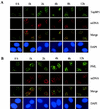
(A) TopBP1 colocalizes in vivo with PML at various time points after IR exposure. SiHa cells were exposed to 15 Gy of IR. Double-color immunofluorescence staining was performed with a monoclonal antibody to TopBP1 (green) and our polyclonal PML antibody (red) at 0, 1, 4, 8, 12, and 36 h after IR treatment. Nuclear DNA in the cells was then counterstained with DAPI (blue). Images were captured by using the Kodak digital imaging system mount on top of a Leica fluorescence microscope. Fluorescent images were recorded and superimposed (Merge), by using the Adobe Photoshop 6.0 software. (B) Colocalization of TopBP1 and PML with Rad 50, Rad9, ATM, Brca1, and BLM after exposure of SiHa cells to 15 Gy of IR. Double-color immunofluorescence staining was performed at 8 h after the SiHa cells were exposed to IR.
FIG. 2.
FIG. 2.
Colocalization of PML and TopBP1 with the ssDNA foci after IR. SiHa cells were labeled with BrdU, as described in Materials and Methods, and exposed to 15 Gy of gamma irradiation. Cells were fixed 0, 1, 2, 4, 8, and 12 h after IR, and double-color immunofluorescence staining was performed with BrdU monoclonal antibody and polyclonal PML antibody (A) or BrdU polyclonal antibody and monoclonal TopBP1 antibody (B), as described in Materials and Methods. Images were recorded as described in the Fig. 1 legend.
FIG. 3.
FIG. 3.
The numbers of PML NBs, IR-induced TopBP1 foci, and ssDNA foci at various time points after IR exposure. Proliferating SiHa cells were exposed to 15 Gy of gamma irradiation, and immunofluorescence staining was performed at 0, 2, 4, 6, 8, and 12 h after irradiation. For detection of IR-induced ssDNA foci, proliferating SiHa cells were incubated with 10 μg of BrdU/ml for 30 h before irradiation. The numbers of PML NBs, TopBP1 foci, and ssDNA foci represent an average of 200 nuclei per time point. (A and B) The percentage of nuclei containing >15 PML NBs and >10 IR-induced TopBP1 foci increased progressively after IR exposure. (C) The percentage of IR-induced ssDNA foci after SiHa cell exposed to IR. Cells were divided into three different groups (i.e., 0 to 20 foci, 21 to 40 foci, and >40 foci) by the number of ssDNA foci formed after IR exposure. The results obtained at each time point represent an average percentages of ssDNA foci in 200 nuclei.
FIG. 4.
FIG. 4.
Coinduction of PML and TopBP1 in vivo. (A) Expression of TopBP1 and PML in response to various dosages of IR in SiHa cells. Cells were treated with the indicated dosages of gamma irradiation, and total proteins were isolated for Western blot analysis 12 h after IR exposure. The same filter was reprobed with anti-α-tubulin antibody to serve as a control. (B) Time course induction of TopBP1 and PML in SiHa cells exposed to 15 Gy of IR. Cells were harvested, total proteins were isolated at the indicated time points, and Western blotting was performed with the respective antibodies. (C to E) Induction of PML significantly increased the expression of TopBP1. (C) PML expression was induced by various concentrations of IFN-α, total protein was isolated after 24 h, and Western blotting was performed with TopBP1 and PML antibodies. (D) PML expression was induced or not induced with 100 μM ZnSO4 and then treated or not treated with 15 Gy of IR. Total proteins were isolated after 24 h, and Western blot analysis was performed as described in Materials and Methods. (E and F) Ad-PML infection increased the expression of TopBP1. SiHa cells were infected with increasing concentrations of Ad-PML and antisense recombinant PML adenovirus (As-PML). Total proteins were isolated after 24 h, and the expression levels of PML and TopBP1 were analyzed by Western blotting.
FIG. 5.
FIG. 5.
PML-dependent upregulation of TopBP1 by IR. (A and D) Effects of inhibition of PML expression by siRNA on IR-induced expression of TopBP1. U2OS cells were transfected with siRNA as described in Materials and Methods and cultured for 48 h. Cells were then exposed to 10 Gy of IR and harvested after an additional 8 h. Total proteins were isolated for Western blot analysis (A) or fixed on slides for immunofluorescence staining (D). (B and F) TopBP1 fails to form IR-induced foci in the PML-deficient NB4 cells and express low levels of TopBP1. NB4 cells with or without treatment with 1 μM ATRA for 72 h were irradiated with 10 Gy of IR. (F) Cells were then collected and immobilized on slides by cytospin, and immunofluorescence staining was performed with TopBP1 monoclonal and PML polyclonal antibodies. Cell nuclei were visualized by staining with DAPI. (B) For Western blot analysis, NB4 cells were treated with ATRA and IR as described above, and total proteins were isolated 12 h after IR. Each lane was loaded with 200 μg of protein, except for the SiHa cells, in which only 100 μg of protein per lane was loaded. (C) IFN fails to induce TopBP1 expression in PML−/− MEFs. PML−/− MEFs and normal MEFs were treated with 2,000 U of IFN-α/ml for 24 h. Total proteins were isolated, and Western blot analysis was performed with PML and TopBP1 antibodies. (E) IFN induced colocalization of PML and TopBP1. SiHa cells treated with 2,000 U of IFN-α/ml or untreated were fixed on slides. Double color immunofluorescence staining was performed with polyclonal PML antibody and monoclonal TopBP1 antibody. (G) Adenovirus-mediated reexpression of PML in PML−/− MEFs induced TopBp1 expression. PML−/− MEFs were infected at a multiplicity of infection of 25 with Ad-PML as described previously (12, 21). Double-color immunofluorescence staining was performed with the PML polyclonal antibody and TopBP1 monoclonal antibody.
FIG. 6.
FIG. 6.
Inducible expression of PML enhanced the stability of TopBP1. (A) Northern blot analysis of TopBP1 expression in the PML-inducible stable cell line. U2OS/PML and the control U2OS/pMEP4 cells were induced with 100 μM ZnSO4 for 16 h. Cells were then exposed to 15 Gy of IR. Total RNA was isolated and Northern blotting was carried out as described in Materials and Methods. (B and C) Inducible PML expression stabilizes TopBP1 protein in U2OS cells. U2OS/PML and U2OS/pMEP4 cells were induced with 100 μM ZnSO4 for 16 h and pulse-labeled with [35S]methionine and [35S]cysteine for 45 min. Immediately after labeling, the cells were irradiated with 15 Gy of IR and chased for different periods of time as indicated. Total cell lysates were isolated and immunoprecipitated with mouse anti-TopBP1 monoclonal antibody (B) or mouse anti-Mre11 monoclonal antibody (C). The precipitated proteins were resolved by SDS-10% PAGE and autoradiographed. The intensities of the protein bands in the autoradiograph were quantified by scanning with the Alpha Innotech gel documentation system. (D) Effects of PML overexpression and IR exposure on cell cycle distribution in U2OS cells. U2OS/PML and U2OS/pMEP4 cells were induced with 100 μM Zn2+ for 16 h and then exposed to 10 Gy of IR. Cells were then continued to culture for the indicated time points. Cell cycle distribution was determined by flow cytometry.
FIG. 7.
FIG. 7.
In vivo association between PML and TopBP1. (A and B) Coimmunoprecipitation of PML and TopBP1 in a U2OS/PML stable cell line. Total protein (800 μg) isolated from the U2OS/PML cells induced with 100 μM of ZnSO4 was used in the immunoprecipitation assay. Coimmunoprecipitation was carried out with the TopBP1 or PML antibody, and then Western blotting was performed with PML or TopBP1 antibody. A nonspecific immunoglobulin G control was also included. (C and D) The N-terminal portion of PML is required for in vivo association with TopBP1. U2OS cells were transfected with pCDNA3/myc-TopBP1 and various pcDNA3/his-PML mutants. At 24 h after transfection, total cell lysates were prepared and immunoprecipitated with nonspecific (i.e., immunoglobulin G) or anti-myc antibody. (D) Precipitated proteins were resolved by SDS-10% PAGE, and Western blot analysis was performed with the His-tagged specific antibody. (C) Expression of the PML mutant plasmids in the cotransfection experiment was confirmed by Western blot analysis. The same filter was reprobed with α-tubulin to serve as an internal control.

Similar articles

Cited by

References

    1. Alcalay, M., L. Thomassoni, E. Colombo, S. Stoldt, F. Grignani, M. Gagioli, L. Szekely, K. Helin, and P. G. Pellicci. 1998. The promyelocytic leukemia gene product (PML) forms stable complexes with the retinoblastoma protein. Mol. Cell. Biol. 18:1084-1093. - PMC - PubMed
    1. Bischof, O., S. H. Kim, J. Irving, S. Beresten, N. A. Ellis, and J. Campisi. 2001. Regulation and localization of the Bloom syndrome protein in response to DNA damage. J. Cell Biol. 153:367-380. - PMC - PubMed
    1. Boisvert, F.-M., M. J. Kruhlak, A. K. Box, M. J. Hendzel, and D. P. Bazett-Jones. 2001. The transcription coactivator CBP is a dynamic component of the promyelocytic leukemia nuclear body. J. Cell Biol. 152:1099-1106. - PMC - PubMed
    1. Carbone, R., M. Pearson, S. Minucci, and P. P. Pelicci. 2002. PML NBs associate with hMre11 complex and p53at sites of irradiation-induced DNA damage. Oncogene 21:1633-1640. - PubMed
    1. Coultas, L., and A. Strasser. 2000. The molecular control of DNA damage-induced cell death. Apoptosis 5:491-507. - PubMed

Publication types

MeSH terms