. 2003 May;111(9):1309-18.
doi: 10.1172/JCI16288.

Estrogen modulates cutaneous wound healing by downregulating macrophage migration inhibitory factor

Affiliations

Estrogen modulates cutaneous wound healing by downregulating macrophage migration inhibitory factor

Gillian S Ashcroft et al. J Clin Invest. 2003 May.

Abstract

Characteristic of both chronic wounds and acute wounds that fail to heal are excessive leukocytosis and reduced matrix deposition. Estrogen is a major regulator of wound repair that can reverse age-related impaired wound healing in human and animal models, characterized by a dampened inflammatory response and increased matrix deposited at the wound site. Macrophage migration inhibitory factor (MIF) is a candidate proinflammatory cytokine involved in the hormonal regulation of inflammation. We demonstrate that MIF is upregulated in a distinct spatial and temporal pattern during wound healing and its expression is markedly elevated in wounds of estrogen-deficient mice as compared with intact animals. Wound-healing studies in mice rendered null for the MIF gene have demonstrated that in the absence of MIF, the excessive inflammation and delayed-healing phenotype associated with reduced estrogen is reversed. Moreover, in vitro assays have shown a striking estrogen-mediated decrease in MIF production by activated murine macrophages, a process involving the estrogen receptor. We suggest that estrogen inhibits the local inflammatory response by downregulating MIF, suggesting a specific target for future therapeutic intervention in impaired wound-healing states.


Figures

Figure 1
Figure 1
MIF localizes to keratinocytes, endothelium, and inflammatory cells during wound healing. (ac) Day 3 wounds from BALB/C mice illustrate suprabasal keratinocyte staining for MIF (b, arrow), endothelial cells (c, arrow), and perivascular inflammatory cell staining (c, green arrows). (a) represents IgG isotype control negative staining (arrow indicates epidermis). All scale bars represent 10 μm. (d) Temporal decrease in wound section immunostaining scores for MIF as compared with normal skin (normal skin scored as 0). Results represent medians (black lines across boxes), boxes represent interquartile ranges, and the bars extending from the boxes indicate the highest and lowest values (n = 6 for each group, *P < 0.05 versus day 3).
Figure 2
Figure 2
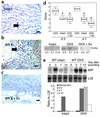
Reduced circulating estrogen leads to enhanced local MIF expression. (ac) Inflammatory cells and endothelium stained for MIF at day 7 after wounding (b, green arrows indicate endothelial cells). Inflammatory cell staining (arrows) for MIF was increased in day-7 wounds of OVX mice (b) as compared with their intact littermates (a). Estrogen (Es) replacement in wild-type OVX mice resulted in a marked reduction in MIF levels (c). All scale bars represent 20 μm. (d) MIF protein levels increased at day 3 (as compared with normal skin, which is scored as 0) and declined by day 14 in the wild-type mice (Western blot, lower panel), with significantly increased levels from days 7–14 in the OVX mice. Results represent medians (black lines across boxes), boxes represent interquartile ranges, and the bars extending from the boxes indicate the highest and lowest values (n = 4, *P < 0.05). Estrogen replacement in OVX mice reversed this increase in MIF. This temporal pattern was confirmed by Western blotting (d, lower panel). By RNase protection assay, MIF mRNA expression peaked at day 3 (e) and had decreased to basal levels by day 14 after wounding in wild-type intact mice. By contrast, MIF expression was more strongly upregulated from days 3–14 after wounding in the wild-type OVX mice. Normal skin is scored as 0, and the housekeeping gene is denoted by L32. For both RNA and protein analyses, four mice per time point were used.
Figure 3
Figure 3
Reduced estrogen has no effect on wound healing in mice null for MIF. Day 3 wounds were wider in OVX wild-type mice (c) with an increased inflammatory response as compared with intact littermates (a). In marked contrast, no differences were observed between wounds from MIF null intact mice (b) and MIF null OVX mice (d). Arrows demarcate wound edges. In (c), the panniculus carnosus muscle is at the extreme edges of the image. Scale bars for ad represent 100 μm. In the OVX MIF null wounds at day 7 (f), increased collagen I deposition was observed as compared with wounds from OVX wild-type mice (e). An absence of staining on parallel sections using an isotype control antibody is shown (g). Scale bars for eg represent 10 μm. Wound areas were significantly greater in OVX wild-type mice than in OVX MIF null mice at day 3 after wounding (h). Results represent means ± SEM (n = 3–5 for each group, *P < 0.05). Wound strength evaluated by disruption of day-3, -5, and -7 wounds in vivo using a BTC1000 device is shown (i). A significant increase in wound strength was observed at days 5 and 7 as compared with day 3. Results represent means ± SEM (n = 4–5 for each group, *P < 0.05 compared to day-3 data). At day 7 after wounding (j), the ultimate breaking strength (mmHg) was significantly reduced in the OVX wild-type mice as compared with OVX MIF null mice and wild-type intact mice. Results represent means ± SEM (n = 4, *P < 0.05).
Figure 4
Figure 4
Reduced inflammation in the MIF null wounds. (a) An absence of MIF leads to reduced wound blood-vessel area (vessel area presented as a percentage of total wound area) at day 7; however, an absence of estrogen has no effect on neovascularization. Results represent means ± SEM (n = 4, *P < 0.05 for comparisons between intact null and intact wild-type or OVX null and OVX wild-type). (b) Mac3 staining illustrates increased inflammatory cell infiltrate in the OVX wild-type wounds as compared with the OVX MIF null wounds at day 3. Scale bars represent 20 μm. Cell numbers per unit area were quantified at day 3 (c). Results represent means ± SEM (n = 7–10 for each group, *P < 0.05). (d) TNF-α protein levels were increased in the wild-type OVX wounds as compared with the MIF null OVX wounds from days 3–14 after wounding (d, graph) by quantification of immunostaining. Normal skin is scored as 0. Results represent medians (black lines across boxes), boxes represent interquartile ranges, and the bars extending from the boxes indicate the highest and lowest values (n = 7, *P < 0.05). All data are taken from OVX mice. Left panels represent day-3 immunostaining, illustrating increased cell numbers positive for TNF-α in the wild-type OVX wounds. Scale bars represent 50 μm.
Figure 5
Figure 5
Treatment of MIF null wounds with TGF-β (200 ng per wound) results in delayed healing as compared with vehicle-treated wounds (a, top panels). Wounds are representative of day 3 after wounding. Arrows demarcate wound edges. Treatment of MIF null wounds with rhMIF results in impaired healing (a, bottom panels) (day 7 after wounding, 1-μg dose) as compared with vehicle (PBS). Scale bar represents 100 μm. Results represent means ± SEM (n = 5, *P < 0.05). (b) Neutralization of MIF in wild-type OVX mice with anti-MIF antibodies leads to improved healing as compared with vehicle alone (day 3 after wounding). Arrows demarcate wound edges, which cannot be visualized in the OVX mice. Graph illustrates wound areas at day 3 after wounding in a dose-response study with increasing doses of anti-MIF antibodies injected at the time of wounding. Scale bar represents 100 μm. Results represent means ± SEM (n = 5, *P < 0.05, **P < 0.01). C, no injection at wound site; PBS, vehicle control.
Figure 6
Figure 6
Murine macrophage MIF production, by LPS-activated cells, is decreased with concurrent estrogen treatment (a). Results represent means ± SEM (n = 10 for each group) *P < 0.05, **P < 0.01, all compared with control media plus LPS-activated macrophages (C+). LPS activation significantly increased MIF production as compared with control media alone (P < 0.05). (b) Immunostaining illustrates that monocytes (CD14+ cells) express both ER-α and ER-β isoforms. The far right panel of b represents a merged image of all three panels (ER-α, ER-β, and CD14). Scale bar represents 10 μm. (c) LPS treatment increases MIF levels from wild-type murine cells and is inhibited by estrogen. In the absence of ER-α (ER-α null), estrogen fails to downregulate LPS-induced MIF production. LPS significantly increased MIF in both wild-type and ER-α null macrophages (P < 0.05). Seeding density of cells is three times higher than in Figure 5a. Results represent means ± SEM (n = 5 for each group, *P < 0.05 as compared with LPS-activated cells). (d) MCF7 ER-expressing cells transfected with a MIF promoter–luciferase reporter construct showed increased activity after LPS activation (a). Estrogen (10–8 M) inhibited this activity. Transfection with pGL3 basic plasmid acted as a negative control (pGL3–ve), and pGL3 control plasmid as a positive control (pGL3+ve). Results represent means ± SEM (n = 3–5 for each group, *P < 0.05 as compared with LPS-activated cells). (e) MCF7 ER-expressing cells transfected with a MIF promoter–luciferase reporter construct showed inhibition of LPS-activated MIF production by estrogen and estrogen agonists (PPT, HPTE) and reversal of estrogenic effects by ER antagonist (ICI). Results represent means ± SEM for three experiments (*P < 0.05 as compared with LPS-activated cells). C, control media alone; +, + LPS treatment for 6 hours; E8, 10–8 M estrogen; E9, 10–9 M estrogen; E10, 10–10 M estrogen; E11, 10–11 M estrogen; RLU, relative light units.

Similar articles

Cited by

References

    1. Ashcroft GS, et al. Secretory leukocyte protease inhibitor (SLPI) mediates non-redundant functions necessary for normal wound healing. Nat. Med. 2000;6:1147–1153. - PubMed
    1. Ashcroft GS, Dodsworth J, Boxtel E, Horan M, Ferguson MWJ. Estrogen accelerates cutaneous wound healing associated with an increase in TGF-β1 levels. Nat. Med. 1997;3:1209–1215. - PubMed
    1. Calvin M, Dyson M, Rymer J, Young SR. The effects of ovarian hormone deficiency on wound contraction in a rat model. Br. J. Obstet. Gynaecol. 1998;105:223–227. - PubMed
    1. Jorgensen O, Schmidt A. Influence of sex hormones on granulation tissue formation and on healing of linear wounds. Acta Chir. Scand. 1962;124:1–10. - PubMed
    1. Pallin B, Ahonen J, Zederfeldt B. Granulation tissue formation in oophorectomized rats treated with female sex hormones: II. Acta Chir. Scand. 1975;141:710–714. - PubMed

Publication types

MeSH terms