In vivo evolution of human immunodeficiency virus type 1 toward increased pathogenicity through CXCR4-mediated killing of uninfected CD4 T cells
- PMID: 12719578
- PMCID: PMC154038
- DOI: 10.1128/jvi.77.10.5846-5854.2003
In vivo evolution of human immunodeficiency virus type 1 toward increased pathogenicity through CXCR4-mediated killing of uninfected CD4 T cells
Abstract
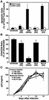
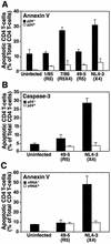
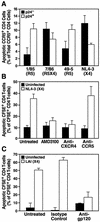
The destruction of the immune system by progressive loss of CD4 T cells is the hallmark of AIDS. CCR5-dependent (R5) human immunodeficiency virus type 1 (HIV-1) isolates predominate in the early, asymptomatic stages of HIV-1 infection, while CXCR4-dependent (X4) isolates typically emerge at later stages, frequently coinciding with a rapid decline in CD4 T cells. Lymphocyte killing in vivo primarily occurs through apoptosis, but the importance of apoptosis of HIV-1-infected cells relative to apoptosis of uninfected bystander cells is controversial. Here we show that in human lymphoid tissues ex vivo, apoptosis of uninfected bystander CD4 T cells is a major mechanism of lymphocyte depletion caused by X4 HIV-1 strains but is only a minor mechanism of depletion by R5 strains. Further, X4 HIV-1-induced bystander apoptosis requires the interaction of the viral envelope glycoprotein gp120 with the CXCR4 coreceptor on CD4 T cells. These results emphasize the contribution of bystander apoptosis to HIV-1 cytotoxicity and suggest that in association with a coreceptor switch in HIV disease, T-cell killing evolves from an infection-restricted stage to generalized toxicity that involves a high degree of bystander apoptosis.
Figures


Similar articles
-
HIV-1 induced bystander apoptosis.Viruses. 2012 Nov 9;4(11):3020-43. doi: 10.3390/v4113020. Viruses. 2012. PMID: 23202514 Free PMC article. Review.
-
Apoptosis of bystander T cells induced by human immunodeficiency virus type 1 with increased envelope/receptor affinity and coreceptor binding site exposure.J Virol. 2004 May;78(9):4541-51. doi: 10.1128/jvi.78.9.4541-4551.2004. J Virol. 2004. PMID: 15078935 Free PMC article.
-
CXCR4 utilization is sufficient to trigger CD4+ T cell depletion in HIV-1-infected human lymphoid tissue.Proc Natl Acad Sci U S A. 1999 Jan 19;96(2):663-8. doi: 10.1073/pnas.96.2.663. Proc Natl Acad Sci U S A. 1999. PMID: 9892690 Free PMC article.
-
Distinct mechanisms of CD4+ and CD8+ T-cell activation and bystander apoptosis induced by human immunodeficiency virus type 1 virions.J Virol. 2005 May;79(10):6299-311. doi: 10.1128/JVI.79.10.6299-6311.2005. J Virol. 2005. PMID: 15858014 Free PMC article.
-
Autophagy and CD4+ T lymphocyte destruction by HIV-1.Autophagy. 2007 Jan-Feb;3(1):32-4. doi: 10.4161/auto.3275. Epub 2007 Jan 14. Autophagy. 2007. PMID: 17012832 Review.
Cited by
-
HIV-1 induced bystander apoptosis.Viruses. 2012 Nov 9;4(11):3020-43. doi: 10.3390/v4113020. Viruses. 2012. PMID: 23202514 Free PMC article. Review.
-
Switching of inferred tropism caused by HIV during interruption of antiretroviral therapy.J Clin Microbiol. 2010 Jul;48(7):2586-8. doi: 10.1128/JCM.02125-09. Epub 2010 May 19. J Clin Microbiol. 2010. PMID: 20484604 Free PMC article.
-
3D Tissue Explant and Single-Cell Suspension Organoid Culture Systems for Ex Vivo Drug Testing on Human Tonsil-Derived T Follicular Helper Cells.Methods Mol Biol. 2022;2380:267-288. doi: 10.1007/978-1-0716-1736-6_22. Methods Mol Biol. 2022. PMID: 34802138
-
Initiation of ART during early acute HIV infection preserves mucosal Th17 function and reverses HIV-related immune activation.PLoS Pathog. 2014 Dec 11;10(12):e1004543. doi: 10.1371/journal.ppat.1004543. eCollection 2014 Dec. PLoS Pathog. 2014. PMID: 25503054 Free PMC article. Clinical Trial.
-
Cyanovirin-N produced in rice endosperm offers effective pre-exposure prophylaxis against HIV-1BaL infection in vitro.Plant Cell Rep. 2016 Jun;35(6):1309-19. doi: 10.1007/s00299-016-1963-5. Epub 2016 Mar 23. Plant Cell Rep. 2016. PMID: 27007716 Free PMC article.
References
-
- Ameisen, J. C., and A. Capron. 1991. Cell dysfunction and depletion in AIDS: the programmed cell death hypothesis. Immunol. Today 12:102-105. - PubMed
-
- Bagasra, O., S. P. Hauptman, H. W. Lischner, M. Sachs, and R. J. Pomerantz. 1992. Detection of human immunodeficiency virus type 1 provirus in mononuclear cells by in situ polymerase chain reaction. N. Engl. J. Med. 326:1385-1391. - PubMed
Publication types
MeSH terms
Substances
Grants and funding
LinkOut - more resources
Full Text Sources
Other Literature Sources
Research Materials

