. 2003 May;23(9):3363-72.
doi: 10.1128/MCB.23.9.3363-3372.2003.

The Grb10/Nedd4 complex regulates ligand-induced ubiquitination and stability of the insulin-like growth factor I receptor

Affiliations

The Grb10/Nedd4 complex regulates ligand-induced ubiquitination and stability of the insulin-like growth factor I receptor

Andrea Vecchione et al. Mol Cell Biol. 2003 May.

Abstract

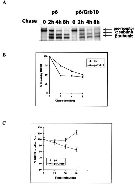
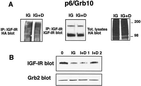
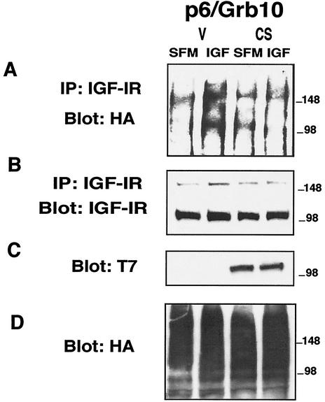
The adapter protein Grb10 belongs to a superfamily of related proteins, including Grb7, -10, and -14 and Caenorhabditis elegans Mig10. Grb10 is an interacting partner of the insulin-like growth factor I receptor (IGF-IR) and the insulin receptor (IR). Previous work showed an inhibitory effect of mouse Grb10 (mGrb10alpha) on IGF-I-mediated mitogenesis (A. Morrione et al., J. Biol. Chem. 272:26382-26387, 1997). With mGrb10alpha as bait in a yeast two-hybrid screen, mouse Nedd4 (mNedd4-1), a ubiquitin protein ligase, was previously isolated as an interacting protein of Grb10 (A. Morrione et al., J. Biol. Chem. 274:24094-24099, 1999). However, Grb10 is not ubiquitinated by Nedd4 in cells. Here we show that in mouse embryo fibroblasts overexpressing Grb10 and the IGF-IR (p6/Grb10), there is a strong ligand-dependent increase in ubiquitination of the IGF-IR compared with that in parental cells (p6). This increased ubiquitination is associated with a shorter half-life and increased internalization of the IGF-IR. The IGF-IR is stabilized following treatment with both MG132 and chloroquine, indicating that both the proteasome and lysosomal pathways mediate degradation of the receptor. Ubiquitination of the IGF-IR likely occurs at the plasma membrane, prior to the formation of endocytic vesicles, as it is insensitive to dansylcadaverine, an inhibitor of early endosome formation in IGF-IR endocytosis. Grb10 coimmunoprecipitates with the IGF-IR and endogenous Nedd4 in p6/Grb10 cells, suggesting the presence of a Grb10/Nedd4/IGF-IR complex. Ubiquitination of the IGF-IR in p6/Grb10 cells is severely impaired by overexpression of a catalytically inactive Nedd4 mutant (Nedd4-CS), which also stabilizes the receptor. Likewise, overexpression of a Grb10 mutant lacking the Src homology 2 (SH2) domain impaired ubiquitination of the IGF-IR in parental p6 and p6/Grb10 cells, indicating that Grb10 binding to Nedd4 is critical for ubiquitination of the receptor. These results suggest a role for the Grb10/Nedd4 complex in regulating ubiquitination and stability of the IGF-IR, and they suggest that Grb10 serves as an adapter to form a bridge between Nedd4 and the IGF-IR. This is the first demonstration of regulation of stability of a tyrosine kinase receptor by the Nedd4 (HECT) family of E3 ligases.


Figures

FIG. 1.
FIG. 1.
Grb10 increases ligand-dependent ubiquitination of the IGF-IR in cells. p6 and p6/Grb10 cells were plated in triplicate onto six-well plates and transiently transfected with the HA-Ub construct. After 24 h, cells were shifted to SFM for an additional 24 h and then stimulated for 10 min with 20 ng of IGF-I/ml in the presence of 20 μM MG132 and 0.4 mM chloroquine to accumulate the ubiquitinated forms. Cell lysates were pooled, and 800 μg was immunoprecipitated (IP) with anti-IGF-IR antibody and blotted with anti-HA antibodies to detect ubiquitinated species (A). This blot was then stripped and reprobed with antiphosphotyrosine antibodies (B) and anti-IGF-IR antibodies to test for the amount of receptor immunoprecipitated (C). Total lysates served as a positive control for HA-Ub transfection (D). Results in panels A to D are representative of three independent experiments. (E) The IGF-IR, but not coprecipitated protein(s), is ubiquitinated in p6/Grb10 cells. After transient transfections as above, lysates of IGF-I-stimulated p6/Grb10 cells were treated with 1% SDS and boiled for 5 min prior to immunoprecipitation with anti-IGF-IR antibodies. After immunoprecipitation, the filter was probed with anti-HA (left) to detect IGF-IR ubiquitination or anti-IGF-IR (center) antibodies (control). Total lysate served as a positive control for HA-Ub transfection (right). (F) IGF-I-induced ubiquitination of the IGF-IR in p6/Grb10 cells is detectable at endogenous levels of ubiquitin. Lysates (2 mg) of IGF-I-stimulated p6/Grb10 cells were immunoprecipitated with anti-IGF-IR antibodies. After immunoprecipitation the filter was probed with antiubiquitin (UB) antibody (left and right) to detect the ubiquitinated IGF-IR. The blot with anti-IGF-IR antibodies (center) is a control. Results in panels E and F are representative of two independent experiments.
FIG. 2.
FIG. 2.
Grb10 is involved in regulating IGF-IR stability and internalization. p6 and p6/Grb10 were serum starved in serum-free Met-Cys-free medium, pulsed with [35S]Met-Cys for 1 h, and chased in SFM supplemented with 20 ng of IGF-I/ml for 0, 2, 4, and 8 h. Cells were then lysed, and 300 μg of protein was immunoprecipitated with anti-IGF-IR antibodies. (A) Immunocomplexes were washed, resolved by SDS-PAGE, dried, and exposed to X-ray film. The IGF-IR is newly synthesized as a proreceptor and then processed into α and β subunits. Results are representative of three independent experiments. (B) Quantitation of IGF-IR using densitometric analysis. The graph was generated with the ImageQuant program. (C) Decreased level of cell surface IGF-IR in p6/Grb10 cells. The level of cell surface IGF-IR in p6 and p6/Grb10 cells was determined by ELISA at different times of IGF-I stimulation, as described in Materials and Methods. IGF-I was used at 200 ng/ml to ensure a saturating concentration of the ligand. The data are the averages of three independent experiments.
FIG. 3.
FIG. 3.
Ligand-stimulated IGF-IR degradation in p6/Grb10 cells is mediated by both the proteasome and lysosomal pathways. (A) p6 or p6/Grb10 cells were serum starved and then stimulated with 20 ng of IGF-I/ml for the indicated times. (B) IGF-IR levels were determined by immunoblotting with anti-IGF-IR polyclonal antibodies. p6/Grb10 cells were stimulated for 20 h with IGF-I alone or supplemented with 20 μM MG132 (MG), 0.4 mM chloroquine (Chl), or both (M+C). The total amount of protein loaded on the gel was monitored with anti-Grb2 monoclonal antibodies (A and B). Results are representative of three independent experiments. Densitometric analysis of the representative experiment was performed with the ImageQuant program, and results are expressed as the percentage of IGF-IR remaining.
FIG. 4.
FIG. 4.
Ubiquitination of the IGF-IR occurs prior to the formation of endocytotic vesicles. (A) p6/Grb10 cells were plated in triplicate onto six-well plates and transiently transfected with HA-Ub. After 24 h, cells were shifted to SFM for an additional 24 h and then stimulated for 10 min with 20 ng of IGF-I/ml in the presence of MG132 and chloroquine (IG). Dansylcadaverine (500 μM) was added at the time of stimulation with IGF-I (IG+D). Cell lysates were pooled, and 800 μg was immunoprecipitated with anti-IGF-IR antibody and blotted with anti-HA antibodies to detect ubiquitinated species. This blot was then stripped and reprobed with anti-IGF-IR antibodies to test for the amount of receptor immunoprecipitated. Total lysates served as positive controls for HA-Ub transfection. (B) p6/Grb10 cells were stimulated for 20 h with IGF-I alone or supplemented with 100 (I+D 1) or 200 (I+D 2) μM dansylcadaverine. IGF-IR levels were determined by Western blotting with anti-IGF-IR polyclonal antibodies. The total amount of protein loaded on the gel was monitored with anti-Grb2 monoclonal antibodies. Results are representative of two independent experiments.
FIG. 5.
FIG. 5.
Grb10 coimmunoprecipitates with the IGF-IR and endogenous Nedd4. Serum-starved p6/Grb10 cells were stimulated with IGF-I for 10 min and lysed, and proteins were immunoprecipitated with anti-Myc antibodies to immunoprecipitate Myc-tagged Grb10 (36, 38). Total lysates were used to show the correct sizes of coprecipitated proteins. In panel E, the third and lower isoform of Grb10 is detected by anti-Grb10 antibodies after longer exposure of the film (not shown). To detect Nedd4 association with the IGF-IR, serum-starved p6 and p6/Grb10 cells were stimulated with IGF-I for 10 min and lysed, and proteins were immunoprecipitated with anti-IGF-IR antibodies.
FIG. 6.
FIG. 6.
Catalytically inactive Nedd4 impairs ubiquitination of the IGF-IR in p6/Grb10 cells. p6/Grb10 cells were transiently cotransfected with either an empty vector (V) or Nedd4(CS) and HA-Ub. IGF-IR was then immunoprecipitated, and levels of its ubiquitination were determined with anti-HA antibodies (A), as described for Fig. 1. (B) Level of the IGF-IR immunoprecipitated; (C) same filter reprobed with anti-T7 antibodies to detect the expression of Nedd4(CS); (D) lysates probed with anti-HA antibodies to ensure equal expression of the HA-Ub plasmid. Results are representative of three independent experiments.
FIG. 7.
FIG. 7.
Catalytically inactive Nedd4(CS) mutant stabilizes the IGF-IR in p6/Grb10 cells. p6/Grb10 cells were transiently transfected with Nedd4(CS). Cells were starved in serum-free Met-Cys-free medium, pulsed with [35S]Met-Cys for 1 h, and chased in SFM supplemented with 20 ng of IGF-I/ml for 8 h. Cells were then lysed, and 300 μg of proteins was immunoprecipitated with anti-IGF-IR antibodies. Immunocomplexes were washed, resolved by SDS-PAGE, dried, and exposed to X-ray film. Densitometric analysis was performed with the ImageQuant program, and results are expressed as the percentage of IGF-IR remaining. (Lower panel) Immunoblot using anti-T7 antibodies to detect the transiently transfected T7-tagged Nedd4(CS) mutant. Results are representative of three independent experiments.
FIG. 8.
FIG. 8.
Overexpression of a Grb10 mutant lacking the SH2 domain impairs ubiquitination of the IGF-IR in p6 and p6/Grb10 cells. p6 and p6/Grb10 cells were transiently cotransfected with either an empty vector (V) or the ΔSH2 mutant and HA-Ub. After IGF-I stimulation, cell lysates were pooled, immunoprecipitated with anti-IGF-IR antibody, and blotted with anti-HA antibodies to detect ubiquitinated species (A and B). This blot was then stripped and reprobed with anti-IGF-IR antibodies (C and D) to determine the amount of receptor immunoprecipitated. Panels E and F show the same filter reprobed with anti-Myc antibodies to detect the expression of Myc-tagged wild-type and mutant Grb10 proteins. In panel F, the endogenous Grb10 is not detectable by anti-Myc antibodies. Total lysates blotted with anti-HA antibodies (G and H) serve as positive controls for HA-Ub transfection. Results are representative of three independent experiments.
FIG. 9.
FIG. 9.
Model for the role of Grb10 as an adapter connecting Nedd4 to the IGF-IR. The Grb10 SH2 domain constitutively binds the C2 domain of Nedd4. Upon ligand stimulation, Grb10 binds the activated IGF-IR through the BPS domain, allowing the formation of a complex that includes the activated IGF-IR, Grb10, and Nedd4. These interactions lead to ligand-mediated and Nedd4-dependent ubiquitination and degradation of the IGF-IR.

Similar articles

Cited by

References

    1. Abriel, H., J. Loffing, J. F. Rebhun, J. H. Pratt, L. Schild, J. D. Horisberger, D. Rotin, and O. Staub. 1999. Defective regulation of the epithelial Na+ channel by Nedd4 in Liddle's syndrome. J. Clin. Investig. 103:667-673. - PMC - PubMed
    1. Aviel, S., G. Winberg, M. Massucci, and A. Ciechanover. 2000. Degradation of the Epstein-Barr virus latent membrane protein 1 (LMP1) by the ubiquitin-proteasome pathway. Targeting via ubiquitination of the N-terminal residue. J. Biol. Chem. 275:23491-23499. - PubMed
    1. Bai, R. Y., T. Jahn, S. Schrem, G. Munzert, K. M. Weidner, J. Y. Wang, and J. Duyster. 1998. The SH2-containing adapter protein GRB10 interacts with BCR-ABL. Oncogene 17:941-948. - PubMed
    1. Beaudenon, S. L., M. R. Huacani, G. Wang, D. P. McDonnell, and J. M. Huibregtse. 1999. Rsp5 ubiquitin-protein ligase mediates DNA damage-induced degradation of the large subunit of RNA polymerase II in Saccharomyces cerevisiae. Mol. Cell. Biol. 19:6972-6979. - PMC - PubMed
    1. Bereziat, V., A. Kasus-Jacobi, D. Perdereau, B. Cariou, J. Girard, and A. F. Burnol. 2002. Inhibition of insulin receptor catalytic activity by the molecular adapter Grb14. J. Biol. Chem. 277:4845-4852. - PubMed

Publication types

MeSH terms

Substances