Chemokine receptor mutant CX3CR1-M280 has impaired adhesive function and correlates with protection from cardiovascular disease in humans
- PMID: 12697743
- PMCID: PMC152935
- DOI: 10.1172/JCI16790
Chemokine receptor mutant CX3CR1-M280 has impaired adhesive function and correlates with protection from cardiovascular disease in humans
Abstract
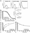
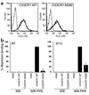
The chemokine receptor CX3CR1 is a proinflammatory leukocyte receptor specific for the chemokine fractalkine (FKN or CX3CL1). In two retrospective studies, CX3CR1 has been implicated in the pathogenesis of atherosclerotic cardiovascular disease (CVD) based on statistical association of a common receptor variant named CX3CR1-M280 with lower prevalence of atherosclerosis, coronary endothelial dysfunction, and acute coronary syndromes. However, the general significance of CX3CR1-M280 and its putative mechanism of action have not previously been defined. Here we show that FKN-dependent cell-cell adhesion under conditions of physiologic shear is severely reduced in cells expressing CX3CR1-M280. This was associated with marked reduction in the kinetics of FKN binding as well as reduced FKN-induced chemotaxis of primary leukocytes from donors homozygous for CX3CR1-M280. We also show that CX3CR1-M280 is independently associated with a lower risk of CVD (adjusted odds ratio, 0.60, P = 0.008) in the Offspring Cohort of the Framingham Heart Study, a long-term prospective study of the risks and natural history of this disease. These data provide mechanism-based and consistent epidemiologic evidence that CX3CR1 may be involved in the pathogenesis of CVD in humans, possibly by supporting leukocyte entry into the coronary artery wall. Moreover, they suggest that CX3CR1-M280 is a genetic risk factor for CVD.
Figures


Comment in
-
The fractalkine receptor CX3CR1 is a key mediator of atherogenesis.J Clin Invest. 2003 Apr;111(8):1118-20. doi: 10.1172/JCI18237. J Clin Invest. 2003. PMID: 12697729 Free PMC article. No abstract available.
Similar articles
-
Opposite effects of CX3CR1 receptor polymorphisms V249I and T280M on the development of acute coronary syndrome. A possible implication of fractalkine in inflammatory activation.Thromb Haemost. 2005 May;93(5):949-54. doi: 10.1160/TH04-11-0735. Thromb Haemost. 2005. PMID: 15886814
-
Atherogenic lipids induce adhesion of human coronary artery smooth muscle cells to macrophages by up-regulating chemokine CX3CL1 on smooth muscle cells in a TNFalpha-NFkappaB-dependent manner.J Biol Chem. 2007 Jun 29;282(26):19167-76. doi: 10.1074/jbc.M701642200. Epub 2007 Apr 23. J Biol Chem. 2007. PMID: 17456471
-
Viral macrophage inflammatory protein-II and fractalkine (CX3CL1) chimeras identify molecular determinants of affinity, efficacy, and selectivity at CX3CR1.Mol Pharmacol. 2004 Dec;66(6):1431-9. doi: 10.1124/mol.104.003277. Epub 2004 Sep 10. Mol Pharmacol. 2004. PMID: 15361546
-
The chemokine CX3CL1 (fractalkine) and its receptor CX3CR1: occurrence and potential role in osteoarthritis.Arch Immunol Ther Exp (Warsz). 2014 Oct;62(5):395-403. doi: 10.1007/s00005-014-0275-0. Epub 2014 Feb 21. Arch Immunol Ther Exp (Warsz). 2014. PMID: 24556958 Free PMC article. Review.
-
Fractalkine/CX3CR1 signalling in chronic pain and inflammation.Curr Pharm Biotechnol. 2011 Oct;12(10):1707-14. doi: 10.2174/138920111798357465. Curr Pharm Biotechnol. 2011. PMID: 21466443 Review.
Cited by
-
Genetic variants in platelet factor 4 modulate inflammatory and platelet activation biomarkers.Circ Cardiovasc Genet. 2012 Aug 1;5(4):412-21. doi: 10.1161/CIRCGENETICS.111.961813. Epub 2012 Jul 4. Circ Cardiovasc Genet. 2012. PMID: 22763266 Free PMC article.
-
Tumor necrosis factor-alpha induces fractalkine expression preferentially in arterial endothelial cells and mithramycin A suppresses TNF-alpha-induced fractalkine expression.Am J Pathol. 2004 May;164(5):1663-72. doi: 10.1016/s0002-9440(10)63725-x. Am J Pathol. 2004. PMID: 15111313 Free PMC article.
-
Monocyte subsets differentially employ CCR2, CCR5, and CX3CR1 to accumulate within atherosclerotic plaques.J Clin Invest. 2007 Jan;117(1):185-94. doi: 10.1172/JCI28549. J Clin Invest. 2007. PMID: 17200718 Free PMC article.
-
The fate of monocytes in atherosclerosis.J Thromb Haemost. 2009 Jul;7 Suppl 1(Suppl 1):28-30. doi: 10.1111/j.1538-7836.2009.03423.x. J Thromb Haemost. 2009. PMID: 19630762 Free PMC article. Review.
-
Fractalkine promotes human monocyte survival via a reduction in oxidative stress.Arterioscler Thromb Vasc Biol. 2014 Dec;34(12):2554-62. doi: 10.1161/ATVBAHA.114.304717. Epub 2014 Oct 30. Arterioscler Thromb Vasc Biol. 2014. PMID: 25359863 Free PMC article.
References
-
- Desai MM, Zhang P, Hennessy CH. Surveillance for morbidity and mortality among older adults—United States, 1995-1996. MMWR CDC Surveill. Summ. 1999;48:7–25. 2. - PubMed
-
- Wilson PW. Metabolic risk factors for coronary heart disease: current and future prospects. Curr. Opin. Cardiol. 1999;14:176–185. - PubMed
-
- Myers RH, Kiely DK, Cupples LA, Kannel WB. Parental history is an independent risk factor for coronary artery disease: the Framingham Study. Am. Heart J. 1990;120:963–969. - PubMed
-
- Ross R. Atherosclerosis—an inflammatory disease. N. Engl. J. Med. 1999;340:115–126. - PubMed
-
- O’Donnell CJ, Levy D. Weighing the evidence for infection as a risk factor for coronary heart disease. Curr. Cardiol. Rep. 2000;2:280–287. - PubMed
Publication types
MeSH terms
Substances
LinkOut - more resources
Full Text Sources
Other Literature Sources
Molecular Biology Databases
Research Materials
Miscellaneous

