. 2003 May;77(9):5415-27.
doi: 10.1128/jvi.77.9.5415-5427.2003.

Recruitment of Tat to heterochromatin protein HP1 via interaction with CTIP2 inhibits human immunodeficiency virus type 1 replication in microglial cells

Affiliations

Recruitment of Tat to heterochromatin protein HP1 via interaction with CTIP2 inhibits human immunodeficiency virus type 1 replication in microglial cells

Olivier Rohr et al. J Virol. 2003 May.

Abstract

The Tat protein of human immunodeficiency virus type 1 (HIV-1) plays a key role as inducer of viral gene expression. We report that Tat function can be potently inhibited in human microglial cells by the recently described nuclear receptor cofactor chicken ovalbumin upstream promoter transcription factor-interacting protein 2 (CTIP2). Overexpression of CTIP2 leads to repression of HIV-1 replication, as a result of inhibition of Tat-mediated transactivation. In contrast, the related CTIP1 was unable to affect Tat function and viral replication. Using confocal microscopy to visualize Tat subcellular distribution in the presence of the CTIPs, we found that overexpression of CTIP2, and not of CTIP1, leads to disruption of Tat nuclear localization and recruitment of Tat within CTIP2-induced nuclear ball-like structures. In addition, our studies demonstrate that CTIP2 colocalizes and associates with the heterochromatin-associated protein HP1alpha. The CTIP2 protein harbors two Tat and HP1 interaction interfaces, the 145-434 and the 717-813 domains. CTIP2 and HP1alpha associate with Tat to form a three-protein complex in which the 145-434 CTIP2 domain interacts with the N-terminal region of Tat, while the 717-813 domain binds to HP1. The importance of this Tat binding interface and of Tat subnuclear relocation was confirmed by analysis of CTIP2 deletion mutants. Our findings suggest that inhibition of HIV-1 expression by CTIP2 correlates with recruitment of Tat within CTIP2-induced structures and relocalization within inactive regions of the chromatin via formation of the Tat-CTIP2-HP1alpha complex. These data highlight a new mechanism of Tat inactivation through subnuclear relocalization that may ultimately lead to inhibition of viral pathogenesis.


Figures

FIG. 1.
FIG. 1.
Effect of CTIP1 and CTIP2 on HIV-1 LTR-driven transcription and on viral replication. (A) Microglial cells were cotransfected with vectors expressing HIV-1 LTR-CAT (1 μg) in the presence of increasing amounts (0.1, 0.5, and 1 μg) of CTIP1 and CTIP2, as indicated. After 2 days, CAT activities were measured and are expressed relative to the value obtained with LTR-CAT alone. Values correspond to an average of at least three independent experiments done in duplicate. Error bars, standard deviations. (B) Cells were cotransfected with HIV-1 pNL4-3 (1.5 μg) and with increasing amounts (0.1, 0.5, and 1 μg) of the indicated CTIP1 and CTIP2 expression vectors. Two days after transfection, samples of the culture supernatant were analyzed for p24 Gag content. Data represent an average of four independent experiments performed in duplicate. They are expressed relative to the value obtained with pNL4-3 alone taken as 1. Depending on the cell confluency, this value varied between 500 and 5,000 pg/ml.
FIG. 2.
FIG. 2.
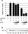
CTIP2, but not CTIP1, inhibits Tat-mediated HIV-1 transcriptional activity. (A) Microglial cells were transfected with HIV-1 LTR-CAT (1 μg) in the presence of vectors expressing Tat (2 ng) and increasing amounts (0.1, 0.5, and 1 μg) of CTIP1 and CTIP2 as indicated. Histograms show CAT activities measured 2 days posttransfection and expressed relative to the value obtained with the LTR-CAT vector alone. Values correspond to an average of at least three independent experiments done in duplicate. Error bars, standard deviations. (B) Western blot analysis of nuclear proteins (20 μg) extracted from cells transfected with the plasmids corresponding to lanes 1 to 8 indicated in panel A. To detect Tat, 1 μg of expression vector was transfected. The blot was probed with monoclonal Tat antibodies.
FIG. 3.
FIG. 3.
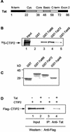
CTIP2 and HIV-1 Tat interact in vitro and in cells. (A) Schematic representation of the domains present in the viral Tat protein. (B) The N-terminal region of Tat interacts with CTIP2. GST pull down assays were performed with 35S-CTIP2 and the indicated GST-Tat fusion proteins. 35S-CTIP2 was translated in vitro and incubated with GST (lane 2) or the indicated full-length and truncated GST-Tat proteins (lanes 3 to 5). After extensive washing, the bound proteins were eluted and analyzed by SDS-polyacrylamide gel electrophoresis. Lane 1, input 35S-CTIP2 (0.2 μl); lanes 2 to 5, GST and GST-Tat incubated with 35S-CTIP2 (10 μl). (C) SDS-PAGE of GST and GST-Tat fusion proteins used in panel B visualized by Coomassie brilliant blue staining. (D) Tat interacts with CTIP2 in vivo. Nuclear protein extracts from microglial cells, previously transfected with CTIP2 in the absence or presence of pCMV-Tat were immunoprecipitated with monoclonal anti-Tat antibodies (lanes 3 and 4). The presence of CTIP2 in the nuclear extracts (lanes 1 and 2) and in the immunoprecipitates (lanes 3 and 4) was detected with anti-Flag antibodies.
FIG. 4.
FIG. 4.
Definition of the in vitro interactions domains of CTIP2 with Tat (A) and HP1α (B). CTIP2 full-length and deletion mutants indicated in the schematic diagram were in vitro translated and used in GST pull down assays to examine their interaction with GST-Tat (A) (lane 3), GST-HP1α (B) (lane 3), and GST as a control (A and B) (lanes 2). Lanes 1, 35S-CTIP2 deletion mutants (0.2 μl); lanes 2 and 3, GST and GST-Tat incubated with the indicated 35S-CTIP2 proteins (10 μl). ND, not determined. Arrows delineate binding interfaces of Tat and HP1α.
FIG. 5.
FIG. 5.
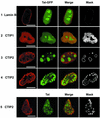
Localization of Tat-GFP and Tat in the presence of CTIP1 and CTIP2 in microglial cell nuclei. Cells were transfected with vectors expressing Tat-GFP (5 ng) in the absence (row 1) or the presence of HA-CTIP1 (row 2) and Flag-CTIP2 (rows 3 and 4). Cells were cotransfected with pCMV-Tat (500 ng) and Flag-CTIP2 (row 5). Cells were fixed 24 h after transfection, incubated with anti-lamin B, and stained with Alexa Fluor 568-conjugated secondary antibodies to detect endogenous lamin B and delineate the nucleus (row 1). To detect CTIP1 and CTIP2, cells were incubated with anti-HA (row 2) and anti-Flag (rows 3, 4, and 5) antibodies and stained with cyanine 3-conjugated secondary antibodies. To detect Tat, cells were incubated with monoclonal Tat antibodies and stained with cyanine 2-conjugated secondary antibodies (row 5). Masks were obtained after selection of the double-labeled pixels in the two-dimensional scatter histograms of gray values constructed from red and green images. Bars: 10 μm. HA-CTIP1 exhibited a diffuse staining pattern, with did not alter the Tat-GFP nucleolar and nuclear fluorescence (row 2). Flag-CTIP2 exhibited a ball-like staining in most of the nuclei. Tat-GFP and Tat were located within these ball-like structures that were distributed randomly in 80 to 90% of the nuclei (rows 3 and 5) or localized at the periphery of the inner nuclear membrane in 10 to 20% of nuclei (row 4). Tat-GFP and Tat colocalize with CTIP2 at the periphery of CTIP2-induced structures (columns 3 and 4).
FIG. 6.
FIG. 6.
CTIP2 colocalizes with HP1α and redistributes Tat-GFP to distinct subnuclear structures within the nucleoplasm or next to the nuclear membrane. Microglial cells were transfected with vectors expressing Tat-GFP (500 ng) in the absence (row 1) or presence of Flag-CTIP2 (rows 2 to 4). Cells were incubated with anti-Flag antibodies and stained with cyanine 5-conjugated secondary antibodies to detect overexpressed CTIP2 (column 1; blue) or incubated with anti-HP1α and stained with Alexa Fluor 568-conjugated secondary antibodies to detect endogenous HP1α (column 3; red). For double immunofluorescence detection, cells were first treated to detect HP1 and then treated to detect CTIP2 (column 4). Masks representing the region of colocalization HP1α/CTIP2 were generated by selecting the double-labeled pixels. Bars: 10 μm. CTIP2 exhibited a staining pattern of distinct structures filled with Tat-GFP (rows 2 and 3). In some nuclei, CTIP2 was peripheral to the nuclear membrane and to ball-like structures of Tat-GFP (row 4). In these nuclei, Tat-GFP recruited at the inner nuclear membrane formed empty ball structures. An arrow points to one of these structures. Note that in all nuclei, HP1α colocalizes with CTIP2 (columns 4 and 5).
FIG. 7.
FIG. 7.
Definition of the CTIP2 in vitro interaction domain of HP1α. The HP1α deletion mutants (indicated in the schematic diagram) were expressed as GST fusion proteins and bound to glutathione-Sepharose beads to examine their interaction with different domains of in vitro translated 35S-labeled full-length CTIP2 and the two indicated deletion mutants. GST and GST-HP1α mutants were incubated with the indicated 35S-CTIP2 proteins (25 μl). Input lane, 35S-CTIP2 (0.2 μl).
FIG. 8.
FIG. 8.
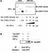
Analysis of the in vitro and in vivo interactions between CTIP2, Tat and HP1α. (A) Analysis of the preferential CTIP2 interaction domain of Tat by GST pull down competition assays. GST-Tat was immobilized on glutathione-Sepharose beads to examine the interaction with the two indicated 35S-labeled CTIP2 deletion mutants, in the absence and in the presence of increasing amounts of 35S-labeled HP1α. Note that HP1α does not bind directly to GST-Tat and that CTIP2 145-434 remains bound to GST-Tat and is not displaced by HP1α. (B) CTIP2 interacts with HP1 in cells only in the presence of CTIP2. Nuclear protein extracts from microglial cells, previously cotransfected with Tat-GFP and HP1 in the absence or presence of CTIP2 were immunoprecipitated with anti-HP1 antibodies and analyzed for Tat-GFP by Western blotting.
FIG. 9.
FIG. 9.
Full-length and 145-434 CTIP2 inhibit HIV-1 LTR-driven transcription and viral replication. (A) Microglial cells were cotransfected with vectors expressing HIV-1 LTR-CAT (1 μg) and Tat (2 ng) in the presence of the indicated CTIP2 deletion mutants (1 μg). CAT assays were performed after 2 days. CAT activities are expressed relative to the value obtained with LTR-CAT alone. Values correspond to an average of at least three independent experiments done in duplicate. (B) Western blot analysis of nuclear proteins extracted from cells transfected for 48 h with the indicated expression vectors used in panel A. The blot was probed with anti-Flag antibodies. Positions of size standards are indicated in kilodaltons. (C) Cells were cotransfected with vectors expressing HIV-1 pNL4-3 (1.5 μg) and the indicated CTIP2 deletion mutants (1 μg). Two days after transfection, samples of the culture supernatant were analyzed for p24 Gag content. Data represent an average of at least three independent experiments performed in duplicate. They are expressed relative to the value obtained with pNL4-3 alone taken as 1, which corresponds to an average of 2000 pg/ml of p24 Gag.
FIG. 10.
FIG. 10.
Tat-GFP distribution is altered in the presence of full-length and 145-434 CTIP2. Microglial cells were transfected with vectors expressing Tat-GFP and the indicated full-length (residues 1 to 813) and deletion mutants of Flag-CTIP2. Nuclei are demarcated by immunolabeling with anti-lamin B antibodies (column 3, red). Cells were incubated with anti-Flag antibodies followed by staining with cyanine 5-conjugated antibodies to detect overexpressed CTIP2 proteins (columns 1 and 4, blue). Bars: 10 μm. Tat-GFP is redistributed within or next to subnuclear structures in nuclei overexpressing full-length and 145-434 CTIP2. In contrast, the nucleoplasmic and nucleolar distribution of Tat-GFP is not altered (compare with Fig. 5, row 1) in cells overexpressing 1-354 and 610-813 CTIP2 proteins in the cytoplasm and nucleus.

Similar articles

Cited by

References

    1. Avram, D., A. Fields, K. Pretty On Top, D. J. Nevrivy, J. E. Ishmael, and M. Leid. 2000. Isolation of a novel family of C(2)H(2) zinc finger proteins implicated in transcriptional repression mediated by chicken ovalbumin upstream promoter transcription factor (COUP-TF) orphan nuclear receptors. J. Biol. Chem. 275:10315-10322. - PMC - PubMed
    1. Bannister, A. J., P. Zegerman, J. F. Partridge, E. A. Miska, J. O. Thomas, R. C. Allshire, and T. Kouzarides. 2001. Selective recognition of methylated lysine 9 on histone H3 by the HP1 chromo domain. Nature 410:120-124. - PubMed
    1. Benkirane, M., R. F. Chun, H. Xiao, V. V. Ogryzko, B. H. Howard, Y. Nakatani, and K. T. Jeang. 1998. Activation of integrated provirus requires histone acetyltransferase. p300 and P/CAF are coactivators for HIV-1 Tat. J. Biol. Chem. 273:24898-24905. - PubMed
    1. Boykins, R. A., R. Mahieux, U. T. Shankavaram, Y. S. Gho, S. F. Lee, I. K. Hewlett, L. M. Wahl, H. K. Kleinman, J. N. Brady, K. M. Yamada, and S. Dhawan. 1999. Cutting edge: a short polypeptide domain of HIV-1-Tat protein mediates pathogenesis. J. Immunol. 163:15-20. - PubMed
    1. Brasher, S. V., B. O. Smith, R. H. Fogh, D. Nietlispach, A. Thiru, P. R. Nielsen, R. W. Broadhurst, L. J. Ball, N. V. Murzina, and E. D. Laue. 2000. The structure of mouse HP1 suggests a unique mode of single peptide recognition by the shadow chromo domain dimer. EMBO J. 19:1587-1597. - PMC - PubMed

Publication types

MeSH terms

LinkOut - more resources