. 2003 May;77(9):5360-9.
doi: 10.1128/jvi.77.9.5360-5369.2003.

Human rhinovirus type 2 is internalized by clathrin-mediated endocytosis

Affiliations

Human rhinovirus type 2 is internalized by clathrin-mediated endocytosis

Luc Snyers et al. J Virol. 2003 May.

Abstract

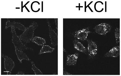
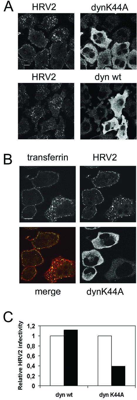
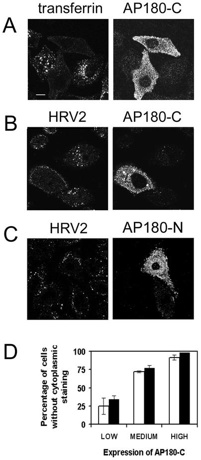
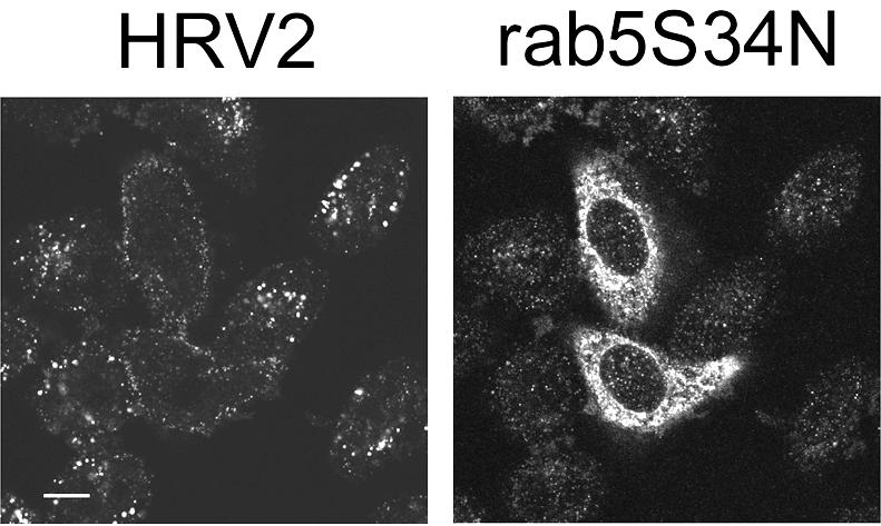
Using several approaches, we investigated the importance of clathrin-mediated endocytosis in the uptake of human rhinovirus serotype 2 (HRV2). By means of confocal immunofluorescence microscopy, we show that K(+) depletion strongly reduces HRV2 internalization. Viral uptake was also substantially reduced by extraction of cholesterol from the plasma membrane with methyl-beta-cyclodextrin, which can inhibit clathrin-mediated endocytosis. In accordance with these data, overexpression of dynamin K44A in HeLa cells prevented HRV2 internalization, as judged by confocal immunofluorescence microscopy, and strongly reduced infection. We also demonstrate that HRV2 bound to the surface of HeLa cells is localized in coated pits but not in caveolae. Finally, transient overexpression of the specific dominant-negative inhibitors of clathrin-mediated endocytosis, the SH3 domain of amphiphysin and the C-terminal domain of AP180, potently inhibited internalization of HRV2. Taken together, these results indicate that HRV2 uses clathrin-mediated endocytosis to infect cells.


Figures

FIG. 1.
FIG. 1.
K+ depletion inhibits HRV2 internalization. HeLa-H1 cells seeded on coverslips were subjected to hypotonic shock for 5 min, washed, and incubated with HRV2 in K+-free medium for 20 min at 34°C. Control cells were treated with the same buffers containing 10 mM K+. Cells were fixed, and HRV2 was visualized by using the anti-HRV2 monoclonal antibody 8F5. Preparations were photographed by using a confocal microscope. Noninfected cells did not show any staining with 8F5. Bar, 10 μm.
FIG. 2.
FIG. 2.
Cholesterol depletion inhibits internalization of HRV2. (A) HeLa-H1 cells in 24-well plates were incubated for 30 min at 34°C with 400 nM bafilomycin (white) or 10 mM MβCD (black) or left untreated (gray). Cells were challenged with HRV2 at 30 PFU/cell for 20 min, washed three times with PBS, and incubated in fresh medium containing the respective drugs for 0, 1, and 3 h at 34°C prior to determination of virus titer, as described in Materials and Methods. Shown are the means and standard errors of two independent experiments. (B) Cells were treated for 30 min with 10 mM MβCD or left untreated as indicated. HRV2 was added, and incubation was continued for 20 min prior to fixation, permeabilization, and immunofluorescence detection of virus. Representative confocal images are shown. Virus is retained on the plasma membrane of MβCD-treated cells. Note that the cells tend to round up as a consequence of the MβCD treatment. Bar, 10 μm.
FIG.3.
FIG.3.
Overexpression of dynK44A inhibits HRV2 uptake in stably transformed HeLa cells. (A) dynK44A or dynwt expression was induced in HeLa cells by incubation without tetracycline. After 48 h, cells were incubated with HRV2 (∼30 PFU/cell) for 20 min at 34°C, fixed, and stained for confocal immunofluorescence detection of virus and dynamin mutants. Cells expressing dynK44A show strongly reduced virus uptake. In these cells, virus clearly accumulates on the plasma membrane. Cells expressing dynwt internalize virus-like untransfected cells. Bar, 10 μm. (B) Cells induced to express HA-tagged dynK44A were incubated with HRV2 and rhodamine-transferrin for 20 min at 34°C, washed, fixed, and stained for immunofluorescence by using anti-HRV2 and anti-HA antibodies. HRV2 (green) and transferrin (red) internalization are blocked in cells overexpressing dynK44A. Note the colocalization (yellow) of virus and transferrin, presumably in early endosomes of the cell that does not express dynK44A. Bar, 10 μm. (C) Cells induced to express HA-dynK44A or HA-dynwt were infected with HRV2 (∼15 PFU/cell) for 20 min, washed, and incubated overnight at 34°C before immunofluorescence detection of virus-producing cells and of the HA tag. For each experiment, a total of ∼250 cells expressing the respective dynamins was observed and the fraction of cells synthesizing virus was determined (black bars). This was divided by the fraction of virus-producing cells that did not express dynamin (white bars).
FIG. 4.
FIG. 4.
Membrane-bound HRV2 is concentrated in coated pits. (A) HeLa-H1 cells were transfected with a GFP-clathrin light-chain A expression plasmid to label coated pits. Two days after transfection, they were incubated with about 30 PFU of HRV2/cell at 4°C for 1 h and processed for confocal immunofluorescence detection of HRV2. Images show the colocalization (yellow) of HRV2 (red) and GFP-clathrin (green) at the plasma membrane of a cell. (B) Same as in panel A, except that cells were incubated for 30 min with 10 mM MβCD prior to incubation with HRV2 for 20 min at 34°C. Note that cells appear rounded due to the action of MβCD. (C) Nontransfected HeLa-H1 cells were incubated with HRV2 for 1 h at 4°C, fixed, permeabilized, and stained for immunofluorescence detection of HRV2 (red) and caveolin-1 (green). Bars, 10 μm.
FIG. 5.
FIG. 5.
HRV2 internalization is blocked by transient overexpression of Amph-SH3. HeLa-H1 cells seeded on coverslips were transfected with an expression plasmid encoding myc-tagged Amph-SH3. Two days after transfection, they were incubated with HRV2, fixed, and permeabilized. HRV2 and Amph-SH3 were detected by using anti-HRV2 monoclonal antibody 8F5 and anti-myc antiserum, respectively, followed by fluorescent conjugates. Cells were examined and photographed by using a confocal microscope. In cells overexpressing Amph-SH3, HRV2 does not accumulate in the perinuclear compartment or in the cytoplasm but remains attached to the plasma membrane. Bar, 10 μM.
FIG. 6.
FIG. 6.
HRV2 internalization is blocked by transient overexpression of AP180-C. (A to C) HeLa-H1 cells seeded on coverslips were transfected with an expression plasmid encoding myc-tagged AP180-C (A and B) or AP180-N (C). Two days after transfection, they were incubated with fluorescent transferrin (A) or HRV2 (B and C), fixed, and permeabilized. HRV2 and AP180-C or AP180-N were detected with anti-HRV2 monoclonal antibody 8F5 and anti-myc antiserum, respectively, followed by fluorescent conjugates. Cells were examined and photographed by using a confocal microscope. In cells overexpressing AP180-C, transferrin and HRV2 internalization are blocked. In cells overexpressing AP180-N, HRV2 internalization is not blocked. Bar, 10 μM. (D) AP180-C expression was rated by visual inspection as being low, medium, or high in fluorescence intensity. The percentage of cells in each group without any cytoplasmic (perinuclear as well as peripheral) fluorescence is given for transferrin and HRV2 (>75 cells/group; mean of three experiments ± standard deviation).
FIG. 7.
FIG. 7.
Rab5 S34N expression reduces uptake of HRV2. HeLa-H1 cells seeded on coverslips were transfected with an expression plasmid encoding myc-tagged Rab5 S34N. Two days after transfection, they were incubated with HRV2, fixed, and permeabilized. HRV2 and Rab5 S34N were detected by using anti-HRV2 monoclonal antibody and anti-myc antiserum, respectively, and fluorescent conjugates. They were examined and photographed by using a confocal microscope. In cells expressing Rab5 S34N, HRV2 does not accumulate in the perinuclear compartment but is retained, at least in part, on the plasma membrane. Bar, 10 μM.

Similar articles

Cited by

References

    1. Anderson, H. A., Y. Chen, and L. C. Norkin. 1996. Bound simian virus 40 translocates to caveolin-enriched membrane domains, and its entry is inhibited by drugs that selectively disrupt caveolae. Mol. Biol. Cell 7:1825-1834. - PMC - PubMed
    1. Basak, S., and H. Turner. 1992. Infectious entry pathway for canine parvovirus. Virology 186:368-376. - PubMed
    1. Bayer, N., D. Schober, M. Huttinger, D. Blaas, and R. Fuchs. 2001. Inhibition of clathrin-dependent endocytosis has multiple effects on human rhinovirus serotype 2 cell entry. J. Biol. Chem. 276:3952-3962. - PubMed
    1. Bayer, N., D. Schober, E. Prchla, R. F. Murphy, D. Blaas, and R. Fuchs. 1998. Effect of bafilomycin A1 and nocodazole on endocytic transport in HeLa cells: implications for viral uncoating and infection. J. Virol. 72:9645-9655. - PMC - PubMed
    1. Blake, K., and S. O'Connell. 1993. Virus culture, p. 81-122. In D. R. Harper (ed.), Virology labfax. Blackwell Scientific Publications, West Smithfield, London, United Kingdom.

Publication types

LinkOut - more resources