. 2003 Apr;23(8):2953-68.
doi: 10.1128/MCB.23.8.2953-2968.2003.

Phosphorylation of serine 303 is a prerequisite for the stress-inducible SUMO modification of heat shock factor 1

Affiliations

Phosphorylation of serine 303 is a prerequisite for the stress-inducible SUMO modification of heat shock factor 1

Ville Hietakangas et al. Mol Cell Biol. 2003 Apr.

Abstract

The heat shock response, which is accompanied by a rapid and robust upregulation of heat shock proteins (Hsps), is a highly conserved protection mechanism against protein-damaging stress. Hsp induction is mainly regulated at transcriptional level by stress-inducible heat shock factor 1 (HSF1). Upon activation, HSF1 trimerizes, binds to DNA, concentrates in the nuclear stress granules, and undergoes a marked multisite phosphorylation, which correlates with its transcriptional activity. In this study, we show that HSF1 is modified by SUMO-1 and SUMO-2 in a stress-inducible manner. Sumoylation is rapidly and transiently enhanced on lysine 298, located in the regulatory domain of HSF1, adjacent to several critical phosphorylation sites. Sumoylation analyses of HSF1 phosphorylation site mutants reveal that specifically the phosphorylation-deficient S303 mutant remains devoid of SUMO modification in vivo and the mutant mimicking phosphorylation of S303 promotes HSF1 sumoylation in vitro, indicating that S303 phosphorylation is required for K298 sumoylation. This finding is further supported by phosphopeptide mapping and analysis with S303/7 phosphospecific antibodies, which demonstrate that serine 303 is a target for strong heat-inducible phosphorylation, corresponding to the inducible HSF1 sumoylation. A transient phosphorylation-dependent colocalization of HSF1 and SUMO-1 in nuclear stress granules provides evidence for a strictly regulated subnuclear interplay between HSF1 and SUMO.


Figures
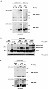
FIG. 1.
FIG. 1.
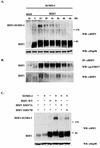
HSF1 undergoes a heat-inducible modification by SUMO-1 and SUMO-2. (A) Human K562 erythroleukemia cells were transiently transfected with either empty vector (mock) or Myc-tagged human HSF1 (HSF1) together with the GFP-fused wild-type or conjugation-defective SUMO-1 (SUMO-1 WT and SUMO-1 GA, respectively), and after 24 h the cells were either left untreated (C) or exposed to heat shock at 42°C for 1 h (HS). Immunoprecipitation was performed with anti-Myc antibody, and the samples were analyzed by immunoblotting with anti-SUMO-1 antibody. The input was blotted with polyclonal anti-human HSF1 antibody. (B) HSF1 WT was cotransfected to K562 cells with either empty vector (mock), His-tagged SUMO-2 and SUMO-1, or GFP-fused SUMO-1. Cells were left untreated (C) or exposed to a 15-min heat shock at 42°C (HS). Samples were analyzed by anti-HSF1 Western blotting. Hsc70 was used as a loading control. (C) K562 cells overexpressing Flag-tagged HSF1 and wild-type GFP-SUMO-1 were subjected to a 1-h heat shock alone (HS), heat shock followed by a 3-h recovery (R), or a 5-h treatment with 20 μM proteasome inhibitor MG132 (MG). Immunoprecipitation was performed with anti-Flag antibody, and the samples were further analyzed as described for panel A. IP, immunoprecipitation; WB, Western blot. Numbers on right of panels A and C and on left of panel B are molecular masses in kilodaltons.
FIG. 2.
FIG. 2.
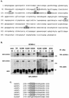
Lysine 298 is a major in vivo sumoylation site on HSF1. (A) The amino acid sequence of HSF1 was analyzed in order to identify possible sumoylation sites corresponding to the consensus SUMO-1 modification sequence. Sequence motifs with perfect consensus are marked with open boxes (K91, K126, and K298), and sites with partial consensus are shaded (K150, K162, and K381). (B) The K→R mutants were coexpressed with wild-type GFP-SUMO-1 in K562 cells and subjected to a 1-h heat shock followed by immunoprecipitation analysis as described for Fig. 1A. The variation in the intensities of sumoylation of the mutants reflects differences between individual experiments. WT, wild type; IP, immunoprecipi- tation; WB, Western blot. Numbers on right are molecular masses in kilodaltons. (C) Alignment of the human HSF1 (hHSF1) SUMO-1 modification sequence in comparison to mouse HSF1 (mHSF1), rat HSF1 (rHSF1), chicken HSF1 (cHSF1), zebra fish D. rerio HSF1 (zHSF1), frog X. laevis HSF1 (XHSF1), and fruit fly D. melanogaster HSF (DrHSF). The SUMO-1 consensus sequence is boxed in the respective species. The alignment was made by using ClustalW multiple sequence alignment.
FIG. 3.
FIG. 3.
Mutation of a phosphorylation site at serine 303 to alanine prevents HSF1 sumoylation. (A) Schematic presentation showing the location of K298 (▿) in the regulatory domain of HSF1 in the proximity of several critical phosphorylation sites (asterisks). The DNA-binding domain (DBD), N-terminal leucine zipper (HR-A/B), regulatory domain (RD), C-terminal leucine zipper (-C), and activation domain (AD) are indicated. (B) Myc-tagged S→A mutants of HSF1 phosphorylation sites were cotransfected with GFP-SUMO-1 into K562 cells followed by a 1-h heat shock, and cell lysates were resolved by SDS-PAGE. Western blot analysis was performed with anti-HSF1 antibody to detect the presence of a high-molecular-weight HSF1 band corresponding to sumoylated HSF1. Equal loading was confirmed by anti-Hsc70 immunoblotting. WT, wild type; WB, Western blot. Numbers on right are molecular masses in kilodaltons.
FIG. 4.
FIG. 4.
S303 is strongly phosphorylated in the wild-type and S307A mutant HSF1. To obtain two-dimensional tryptic phosphopeptide maps, wild-type (WT), S303A, S307A, or S303/7A HSF1 was transfected into K562 cells, after which the cells were in vivo labeled with [32P]orthophosphate and exposed to a heat shock (42°C, 1 h). The labeled HSF1 was digested with trypsin and resolved by two-dimensional mapping. The white arrow indicates phosphopeptide phosphorylated on both S303 and S307. The black arrow indicates phosphopeptides phosphorylated either on S303 or on S307. The starting points are indicated as “x.”
FIG. 5.
FIG. 5.
Characterization of an antibody specific to phosphorylated S303/7. (A) K562 cells were subjected to a heat shock (HS; 1 h at 42°C) or left untreated (C), and cell lysates were resolved by SDS-PAGE and blotted with anti-p-S303/7-HSF1 antibody. The specificity was verified by preincubating the antibody with a 500 M excess of free phosphopeptide (right panel). (B) K562 cells were transfected with Myc-tagged wild-type HSF1, S303A, S307A, or S303/7A, and the cells were subjected to a 1-h heat shock at 42°C. HSF1 was immunoprecipitated with anti-Myc antibody, and the immunoprecipitates were resolved by SDS-PAGE and blotted with anti-p-S303/7-HSF1 antibody. Equal amounts of HSF1 were controlled by blotting the membranes with anti-HSF1 antibody. (C) hsf1−/− MEFs were transfected with an empty plasmid (mock), wild-type HSF1, or K298R mutant. Cells were exposed to a 1-h heat shock, and cell lysates were resolved by SDS-PAGE and blotted with anti-p-S303/7, anti-HSF1, and anti-Hsc70 antibodies. (D) K562 cells were treated as indicated, and the cell lysates were preincubated with preimmune serum (Pre) or antibodies specific to phospho-S303/7-HSF1 or total HSF1. The HSE-binding HSF1 complex was analyzed by gel mobility shift assay. CHBA, constitutive HSE-binding activity; NS, nonspecific protein-DNA interaction; WT, wild type; IP, immunoprecipitation; WB, Western blot.
FIG. 6.
FIG. 6.
Phosphorylation of S303/7 occurs concomitantly with sumoylation of HSF1. (A) K562 cells were cotransfected with Myc-tagged wild-type HSF1 and GFP-SUMO-1 and heat shocked for the indicated time periods. Cell lysates were resolved by SDS-PAGE and analyzed by immunoblotting with anti-HSF1 antibody to determine the level of HSF1 sumoylation. Equal loading was confirmed by anti-Hsp90 immunoblotting. (B) HSF1 was immunoprecipitated from the same samples as for panel A with a monoclonal anti-HSF1 antibody, and the immunoprecipitates were resolved by SDS-PAGE and immunoblotted with anti-p-S303/7-HSF1 and anti-HSF1 antibodies. (C) Myc-tagged wild-type HSF1, S303/7A, or S303/7D was cotransfected with GFP-SUMO-1 into K562 cells followed by a 15-min heat shock at 42°C. Cell lysates were resolved by SDS-PAGE and analyzed by immunoblotting with anti-HSF1 antibody. Equal loading was confirmed by anti-Hsp90 immunoblotting. WT, wild type; WB, Western blot; IP, immunoprecipitation. Numbers on right of panels A and C are molecular masses in kilodaltons.
FIG. 7.
FIG. 7.
HSF1 S303/7D is preferentially sumoylated in vitro. In vitro-translated, 35S-labeled HSF1 mutants and androgen receptor (AR) were incubated with purified GST-SUMOGG-1 (SUMO-1) or GST-SAE1/GST-SAE2 (SAE1/SAE2) in the presence or absence of GST-Ubc9 as indicated. Reaction products were resolved by SDS-PAGE, and the signal was visualized by fluorography. Sumoylation is detected as the appearance of slower-migrating bands. WT, wild type.
FIG. 8.
FIG. 8.
The wild-type and K298R HSF1 rescue the heat shock response equally well in hsf1−/− MEFs. (A) Gel mobility shift assay (upper panel) was performed on hsf1−/− MEFs transfected with the wild-type or K298R mutant HSF1 and left untreated (C) or subjected to a 1-h heat shock (HS). The input lysates were resolved by SDS-PAGE and blotted with anti-HSF1 and anti-Hsc70 antibodies (lower panels). (B) hsf1−/− MEFs were transfected with empty vector (mock) or the wild-type or K298R mutant HSF1 and left untreated (C) or subjected to a 1-h heat shock (HS) or a 1-h heat shock followed by a 3-h recovery (R). Induction of the heat shock response was analyzed by Western blotting with anti-HSF1 and anti-Hsp70 antibodies. Anti-Hsc70 antibodies were used for loading control. Lack of Hsp70 induction in the absence of HSF1 (mock) is shown in the right panel. WT, wild type; WB, Western blot; CHBA, constitutive HSE-binding activity.
FIG. 9.
FIG. 9.
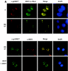
HSF1 and SUMO-1 transiently form cogranules at the onset of the heat shock response. (A) (Upper panel) HeLa cells were transiently transfected with HSF1-Myc and heat shocked for 15 min at 42°C or left untreated (C). Methanol-fixed cells were double stained by using anti-p-S303/7 antibody to detect phosphorylated HSF1 and anti-Myc antibody to localize total HSF1. Immunostaining was analyzed by fluorescence microscopy. Colocalization can be seen as yellow in the merged image. DAPI was used for nuclear staining. (Lower panel) HeLa cells were transiently transfected with HSF1-Myc and heat shocked for 15 min at 42°C. Cells were double stained with anti-p-S303/7 and anti-HSF1 antibodies in the presence or absence of 150 μg of recombinant human HSF1 (rHSF1) (17)/ml. (B) HeLa cells were transiently transfected with wild-type HSF1-Myc and GFP-SUMO-1 and heat shocked at 42°C for indicated times or left untreated (C). HSF1 was detected by using monoclonal anti-Myc antibody combined with a red fluorescent secondary antibody, and GFP-SUMO-1 was visualized through the green channel. (C) HeLa cells were transiently transfected with S303A or K298R HSF1 mutants together with GFP-SUMO-1. Cells were heat shocked at 42°C for 15 min and analyzed as described above.
FIG. 9.
FIG. 9.
HSF1 and SUMO-1 transiently form cogranules at the onset of the heat shock response. (A) (Upper panel) HeLa cells were transiently transfected with HSF1-Myc and heat shocked for 15 min at 42°C or left untreated (C). Methanol-fixed cells were double stained by using anti-p-S303/7 antibody to detect phosphorylated HSF1 and anti-Myc antibody to localize total HSF1. Immunostaining was analyzed by fluorescence microscopy. Colocalization can be seen as yellow in the merged image. DAPI was used for nuclear staining. (Lower panel) HeLa cells were transiently transfected with HSF1-Myc and heat shocked for 15 min at 42°C. Cells were double stained with anti-p-S303/7 and anti-HSF1 antibodies in the presence or absence of 150 μg of recombinant human HSF1 (rHSF1) (17)/ml. (B) HeLa cells were transiently transfected with wild-type HSF1-Myc and GFP-SUMO-1 and heat shocked at 42°C for indicated times or left untreated (C). HSF1 was detected by using monoclonal anti-Myc antibody combined with a red fluorescent secondary antibody, and GFP-SUMO-1 was visualized through the green channel. (C) HeLa cells were transiently transfected with S303A or K298R HSF1 mutants together with GFP-SUMO-1. Cells were heat shocked at 42°C for 15 min and analyzed as described above.

Similar articles

Cited by

References

    1. Appella, E., and C. W. Anderson. 2001. Post-translational modifications and activation of p53 by genotoxic stresses. Eur. J. Biochem. 268:2764-2772. - PubMed
    1. Ben-Neriah, Y. 2002. Regulatory functions of ubiquitination in the immune system. Nat. Immunol. 3:20-26. - PubMed
    1. Bies, J., J. Markus, and L. Wolff. 2002. Covalent attachment of the SUMO-1 protein to the negative regulatory domain of the c-Myb transcription factor modifies its stability and transactivation capacity. J. Biol. Chem. 277:8999-9009. - PubMed
    1. Chu, B., F. Soncin, B. D. Price, M. A. Stevenson, and S. K. Calderwood. 1996. Sequential phosphorylation by mitogen-activated protein kinase and glycogen synthase kinase 3 represses transcriptional activation by heat shock factor-1. J. Biol. Chem. 271:30847-30857. - PubMed
    1. Chu, B., R. Zhong, F. Soncin, M. A. Stevenson, and S. K. Calderwood. 1998. Transcriptional activity of heat shock factor 1 at 37°C is repressed through phosphorylation on two distinct serine residues by glycogen synthase kinase 3 and protein kinases Cα and Cζ. J. Biol. Chem. 273:18640-18646. - PubMed

Publication types

MeSH terms