

The barrier-to-autointegration factor is a component of functional human immunodeficiency virus type 1 preintegration complexes
- PMID: 12663813
- PMCID: PMC152146
- DOI: 10.1128/jvi.77.8.5030-5036.2003
The barrier-to-autointegration factor is a component of functional human immunodeficiency virus type 1 preintegration complexes
Abstract
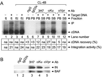
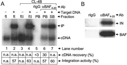
Retroviral integration in vivo is mediated by preintegration complexes (PICs) derived from infectious virions. In addition to the integrase enzyme and cDNA substrate, PICs contain a variety of viral and host cell proteins. Whereas two different cell proteins, high-mobility group protein A1 (HMGA1) and the barrier-to-autointegration factor (BAF), were identified as integration cofactors based on activities in in vitro PIC assays, only HMGA1 was previously identified as a PIC component. By using antibodies against known viral and cellular PIC components, we demonstrate here functional coimmunoprecipitation of endogenous BAF protein with human immunodeficiency virus type 1 (HIV-1) PICs. Since integrase protein and integration activity were also coimmunoprecipitated by anti-BAF antibodies, we conclude that BAF is a component of HIV-1 PICs. These data are consistent with the model that BAF functions as an integration cofactor in vivo.
Figures


Similar articles
-
Barrier-to-autointegration factor BAF binds p55 Gag and matrix and is a host component of human immunodeficiency virus type 1 virions.J Virol. 2003 Dec;77(24):13084-92. doi: 10.1128/jvi.77.24.13084-13092.2003. J Virol. 2003. PMID: 14645565 Free PMC article.
-
The barrier-to-autointegration protein is a host factor for HIV type 1 integration.Proc Natl Acad Sci U S A. 1998 Dec 22;95(26):15270-4. doi: 10.1073/pnas.95.26.15270. Proc Natl Acad Sci U S A. 1998. PMID: 9860958 Free PMC article.
-
Regulatory mechanisms by which barrier-to-autointegration factor blocks autointegration and stimulates intermolecular integration of Moloney murine leukemia virus preintegration complexes.J Virol. 2002 Dec;76(23):12376-80. doi: 10.1128/jvi.76.23.12376-12380.2002. J Virol. 2002. PMID: 12414981 Free PMC article.
-
Cellular co-factors of HIV-1 integration.Trends Biochem Sci. 2006 Feb;31(2):98-105. doi: 10.1016/j.tibs.2005.12.002. Epub 2006 Jan 5. Trends Biochem Sci. 2006. PMID: 16403635 Review.
-
Nuclear import of the pre-integration complex (PIC): the Achilles heel of HIV?Curr Drug Targets. 2003 Jul;4(5):409-29. doi: 10.2174/1389450033490984. Curr Drug Targets. 2003. PMID: 12816349 Review.
Cited by
-
Emerging drug targets for antiretroviral therapy.Drugs. 2005;65(13):1747-66. doi: 10.2165/00003495-200565130-00002. Drugs. 2005. PMID: 16114975 Review.
-
Chromosomal integration of LTR-flanked DNA in yeast expressing HIV-1 integrase: down regulation by RAD51.Nucleic Acids Res. 2006;34(21):6215-24. doi: 10.1093/nar/gkl843. Epub 2006 Nov 7. Nucleic Acids Res. 2006. PMID: 17090598 Free PMC article.
-
Immunity of replicating Mu to self-integration: a novel mechanism employing MuB protein.Mob DNA. 2010 Feb 1;1(1):8. doi: 10.1186/1759-8753-1-8. Mob DNA. 2010. PMID: 20226074 Free PMC article.
-
Barrier-to-autointegration factor BAF binds p55 Gag and matrix and is a host component of human immunodeficiency virus type 1 virions.J Virol. 2003 Dec;77(24):13084-92. doi: 10.1128/jvi.77.24.13084-13092.2003. J Virol. 2003. PMID: 14645565 Free PMC article.
-
Integrase and integration: biochemical activities of HIV-1 integrase.Retrovirology. 2008 Dec 17;5:114. doi: 10.1186/1742-4690-5-114. Retrovirology. 2008. PMID: 19091057 Free PMC article. Review.
References
-
- Bowerman, B., P. O. Brown, J. M. Bishop, and H. E. Varmus. 1989. A large nucleoprotein complex mediates the integration of retroviral DNA. Genes Dev. 3:469-478. - PubMed
-
- Brown, P. O., B. Bowerman, H. E. Varmus, and J. M. Bishop. 1987. Correct integration of retroviral DNA in vitro. Cell 49:347-356. - PubMed
-
- Bukrinsky, M. I., N. Sharova, T. L. McDonald, T. Pushkarskaya, W. G. Tarpley, and M. Stevenson. 1993. Association of integrase, matrix, and reverse transcriptase antigens of human immunodeficiency virus type 1 with viral nucleic acids following acute infection. Proc. Natl. Acad. Sci. USA 90:6125-6129. - PMC - PubMed
-
- Bustin, M. 2001. Revised nomenclature for high mobility group (HMG) chromosomal proteins. Trends Biochem. Sci. 26:152-153. - PubMed
-
- Cai, M., Y. Huang, R. Zheng, S.-Q. Wei, R. Ghirlando, M. S. Lee, R. Craigie, A. M. Gronenborn, and G. M. Clore. 1998. Solution structure of the cellular factor BAF responsible for protecting retroviral DNA from autointegration. Nat. Struct. Biol. 5:903-909. - PubMed
Publication types
MeSH terms
Substances
Grants and funding
LinkOut - more resources
Full Text Sources
Molecular Biology Databases
Research Materials
Miscellaneous
