. 2003 Apr;77(8):4489-501.
doi: 10.1128/jvi.77.8.4489-4501.2003.

Constitutive inositol phosphate formation in cytomegalovirus-infected human fibroblasts is due to expression of the chemokine receptor homologue pUS28

Affiliations

Constitutive inositol phosphate formation in cytomegalovirus-infected human fibroblasts is due to expression of the chemokine receptor homologue pUS28

Rosalba Minisini et al. J Virol. 2003 Apr.

Abstract

An open reading frame (ORF), US28, with homology to mammalian chemokine receptors has been identified in the genome of human cytomegalovirus (HCMV). Its protein product, pUS28, has been shown to bind several human CC chemokines, including RANTES, MCP-1, and MIP-1 alpha, and the CX(3)C chemokine fractalkine with high affinity. Addition of CC chemokines to cells expressing pUS28 was reported to cause a pertussis toxin-sensitive increase in the concentration of cytosolic free Ca(2+). Recently, pUS28 was shown to mediate constitutive, ligand-independent, and pertussis toxin-insensitive activation of phospholipase C via G(q/11)-dependent signaling pathways in transiently transfected COS-7 cells. Since these findings are not easily reconciled with the former observations, we analyzed the role of pUS28 in mediating CC chemokine activation of pertussis toxin-sensitive G proteins in cell membranes and phospholipase C in intact cells. The transmembrane signaling functions of pUS28 were studied in HCMV-infected cells rather than in cDNA-transfected cells. Since DNA sequence analysis of ORF US28 of different laboratory and clinical strains had revealed amino acid sequence differences in the amino-terminal portion of pUS28, we compared two laboratory HCMV strains, AD169 and Toledo, and one clinical strain, TB40/E. The results showed that infection of human fibroblasts with all three HCMV strains led to a vigorous, constitutively enhanced formation of inositol phosphates which was insensitive to pertussis toxin. This effect was critically dependent on the presence of the US28 ORF in the HCMV genome but was independent of the amino acid sequence divergence of the three HCMV strains investigated. The constitutive activity of pUS28 is not explained by expression of pUS28 at high density in HCMV-infected cells. The pUS28 ligands RANTES and MCP-1 failed to stimulate binding of guanosine 5'-O-(3-[(35)S]thiotriphosphate to membranes of HCMV-infected cells and did not enhance constitutive activation of phospholipase C in intact HCMV-infected cells. These findings raise the possibility that the effects of CC chemokines and pertussis toxin on G protein-mediated transmembrane signaling previously observed in HCMV-infected cells are either independent of or not directly mediated by the protein product of ORF US28.


Figures
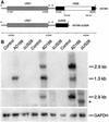
FIG. 1.
FIG. 1.
Construction of the deletion mutant HCMV AD169-ΔUS28. (A) Schematic representation of the protocol. The region extending from the putative TATA box located in the 5′ region of US27 to the putative site of polyadenylation in the 3′ region of US28 is shown (84, 87). The coding regions of US27 and US28 are shown as horizontal bars. The putative TATA boxes and the polyadenylation signal are marked by solid and open arrows, respectively. The noncoding region between the coding regions of US27 and US28 lacks a polyadenylation signal (87). Nucleotides 219,431 to 220,161 of US28 were removed from a plasmid carrying the ORFs US27, US28, and US29, as well as parts of US26 and US30 of HCMV AD169, resulting in an in-frame deletion of residues 78 to 321 of pUS28. Prediction of the transmembrane regions of pUS28 according to the method of Baldwin et al. (6) (not shown) suggests that the residual ΔUS28 protein, if expressed, consists of the extracellular amino terminus, the first transmembrane domain, the first intracellular loop, 8 of 18 residues constituting the second transmembrane domain, and 33 of 62 residues constituting the intracellular carboxyl-terminal domain. It is unlikely that the first intracellular loop and the distal part of the carboxyl terminus of G protein-coupled receptors are involved in G protein activation (32, 48). The nucleotide numbering corresponding to EMBL/GenBank accession no. X17403 is shown (see Materials and Methods for details). (B) Expression of mRNAs encoding pUS27 and/or pUS28 in HCMV-infected fibroblasts. HFF were infected with either wild-type HCMV AD169 or AD169-ΔUS28. Total RNA was prepared from the cells 24, 48, and 72 h p.i., fractionated (10 μg/lane) by denaturating formaldehyde-agarose (1% [wt/vol]) gel electrophoresis, transferred to a nylon membrane, and hybridized with radiolabeled cDNA probes corresponding either to the deleted NotI/KpnI fragment of ORF US28 (nt 219,426 to 220,161) (top), to ORF US27 (nt 217,904 to 218,992) (middle), or to rat GAPDH (bottom). The positions of the bicistronic transcript encoding both pUS27 and pUS28 (2.9 kb), the transcript encoding pUS28 (1.3 kb), the bicistronic transcript carrying the deletion in the US28 coding region (asterisk), and the mRNA encoding GAPDH (GAPDH) are indicated.
FIG. 2.
FIG. 2.
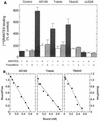
Binding of 125I-RANTES to HCMV-infected human fibroblasts. (A) Specificity of 125I-RANTES binding. HFF were infected as indicated either with the laboratory HCMV strains AD169 and Toledo, the clinical isolate TB40/E, or the deletion mutant AD169-ΔUS28. Uninfected cells (Control) were analyzed for comparison. The cells were incubated with 28 pM 125I-RANTES. The incubation was performed in the absence (−) or presence (+) of unlabeled RANTES or fractalkine (both at 10 nM) to discriminate specific from nonspecific binding. 125I-RANTES binding is given as a percentage of the binding to uninfected cells. The results correspond to the means plus standard deviations of triplicate determinations. (B) 125I-RANTES saturation binding. HFF were infected as indicated with the HCMV strains AD169, Toledo, or TB40/E. The infected cells were incubated with 28 pM 125I-RANTES in the absence or presence of unlabeled RANTES (10 pM to 10 nM). Uninfected cells were incubated in parallel with 28 pM 125I-RANTES in the absence of unlabeled RANTES to determine the amount of nonspecific binding. The experimental data were transformed according to the method of Scatchard (72) and subjected to linear regression analysis. The equilibrium dissociation constants (KD) were 0.53, 0.73, and 0.47 nM, and the maximum numbers of binding sites were 612, 715, and 544 pM, corresponding to 1.5 × 10⁶, 1.8 × 10⁶, and 1.4 × 10⁶ binding sites/cell for cells infected with AD169, Toledo, and TB40/E, respectively.
FIG. 3.
FIG. 3.
Agonist-stimulated binding of [35S]GTP[S] to membranes of HCMV-infected human fibroblasts. (A) HFF were infected with the indicated strain of wild-type HCMV (AD169, TB40/E, or Toledo) or AD169-ΔUS28. Uninfected cells (Control) were analyzed for comparison. The cells were homogenized, and aliquots of the particulate fractions prepared from the postnuclear supernatants (10 μg of protein/sample) were incubated in the absence (−) or presence (+) of 200 nM RANTES or 10 μM LPA with 0.34 nM [35S]GTP[S]. The samples were analyzed for bound [35S]GTP[S] by rapid filtration and scintillation counting. (B) Effect of PTX on agonist-stimulated binding of [35S]GTP[S]. HFF were infected with wild-type AD169 or AD169-ΔUS28. Uninfected cells (Control) were analyzed for comparison. The cells were pretreated as indicated for 16 h by incubation with PTX (100 ng/ml) or carrier. The cells were then homogenized and subjected to subcellular fractionation. Binding of [35S]GTP[S] to the particulate fractions was determined as described for panel A. The results correspond to the means plus standard deviations of triplicate determinations.
FIG. 4.
FIG. 4.
Stimulation of inositol phosphate formation in HCMV-infected fibroblasts. (A) Effect of chemokines. HFF were infected at an MOI of 1 with HCMV AD169. Five hours after infection, the cells were radiolabeled by incubation in inositol-free medium supplemented with 3 μCi of [3H]inositol/ml. Forty-eight hours after infection, the radiolabeled cells were incubated for 90 s in the absence (−) or presence (+) of 100 nM RANTES, 100 nM MCP-1, or 100 nM fractalkine and then analyzed for the accumulation of [3H]inositol phosphates as described in Materials and Methods. Uninfected cells (Control) were analyzed for comparison. The data are presented as percentages of the [3H]inositol phosphate formation measured in uninfected fibroblasts in the absence of chemokines. (B) Dependence of increased inositol phosphate formation on pUS28. HFF were infected as indicated with HCMV AD169, AD169-ΔUS28, or AD169-ΔUS27. Uninfected control cells (Co.) were analyzed for comparison. Radiolabeling of cells with [3H]inositol and analysis of [3H]inositol phosphate formation was done as described for panel A. The data are presented as percentages of the [3H]inositol phosphate formation measured in uninfected fibroblasts. In both panels, the results correspond to the means plus standard deviations of triplicate determinations.
FIG. 5.
FIG. 5.
Dependence of increased production of inositol phosphates in HCMV-infected fibroblasts on time of infection. HFF were seeded in six-well plates and infected at an MOI of 1 with HCMV AD169. The cells were then radiolabeled by incubation in inositol-free medium supplemented with 3 μCi of myo-[2-3H]inositol/ml. After the indicated times of infection, inositol phosphate accumulation was measured. The data are presented as percentages of the [3H]inositol phosphate formation measured in uninfected control cells (Co.) at hour 48 of the infection protocol. The results correspond to the means plus standard deviations of triplicate determinations. (Inset) Effect of PAA on the expression of mRNA encoding pUS27 and/or pUS28 in HCMV-infected fibroblasts. HFF were infected with HCMV AD169 and treated (+) with PAA (250 μg/ml). Uninfected cells were analyzed for comparison. Total RNA was prepared from the cells 24, 48, and 72 h p.i., fractionated (10 μg/lane) by denaturating formaldehyde-agarose (1% [wt/vol]) gel electrophoresis, transferred to a nylon membrane, and hybridized with a radiolabeled cDNA probe encoding pUS28. The positions of the bicistronic transcript encoding both pUS27 and pUS28 (2.9 kb) and of the transcript encoding pUS28 (1.3 kb) are indicated.
FIG. 6.
FIG. 6.
Dependence of increased production of inositol phosphates in HCMV-infected fibroblasts on MOI and HCMV strain. (A) MOI. HFF were infected as indicated at increasing MOI with either wild-type HCMV AD169 or AD169-ΔUS28. After 5 h of infection, the medium was exchanged and the infected cells were labeled for 43 h in inositol-free medium supplemented with 3 μCi of [3H]inositol/ml. Forty-eight hours after infection, inositol phosphate accumulation was measured. The data are presented as percentages of the [3H]inositol phosphate formation measured in uninfected control cells (Co.). (B) Virus strain. HFF were infected as indicated at increasing MOI with HCMV AD169, TB40/E, or Toledo. After 5 h of infection, the medium was exchanged and the infected cells were labeled for 43 h in inositol-free medium supplemented with 3 μCi of [3H]inositol/ml. Forty-eight hours after infection, inositol phosphate accumulation was measured. The data are presented as percentages of the [3H]inositol phosphate formation measured in uninfected control cells (Co.). The results correspond to the means plus standard deviations of triplicate determinations.
FIG. 7.
FIG. 7.
Effect of incubation medium on enhanced inositol phosphate production in HCMV-infected fibroblasts. HFF were seeded in six-well plates and infected with either wild-type AD169 or AD169-ΔUS28. After 5 h of infection, the medium was exchanged and the cells were labeled for 43 h in inositol-free medium supplemented with 3 μCi of [3H]inositol/ml. Thereafter, the medium (1 ml/well) was removed by aspiration, and the remaining cell layer was washed three times with 2 ml of prewarmed DMEM without supplements. Immediately after the addition of fresh medium containing 10 mM LiCl to the washed cells (1 ml/well), the medium was supplemented (+) with RANTES (100 nM final concentration) or carrier. Following incubation for 90 s, inositol phosphate formation was determined. The results correspond to the means plus standard deviations of triplicate determinations.

Similar articles

Cited by

References

    1. Adams, J. W., Y. Sakata, M. G. Davis, V. P. Sah, Y. Wang, S. B. Liggett, K. R. Chien, J. H. Brown, and G. W. Dorn. 1998. Enhanced Gαq signaling: a common pathway mediates cardiac hypertrophy and apoptotic heart failure. Proc. Natl. Acad. Sci. USA 95:10140-10145. - PMC - PubMed
    1. Albrecht, T., M. Nachtigal, S. C. St Jeor, and F. Rapp. 1976. Induction of cellular DNA synthesis and increased mitotic activity in Syrian hamster embryo cells abortively infected with human cytomegalovirus. J. Gen. Virol. 30:167-177. - PubMed
    1. Albrecht, T., M. P. Fons, I. Boldogh, S. AbuBakar, C. Z. Deng, and D. Millinoff. 1991. Metabolic and cellular effects of human cytomegalovirus infection. Transplant. Proc. 23(Suppl. 3):48-55. - PubMed
    1. Allen, L. F., R. J. Lefkowitz, M. G. Caron, and S. Cotecchia. 1991. G-protein-coupled receptor genes as protooncogenes: constitutively activating mutation of the α1B-adrenergic receptor enhances mitogenesis and tumorigenicity. Proc. Natl. Acad. Sci. USA 88:11354-11358. - PMC - PubMed
    1. Althoefer, H., P. Eversole-Cire, and M. I. Simon. 1997. Constitutively active Gαq and Gα13 trigger apoptosis through different pathways. J. Biol. Chem. 272:24380-24386. - PubMed

Publication types