. 2003 Mar;14(3):1158-71.
doi: 10.1091/mbc.02-06-0099.

Hic-5 communicates between focal adhesions and the nucleus through oxidant-sensitive nuclear export signal

Affiliations

Hic-5 communicates between focal adhesions and the nucleus through oxidant-sensitive nuclear export signal

Motoko Shibanuma et al. Mol Biol Cell. 2003 Mar.

Abstract

hic-5 was originally isolated as an H(2)O(2)-inducible cDNA clone whose product was normally found at focal adhesions. In this study, we found that Hic-5 accumulated in the nucleus in response to oxidants such as H(2)O(2). Other focal adhesion proteins including paxillin, the most homologous to Hic-5, remained in the cytoplasm. Mutation analyses revealed that the C- and N-terminal halves of Hic-5 contributed to its nuclear localization in a positive and negative manner, respectively. After the finding that leptomycin B (LMB), an inhibitor of nuclear export signal (NES), caused Hic-5 to be retained in the nucleus, Hic-5 was demonstrated to harbor NES in the N-terminal, which was sensitive to oxidants, thereby regulating the nuclear accumulation of Hic-5. NES consisted of a leucine-rich stretch and two cysteines with a limited similarity to Yap/Pap-type NES. In the nucleus, Hic-5 was suggested to participate in the gene expression of c-fos. Using dominant negative mutants, we found that Hic-5 was actually involved in endogenous c-fos gene expression upon H(2)O(2) treatment. Hic-5 was thus proposed as a focal adhesion protein with the novel aspect of shuttling between focal adhesions and the nucleus through an oxidant-sensitive NES, mediating the redox signaling directly to the nucleus.


Figures
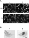
Figure 1
Figure 1
Nuclear accumulation of endogenous Hic-5 in the presence of H2O2. C3H10T1/2 (A) and MC3T3 (B) cells were treated with (+) or without (−) 1.5 mM (A) or 0.5 mM (B) H2O2 for 60 min. After fixation, the cells were processed for coimmunostaining with the antibody to Hic-5 and vinculin. The localization of the proteins was examined using a microscope (A) or a laser scanning confocal microscope (B). Bars, 20 (A) and 10 (B) μm.
Figure 2
Figure 2
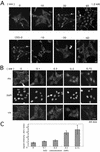
Time- and dose-dependent nuclear accumulation of Hic-5 in response to H2O2. (A) MEF#43/Tet-Off/LD1 mhic-5 induced to express the HA-tagged Hic-5 by removal of doxycycline (−Tet) was exposed to H2O2 (1.0 mM) for the period indicated (upper row). In the lower row, after 30 min exposure to H2O2, the medium was replaced with the conditioned medium without H2O2, and the cells were further incubated for 0, 10, 30, and 60 min. The cells were then fixed and immunolabeled with the antibody to HA tag. (B) Cells were exposed to H2O2 at the indicated doses for 30 min, and then coimmunstained with the antibody to HA tag (Hic) and vinculin (vin). DNA was stained with DAPI for localizing the nuclei. Bars, 20 μm. (C) The extent of the nuclear localization was evaluated on the image of (B, Hic) by measuring the cytoplasmic and nuclear signal intensity of individual cells with AquaCosmos image acquisition and analysis system (Hamamatsu Photonics; K. K. Hamamatsu, Japan). The values were mean ± SD obtained from at least six cells.
Figure 3
Figure 3
Nuclear accumulation of Hic-5 induced by sulfhydryl reagents and subcellular localization of Hic-5, paxillin, and their chimeric proteins in the presence or absence of H2O2. (A) MEF#43/Tet-Off/LD1 mhic-5 (−Tet) was exposed to 20 μM N-ethylmaleimide (NEM) or 2.0 mM diethyl maleate (DiM) for 30 min and then processed as in Figure 2. (B) MC3T3 cells were untreated (−) or treated with TGFβ1 (5 ng/ml for 90 min) as described previously (Ohba et al., 1994), DiM (2.0 mM for 90 min), and LMB (10 ng/ml for 6h), and processed for immunostaining with the antibody to Hic-5 as in Figure 1. (C) C3H10T1/2 cells were transfected with the plasmids, pCG-mhic-5, pcDNA3.1A-hhic-5 and pCG-pax, for expressing the indicated tagged-proteins, HA-mHic-5, hHic-5-myc, and HA-pax, respectively. After 24 h, the cells were treated with (+) or without (−) 1.5 mM H2O2 for 60 min, and then the expressed proteins were visualized with the antibodies to the tags. The nuclei were located by staining the DNA with DAPI (DAPI). (D) The expression plasmids for the chimeric proteins, pCG-hic/pax (Hic/pax) and pCG-pax/hic (pax/Hic), were introduced into C3H10T1/2 cells, and immunostaining with the antibody to HA tag was performed as in B. Bars, 20 μm.
Figure 4
Figure 4
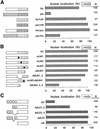
The nuclear localization of Hic-5 variants either in the presence or absence of H2O2. (A) Wild-type, LIM-only regions or the chimeric proteins of Hic-5 and paxillin were expressed from the expression plasmids, pcDNA3.1A-hhic-5 (Hic), pCG-pax (pax), pcDNA3.1A-hhicLIM (Hic/LIM), pCG-paxLIM (pax/LIM), pCG-hic/pax (Hic/pax), and pCG-pax/hic (pax/Hic), respectively, in C3H10T1/2 cells. Their localization was examined by immunostaining with antibodies to the tags as in Figure 5 after treatment with 1.0 mM H2O2 for 60 min. The nuclear localization was scored as positive and shown as a percentage when the signal in the nucleus was more intense than that in the cytoplasm or the nucleus and the cytoplasm were evenly labeled. N was the total number of cells scored in three independent transfections. (B) A series of LIM mutants of Hic-5 was expressed, and their localization was examined as in A. The plasmids introduced into the cells were pCG-LD1 mhic (WT),/mL1 (mLIM1),/mL2 (mLIM2),/mL3 (mLIM3),/delL4 (delLIM4),/delL1,2 (delLIM1,2),/ml3-delL4 (mLIM3-delLIM4), and/delL2,3,4 (delLIM2,3,4). The squares indicate LIM domains; open squares for the wild-type and closed squares for mutated versions. (C) The N-terminal deletion mutants of Hic-5 were expressed, and their nuclear localization was examined as in A and B but without the treatment with H2O2. The expression plasmids used here were pCG-hhic-5 (WT), pCG-delLD1–2hhic-5 (delLD1,2), pCG-delLD3–4hhic-5 (delLD3,4), pCG-delLD3hhic-5 (delLD3), and pCG-hhicLIM (LIM). The open circles represent a LD motif.
Figure 5
Figure 5
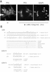
Nuclear accumulation induced by LMB and the sequences of LD motifs as the assumptive NES of Hic-5. (A) MEF#43/Tet-Off/LD1 mhic-5 (−Tet) was exposed to LMB (10 ng/ml) for 2 h, and then processed as in Figure 2. Bar, 20 μm. (B) The amino acid sequences of the LD motifs (LD2, 3, 4) of mouse and human Hic-5 are shown along with the corresponding LD of paxillin. The sequences around LD2 are also shown, extended to contain the two cysteines marked by asterisks. The leucine residues in each LD motif are overlined. The sequences under the wild-type were those of the mutants in which leucine or cysteine residues were replaced as indicated.
Figure 6
Figure 6
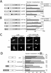
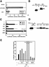
Identification and characterization of oxidant-sensitive NES containing leucine and cysteine residues in the N-terminal region of Hic-5. (A and B) The variants of mouse LD1-null form of Hic-5 in which leucine or cysteine residues were substituted as shown in Figure 5B were expressed in C3H10T1/2 cells from pCG-mhic-5 (WT hic) and its derivatives (mLD2, mLD3, mLD4, mCf/N, mCl/S, and mCfl/NS), and then visualized as in Figure 3C after treatment with (H2O2+) or without (H2O2 −) 1.0 mM H2O2 for 60 min. The nuclear localization of the variant proteins was evaluated as in Figure 4 and graphed (mean ± SD derived from a series of experiments repeated more than five times, in each of which around one hundred cells were examined). The significance of the differences was assessed by t test. P values with no mark were for each treatment and those marked with asterisks were for each mutation. The cartoon outlined the protein structure examined, in which open and closed circles indicate wild-type and mutant LD motifs, and the substitutions of the cysteines are also indicated. (C) The LIM and (CC-LD3)LIM were expressed from pCG-paxLIM and its derivatives (MATERIALS AND METHODS), and the cells were exposed to 2.0 mM DiM for 60 min. The control (−) and DiM-exposed cells (DiM) were then immunolabeled with the antibody to HA tag (HA). The nuclei were located by staining the DNA with DAPI (DAPI). Bar, 20 μm. (D) The fusion proteins as illustrated were expressed from respective expression plasmids based on pCG-paxLIM (MATERIALS AND METHODS), and then visualized as in C under untreated conditions (−), or after exposure to 1.0 mM H2O2 (H2) or 2.0 mM DiM (Di) for 60 min. The nuclear localization was scored as nuclear (+) and nuclear and cytoplasmic (+/−). LIM; LIM region of paxillin, L or LD3; amino acids 131–154 of mouse LD1-null Hic including the LD3 motif, CC-LD3; residues 41–154 of the Hic-5 including the two cysteines and the LD3 motif, Hic-5(N); 1–227 of human Hic-5.
Figure 7
Figure 7
The ability of the nuclear localized Hic-5 to transactivate the c-fos gene, for which LIM1 and 4 were required. (A) The reporter plasmid of c-fos and the effector plasmid expressing Hic-5 or paxillin were transiently cotransfected into C2C12 cells together with an internal control plasmid, pRL/CMV. The effector plasmids used here were pCG-LD1 mhic-5 (LD1mHic) and pCG-pax (pax), respectively, which were either the wild-type (NLS−) or nuclear-targeted (NLS+). Luciferase activities were determined as outlined in MATERIALS AND METHODS and shown as a ratio to the controls transfected with the pCG vector (vector) after normalization with the internal control. Bars represent the mean ± SD from at least three independent experiments. (B) pCG vector (−), pCG-LD1 mhic-5 (Hic/LD1+) and pCG-pax (pax) with (+) or without (−) NLS were transfected into C2C12, and 24 h later the cells were lysed and processed for Western blotting. The expressed proteins were detected with the antibody to HA tag. (C) The wild-type and the LIM mutants of Hic-5 were expressed as effectors from pCG-LD1 mhic (WT),/mL1–3 (mL1–3),/delL4 (delL4) with (NLS+) or without NLS (NLS−) in the cells with the c-fos reporter, and a luciferase assay was performed as in A. (D) The expression plasmids of the LIM mutants used in the above assay of C were transfected into C2C12 cells, and 24 h later, Western blotting was carried out using the antibody to HA tag. (E) The c-fos reporter was introduced into the cells with combinations of plasmids at various ratios of the wild-type (1:) and each LIM mutant (:1–3) as indicated, and a luciferase assay was carried out as in A and C. Each assay was done in duplicate. The experiments were repeated three times and similar results were obtained.
Figure 8
Figure 8
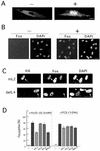
Involvement of Hic-5 in endogenous c-fos induction by H2O2. (A and B) After serum deprivation for >24 h, TIG-7 cells were treated with (+) or without (−) 0.4 mM H2O2 for 5 h, and endogenous Hic-5 (A) or c-Fos (B) was immunolabeled with specific antibodies. DAPI was used to stain DNA. Bars, 10 (A) and 20 (B) μm. (C) (D) The same expression plasmids for the nuclear localized type of LIM mutants used in Figure 7C were first introduced into TIG-7 cells, and then, the cells were serum-deprived and exposed to the stimuli for 5h. Subsequent to fixation, coimmunostaining was performed with the antibodies to HA tag and Fos, along with DAPI. The immunolabels with antibody to HA identify the cells expressing the mutants. Among the cells expressing the indicated LIM mutants, those expressing Fos were enumerated microscopically, and the counts are shown as percentages in D. The experiment was repeated three times, and for each mutant, around one hundred cells were examined in total. In C, representative results of the immunostaining are shown in the cases of mL2 and delL4 expression in the cells treated with 0.5 mM H2O2. Bar, 20 μm.
Figure 9
Figure 9
Alignments of the NES sequences. (A) The NESs with cysteine residues were aligned. (B) (C) The NESs found among the LIM protein family were aligned and compared with NES of Rev. The consensus proposed as the Rev-type NES is shown at the bottom. The amino acids emphasized by open letters and filled boxes were those whose importance was demonstrated experimentally. The large group hydrophobic amino acids are shaded.

Similar articles

Cited by

References

    1. Alpin A, Juliano RL. Regulation of nucleocytoplasmic trafficking by cell adhesion receptors and he cytoskeleton. J Cell Biol. 2001;155:187–191. - PMC - PubMed
    1. Aoto H, Sasaki H, Ishino M, Sasaki T. Nuclear translocation of cell adhesion kinase beta/praline-rich tyrosine kinase 2. Cell Struct Funct. 2002;27:47–61. - PubMed
    1. Brown MC, Curtis MS, Turner CE. Paxillin LD motifs may define a new family of protein recognition domains. Nature Struct Biol. 1998;5:677–678. - PubMed
    1. Crawford D, Zbinden I, Amstad P, Cerutti P. Oxidant stress induces the protooncogene c-fos and c-myc in mouse epidermal cells. Oncogene. 1988;3:27–32. - PubMed
    1. Dawid IB, Toyama R, Taira M. LIM domain proteins. CR Acad Sci Iii. 1995;318:295–306. - PubMed

Publication types

MeSH terms

Substances