. 2003 Mar;111(5):749-58.
doi: 10.1172/JCI16924.

c-Fms and the alphavbeta3 integrin collaborate during osteoclast differentiation

Affiliations

c-Fms and the alphavbeta3 integrin collaborate during osteoclast differentiation

Roberta Faccio et al. J Clin Invest. 2003 Mar.

Abstract

beta(3) integrin-null osteoclasts are dysfunctional, but their numbers are increased in vivo. In vitro, however, the number of beta(3)(-/-) osteoclasts is reduced because of arrested differentiation. This paradox suggests cytokine regulation of beta(3)(-/-) osteoclastogenesis differs in vitro and in vivo. In vitro, additional MCSF, but not receptor activator of NF-kappaB ligand (RANKL), completely rescues beta(3)(-/-) osteoclastogenesis. Similarly, activation of extracellular signal-regulated kinases (ERKs) and expression of c-Fos, both essential for osteoclastogenesis, are attenuated in beta(3)(-/-) preosteoclasts, but completely restored by additional MCSF. In fact, circulating and bone marrow cell membrane-bound MCSFs are enhanced in beta(3)(-/-) mice, correlating with the increase in the osteoclast number. To identify components of the MCSF receptor that is critical for osteoclastogenesis in beta(3)(-/-) cells, we retrovirally transduced authentic osteoclast precursors with chimeric c-Fms constructs containing various cytoplasmic domain mutations. Normalization of osteoclastogenesis and ERK activation, in beta(3)(-/-) cells, uniquely requires c-Fms tyrosine 697. Finally, like high-dose MCSF, overexpression of c-Fos normalizes the number of beta(3)(-/-) osteoclasts in vitro, but not their ability to resorb dentin. Thus, while c-Fms and alpha(v)beta(3) collaborate in the osteoclastogenic process via shared activation of the ERK/c-Fos signaling pathway, the integrin is essential for matrix degradation.


Figures

Figure 1
Figure 1
High-dose MCSF rescues β3–/– osteoclastogenesis. BMMs derived from β3+/+ or β3–/– mice were cultured in RANKL and either 10 ng/ml (a) or 100 ng/ml (b) MCSF for 3, 7, or 10 days, fixed in 4% paraformaldehyde, and stained for TRAP activity. Within 3 days, β3+/+ BMMs in low-dose MCSF developed into mononuclear and binuclear TRAP-expressing cells (pOCs). Characteristic multinucleated OCs appeared within 7-10 days. In contrast, β3–/– BMMs failed to normally form pre-OCs and to differentiate into typical OCs within this time frame. When β3–/– and β3+/+ BMMs were cultured in high-dose (100 ng/ml) MCSF, their differentiation into mature OCs was similar at days 7 and 10. Indicated are the numbers of binucleated pre-OCs (pOCs) (day 3) or multinucleated OCs (days 7 and 10) per well, from three independent experiments. ×4. ND, not determined.
Figure 2
Figure 2
The capacity of high-dose MCSF to increase β3–/– OC number is not due to accelerated proliferation or arrested apoptosis. β3+/+ or β3–/– BMMs were cultured with increasing concentrations of MCSF in the absence (a) or presence (b) of 100 ng/ml RANKL. After 3 days, proliferation was assessed by MTT assay. Regardless of genotype, the proliferative rates of the cells were similar at each concentration of cytokine. β3+/+ or β3–/– BMMs were maintained in culture in low-dose (10 ng/ml, c) or high-dose (100 ng/ml, d) MCSF, alone (Mφ) or with 100 ng/ml RANKL, the latter added at initiation of culture (d4) or after 2 days (d2). Cytokines were present during the entire 4-day culture period in half the wells (black bars) and were withdrawn from the other half during the last 24 hours to induce apoptosis (white bars), the magnitude of which was determined by ELISA. The apoptotic rate of β3–/– cells was diminished in low-dose MCSF but indistinguishable from WT in high concentrations of the cytokine.
Figure 3
Figure 3
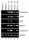
High-dose MCSF promotes expression of osteoclastogenic markers in β3–/– pre-OCs. BMMs derived from β3+/+ or β3–/– mice were cultured with RANKL and either low-dose (10 ng/ml; LD) or high-dose (100 ng/ml; HD) MCSF. After 3 days, RNA was extracted, and expression of the mRNA of osteoclastogenic markers cathepsin K, TRAP, MITF, MMP-9, and calcitonin receptor was measured by RT-PCR. Untreated β3+/+ (lane 1) and β3–/– (lane 2) BMMs, serving as negative control, failed to express these mRNA species, whereas they were abundant in β3+/+ OCs regardless of ambient MCSF (lanes 3 and 4). Compared with those of their WT counterparts, TRAP mRNA levels were decreased twofold, cathepsin K mRNA threefold, MMP-9 mRNA threefold, MITF mRNA fivefold, and calcitonin receptor (R) mRNA fivefold in β3–/– cells treated with low-dose MCSF (lane 5). Reflecting its capacity to rescue β3–/– osteoclastogenesis, high-dose MCSF substantially enhanced these mRNAs in mutant cells (lane 6).
Figure 4
Figure 4
MCSF is increased in β3–/– mice in vivo. Circulating and bone-microenvironment MCSF levels from three β3–/– and three β3+/+ mice were measured by ELISA (a) or Western blot (b and c). (a) Serum MCSF levels analyzed by ELISA in β3–/– mice were significantly increased relative to those of β3+/+ counterparts. *P < 0.05. (b) Marrow cells isolated from the same β3+/+ and β3–/– mice were lysed and subjected to immunoblot using an anti-MCSF mAb. Representative gels demonstrate a 22-kDa band, corresponding to membrane-residing MCSF, in both circumstances but was three times higher in β3–/– cells. Actin served as loading control. (c) Densitometric analysis of ratio of membrane-residing MCSF relative to actin in bone marrow cells derived from three mice of each genotype. *P < 0.005.
Figure 5
Figure 5
Adhesion-induced ERK activation is defective in β3–/– pre-OCs. β3+/+ or β3–/– BMMs were maintained for 3 days in low-dose MCSF and 100 ng/ml RANKL. The cells were detached and replated in OPN-coated dishes. Nonadherent cells were removed, and attached pre-OCs were lysed at the indicated times. Equal amounts of total protein were immunoblotted with an antibody to phospho-ERK (p-ERK) or β-actin. ERK phosphorylation maximized within 15 minutes of attachment of β3+/+ pre-OCs and was sustained for at least 120 minutes. While ERK phosphorylation also maximized within 15 minutes of attachment, the signal rapidly dissipated in the absence of the integrin.
Figure 6
Figure 6
High-dose MCSF rescues ERK activation in β3–/– pre-OCs. Pre-OCs derived from BMMs treated with RANKL and low-dose MCSF for 3 days were starved for 2 hours in serum-free medium and exposed to 10 or 100 ng/ml MCSF, with time. ERK activation was detected in total cell lysates using an anti–phospo-ERK antibody. β-Actin served as loading control. In the presence of low-dose MCSF, ERK activation was more pronounced and sustained longer in β3+/+ than in β3–/– pre-OCs. In the presence of high-dose MCSF, however, ERK phosphorylation is indistinguishable in both cell types.
Figure 7
Figure 7
c-FmsY697 is specifically required for osteoclastogenesis in the absence of αvβ3. (a) Equal numbers of β3+/+ and β3–/– BMMs were retrovirally transduced with vector alone, nonmutated EpoR/c-Fms (WT), or EpoR/c-Fms carrying individual tyrosine-to-phenylalanine point mutation. Cells were selected in puromycin for 3 days and exposed for 3 days to RANKL and low-dose MCSF. The cells were then placed in serum-free medium for 2 hours, exposed to 25 U/ml Epo for varying times, and lysed. Equivalent expression of c-Fms and mutated forms of EpoR/c-Fms was established by immunoblot using a C-terminal anti–c-Fms antibody. (b) Puromycin-selected cells were cultured for 7 days in the optimal osteoclastogenic concentration of Epo (25 ng/ml) and RANKL (100 ng/ml). Osteoclastogenesis was measured by TRAP assay and expressed as a percentage of the value obtained with control (WT) EpoR/c-Fms transductants. While EpoR/c-FmsY697F was as effective as control in inducing β3+/+ osteoclastogenesis, the same mutant dampened the process 3.5-fold in β3–/– cells. In contrast, EpoR/c-FmsY721F did not impact the osteoclastogenic process. *P < 0.001 vs. vector; **P < 0.05 vs. vector.
Figure 8
Figure 8
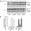
c-FmsY697 is specifically required for sustained ERK phosphorylation in β3–/– pre-OCs. (a) BMMs from β3+/+ and β3–/– mice, transfected with the control EpoR/c-Fms (WT) or with the indicated EpoR/c-Fms mutants and selected in puromycin for 3 days, were exposed for 3 days to RANKL and low-dose MCSF. Pre-OCs were maintained for 2 hours in serum-free medium and exposed to 25 U/ml Epo, for the indicated times. ERK activation in response to Epo was detected in total cell lysates using an anti–phospo-ERK antibody. Only one EpoR/c-Fms mutant, Y697F, differentially affected the ERK signal in β3+/+ and β3–/– cells. Specifically, while it did not impact β3+/+ cells, this mutation completely abrogated the prolonged ERK activation in β3–/– OCs. (b) Samples used in a were immunoblotted with an anti-ERK mAb as loading control.
Figure 9
Figure 9
High-dose MCSF is required to activate RSK and c-Fos in β3–/– pre-OCs. β3+/+ and β3–/– BMMs exposed to RANKL and low-dose MCSF for 3 days were placed in serum-free medium for 2 hours and then exposed to 10 or 100 ng/ml MCSF, for the indicated times. (a and b) RSK activation was assessed by immunoblot of its phosphorylated species (p-RSK). Total RSK served as loading control. RSK phosphorylation was attenuated in β3–/– pre-OCs cultured in low-dose MCSF (a), but not in those cultured in high-dose MCSF (b). (c and d) c-Fos expression was detected by immunoblot in pre-OCs treated for 30 or 60 minutes with low- or high-dose MCSF. In β3–/– pre-OCs, c-Fos expression was induced only when cells were treated with high-dose MCSF. β-Actin served as loading control.
Figure 10
Figure 10
c-Fos overexpression by β3–/– cells rescues osteoclastogenesis but not matrix resorption. β3+/+ BMMs and β3–/– BMMs retrovirally transduced with pBabe vector (MOCK) or pBabe/c-Fos were selected in puromycin for 5 days, and resistant cells were used in the indicated experiments. (a) An equal number of BMMs were plated in 96-well plates and cultured in the presence of RANKL and low-dose MCSF. After 7 days, cells were stained for TRAP activity. While osteoclastogenesis remained arrested in MOCK-transduced β3–/– cells, those overexpressing c-Fos generated OCs indistinguishable from WT. (b) Equal amounts of protein were loaded in each lane, and c-Fos content was assessed by immunoblot. β3–/– OCs retrovirally transduced with pBabe/c-Fos vector expressed the same level of c-Fos protein as did β3+/+ cells (arrow). (c) β3+/+, MOCK β3–/–, and c-Fos β3–/– pre-OCs were plated on dentin slices with RANKL and low-dose MCSF. MOCK β3–/– cells were also cultured with RANKL and high-dose MCSF. After four days, dentin slices were stained for TRAP activity (+ Cells), or the cells were removed to visualize resorptive pits (– Cells). β3+/+ cells differentiated into OCs with a characteristic resorptive phenotype and excavated many large, well-demarcated lacunae. MOCK-transduced β3–/– cells formed few OCs in the presence of low-dose MCSF and generated poorly defined, small pits. High-dose MCSF and c-Fos overexpression yielded numerous multinucleated TRAP-expressing OCs that exhibited a nonresorbing phenotype and also generated poorly defined, small pits. Indicated are the mean numbers of pits ± SEM from three different fields per variable. ×10.

Similar articles

Cited by

References

    1. Hamilton JA. CSF-1 signal transduction. J. Leukoc. Biol. 1997;62:145–155. - PubMed
    1. Felix R, et al. Impairment of macrophage colony-stimulating factor production and lack of resident bone marrow macrophages in the osteopetrotic op/op mouse. J. Bone Miner. Res. 1990;5:781–789. - PubMed
    1. Wiktor-Jedrzejczak W, et al. Total absence of colony-stimulating factor 1 in the macrophage-deficient osteopetrotic (op/op) mouse. Proc. Natl. Acad. Sci. U. S. A. 1990;87:4828–4832. - PMC - PubMed
    1. Feng X, et al. Tyrosines 559 and 807 in the cytoplasmic tail of the m-csf receptor play distinct roles in osteoclast differentiation and function. Endocrinology. 2002;143:4868–4874. - PubMed
    1. van der Flier A, Sonnenberg A. Function and interactions of integrins. Cell Tissue Res. 2001;305:285–298. - PubMed

Publication types

MeSH terms