Comparative Study
. 2003 Mar;23(6):2109-22.
doi: 10.1128/MCB.23.6.2109-2122.2003.

A novel RING finger protein, human enhancer of invasion 10, alters mitotic progression through regulation of cyclin B levels

Affiliations
Comparative Study

A novel RING finger protein, human enhancer of invasion 10, alters mitotic progression through regulation of cyclin B levels

Garabet G Toby et al. Mol Cell Biol. 2003 Mar.

Abstract

The process of cellular morphogenesis is highly conserved in eukaryotes and is dependent upon the function of proteins that are centrally involved in specification of the cell cycle. The human enhancer of invasion clone 10 (HEI10) protein was identified from a HeLa cell library based on its ability to promote yeast agar invasion and filamentation. Through two-hybrid screening, the mitotic cyclin B1 and an E2 ubiquitin-conjugating enzyme were isolated as HEI10-interacting proteins. Mutation of the HEI10 divergent RING finger motif (characteristic of E3 ubiquitin ligases) and Cdc2/cyclin binding and phosphorylation sites alter HEI10-dependent yeast phenotypes, including delay in G(2)/M transition. In vertebrates, the addition of HEI10 inhibits nuclear envelope breakdown and mitotic entry in Xenopus egg extracts. Mechanistically, HEI10 expression reduces cyclin B levels in cycling Xenopus eggs and reduces levels of the cyclin B ortholog Clb2p in yeast. HEI10 is itself a specific in vitro substrate of purified cyclin B/cdc2, with a TPVR motif as primary phosphorylation site. Finally, HEI10 is itself ubiquitinated in egg extracts and is also autoubiquitinated in vitro. These and other points lead to a model in which HEI10 defines a divergent class of E3 ubiquitin ligase, functioning in progression through G(2)/M.


Figures

FIG. 1.
FIG. 1.
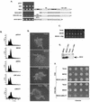
HEI10 profile in yeast. (A) Expression of HEI10 in yeast induces agar invasion. HEI10 was expressed in yeast under the control of the GAL1 promoter from the vector pJG4-4. Yeast expressing HEI10 remain attached to the agar after exposure to vigorous blasts of water. (B) Budding patterns of yeast expressing HEI10. Yeast were stained with Calcofluor to visualize their previous budding sites. Yeast were grouped according to budding pattern under unipolar (bud scars clustered at one pole), bipolar (bud scars at opposite poles), and random budding. (C) Filamentation phenotype of yeast expressing pJG4-4, HEI10/pJG4-4, or HEF1/pJG4-4 after culture on SLAHGR medium for 24 h. (D) Bud emergence in HEI10-expressing yeast occurs simultaneously in mother and daughter, a characteristic of pseudohyphal growth. HEI10-expressing yeast cells were streaked onto single cells on SLAHGR plates, grown for 18 h, and imaged at 1-h time intervals (magnification, ×28). Arrows indicate bud emergence sites. (E) HEI10 expression leads to accumulation in G2. A FACS profile of yeast expressing pJG4-4 versus HEI10 is shown. (F) HEI10 fails to activate the FLO11-LacZ reporter. Yeast cells expressing pJG4-4, HEI10, or HEF1, together with a FLO11pro-LacZ reporter, were plated on uninducing (glucose) or inducing (galactose) media containing X-Gal (5-bromo-4-chloro-3-indolyl-β-d-galactopyranoside) to assess reporter activation.
FIG. 2.
FIG. 2.
Structure of the HEI10 protein and HEI10 RNA expression. (A) HEI10 encodes an 277-aa open reading frame. A diagram of HEI10 domains is presented, with structural motifs as described in Results. (B) HEI10 transcript is expressed to different levels in a number of human tissues. (C) Sequence alignment of HEI10 candidate RING domain versus other RING fingers. The c-Cbl RING finger is presented as a reference C3HC4 RING consensus sequence, with core cysteines indicated in green. Deviation of HEI10 orthologs from five different vertebrate species from the c-Cbl RING consensus residues is indicated in red. Other RING fingers from proteins with confirmed E3 ligase activity are presented as context; these include the proteins HHAR1, Doa10, TEB4, HRD1, and Parkin.
FIG. 3.
FIG. 3.
Structure-function relationship of HEI10 domains and budding phenotypes. (A) HEI10 and HEI10 mutants assessed are shown (diagram on right). The ability of HEI10 and each derivative to induce agar invasion and aggregation is shown; each patch represents an independent transformant. For this assay, HEI10 or HEI10 mutants were expressed under the control of the GAL1 promoter and were grown on glucose or galactose and then washed vigorously with water. (B) FACS profiles of the yeast strains shown in panel A. (C) HEI10's ability to induce invasion on rich media is impaired upon coexpression of Clb2p. Yeast express proteins are as indicated; the wash assay was as described for panel A, with each patch representing an individual transformant. (D) HEI10 induction of filamentation on low-nitrogen media is impaired by coexpression with Clb2p. Filamentation assay was as shown in Fig. 1C; a representative colony is shown. (E) Western blot analysis of HEI10 expression in yeast cells in the presence or absence of Clb2p. (F) Growth on rich medium with galactose for yeast-expressing vector, HEI10, or HEI10-Δ200--277, together with Clb2p or a Clb2p-Δbox mutant. Each patch represents an individual transformant.
FIG. 4.
FIG. 4.
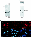
Expression of endogenous HEI10. (A) Western visualization of HEI10 in MCF-7 or U20S cells with anti-HEI10 antibodies (left) or with antibodies plus blocking peptide (right panel). (B) Immunofluorescence analysis of MCF-7 cells show that HEI10 with the antibody is predominantly a nuclear protein (left panel); in contrast, staining with the peptide-blocked anti-HEI10 shows only weak cytoplasmic staining (center panel). In cells undergoing mitosis (right panels), HEI10 signal colocalizes with DAPI-stained DNA (brightness adjusted versus other images). (C) Cell fractionation of MCF-7 lysates. HEI10 is present in the nuclear fraction of MCF-7 lysates (N) but not in the cytosolic fraction (C). A Western blot of the same lysates with antipaxillin, a cytosolic protein, is shown as a control for the purity of the fractions. The 19-kDa band is predominantly present in the cytosolic fraction.
FIG. 5.
FIG. 5.
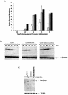
HEI10 expression limits mitotic entry and limits cyclin B-Clb2p accumulation. (A) Nuclear envelope breakdown of Xenopus extracts after sperm addition in the presence of GST-HEI10 (open bars), buffer (shaded bars), or GST (solid bars). (B) Egg lysates shown after incubation for the times indicated after sperm addition in the presence of GST, GST-HEI10, or GST-HEI10-ΔRING were used for Western analysis with anti-cyclin B (top) or tubulin (bottom). (C) Western analysis of Clb2p-HA levels in yeast transformed with pJG4-4 vector, HEI10, or HEI10-ΔRING. The anti-tubulin control (TUB2) is shown at the bottom.
FIG. 6.
FIG. 6.
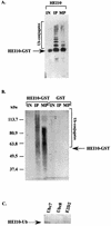
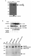
HEI10 and ubiquitination. (A) Bacterially purified GST-HEI10 is shown as an input species (IN) or after incubation with synchronized interphase (IP) or M-phase (MP) Xenopus egg extract after visualization with anti-HEI10. (B) GST-HEI10 or GST bacterially purified proteins, after incubation with interphase (IP) or mitotic (MP) extract, visualized with anti-ubiquitin antibody. (C) Purified HEI10 was incubated in an in vitro ubiquitination cocktail containing either UbcH7, UbcH8, or E2D2 as E2 ubiquitin-conjugating enzyme and then visualized with to anti-ubiquitin antibody.
FIG. 7.
FIG. 7.
HEI10 is phosphorylated in vitro by cyclin B1/Cdc2. (A) Autoradiograph showing increased incorporation of radiolabeled [γ-32P]ATP in HEI10-GST with an increased amount of cyclin B-cdc2 (lanes 2 and 3). (B) Upon incubation with increasing amounts of cyclin B-cdc2, HEI10 mobility was decreased (lanes 2, 3, and 4). The mobility was increased if λ phosphatase was added to the reaction (lane 5). A band with a size similar to that of HEI10-GST was visualized by Western blot by using antiphosphothreonine-specific antibodies when HEI10-GST was incubated with increasing amounts of cyclin B-cdc2 (lanes 2, 3, and 4). (C) Autoradiograph showing in vitro phosphorylation of HEI10 and HEI10 mutants. Phosphorylation was abrogated in HEI10-Δ200-277, whereas it was strikingly reduced in upon mutation of TP221,222. The arrows on the left show the predicted mobilities of the various proteins.

Similar articles

Cited by

References

    1. Adams, P. D., W. R. Sellers, S. K. Sharma, A. D. Wu, C. M. Nalin, and W. G. J. Kaelin. 1996. Identification of a cyclin-cdk2 recognition motif present in substrates and p21-like cyclin-dependent kinase inhibitors. Mol. Cell. Biol. 16:6623-6633. - PMC - PubMed
    1. Ahn, S. H., A. Acurio, and S. J. Kron. 1999. Regulation of G2/M progression by the STE mitogen-activated protein kinase pathway in budding yeast filamentous growth. Mol. Biol. Cell 10:3301-3316. - PMC - PubMed
    1. Ahn, S. H., B. T. Tobe, J. N. Fitz Gerald, S. L. Anderson, A. Acurio, and S. J. Kron. 2001. Enhanced cell polarity in mutants of the budding yeast cyclin-dependent kinase Cdc28p. Mol. Biol. Cell 12:3589-3600. - PMC - PubMed
    1. Ardley, H. C., N. G. Tan, S. A. Rose, A. F. Markham, and P. A. Robinson. 2001. Features of the parkin/ariadne-like ubiquitin ligase, HHARI, that regulate its interaction with the ubiquitin-conjugating enzyme, Ubch7. J. Biol. Chem. 276:19640-19647. - PubMed
    1. Bischoff, J. R., P. N. Friedman, D. R. Marshak, C. Prives, and D. Beach. 1990. Human p53 is phosphorylated by p60-cdc2 and cyclin B-cdc2. Proc. Natl. Acad. Sci. USA 87:4766-4770. - PMC - PubMed

Publication types

MeSH terms