. 2003 Mar;23(6):1885-95.
doi: 10.1128/MCB.23.6.1885-1895.2003.

Human cytomegalovirus pp71 stimulates cell cycle progression by inducing the proteasome-dependent degradation of the retinoblastoma family of tumor suppressors

Affiliations

Human cytomegalovirus pp71 stimulates cell cycle progression by inducing the proteasome-dependent degradation of the retinoblastoma family of tumor suppressors

Robert F Kalejta et al. Mol Cell Biol. 2003 Mar.

Abstract

The oncoproteins of the DNA tumor viruses, adenovirus E1A, simian virus 40 T antigen, and papillomavirus E7, each interact with the retinoblastoma family of tumor suppressors, leading to cell cycle stimulation, apoptosis induction, and cellular transformation. These proteins utilize a conserved LXCXE motif, which is also found in cellular proteins, to target the retinoblastoma family. Here, we describe a herpesvirus protein that shares a subset of the properties of the DNA tumor virus oncoproteins but maintains important differences as well. The human cytomegalovirus pp71 protein employs an LXCXD motif to attack the retinoblastoma family members and induce DNA synthesis in quiescent cells. pp71 binds to and induces the degradation of the hypophosphorylated forms of the retinoblastoma protein and its family members p107 and p130 in a proteasome-dependent manner. However, pp71 does not induce apoptosis and fails to transform cells. Thus, the similarities and differences in comparison to E1A, T antigen, and E7 make pp71 an interesting new tool with which to further dissect the role of the retinoblastoma/E2F pathway in cellular growth control and carcinogenesis.


Figures

FIG. 1.
FIG. 1.
HCMV pp71 induces DNA synthesis in quiescent cells. Quiescent Rat-1 cells were mock infected (filled histograms) or infected with ADtrack-based recombinant adenoviruses expressing either pp71 (gray) or pp65 (black), harvested at the indicated time after infection (in hours) and analyzed for DNA content by flow cytometry. Similar expression levels of pp71 and pp65 were confirmed by Western blotting (data not shown). M, mock; PI, propidium iodide.
FIG. 2.
FIG. 2.
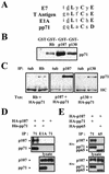
pp71 binds to the Rb family in vitro and in vivo. (A) Comparison of viral LXCXE sequences. Conserved residues are in bold. This motif is generally preceded by an acidic residue (underlined). (B) Glutathione-Sepharose beads preloaded with GST or GST fusions to the indicated Rb family members were incubated with lysates of U-2 OS cells transfected with pCGN71 and then processed for immunoblotting with an HA-specific antibody. (C) U-2 OS cells were transfected (Txn) with expression plasmids (pSG5-pp71, pCMV-Rb, pCMV-p107, and pCMV-p130) for the indicated proteins. After 24 h in 0.1% serum-containing medium, lysates were prepared and subjected to immunoprecipitation (IP) with the indicated rabbit polyclonal antisera. Bound pp71 was detected with the HA antibody. HC, antibody heavy chain; tub, tubulin. (D) U-2 OS cells were transfected (+) with expression plasmids (pSG5-His-pp71 and pHAp107) for the indicated proteins. After 24 h in 0.1% serum-containing medium, lysates were prepared and subjected to immunoprecipitation (IP) with either a cocktail of the five pp71 mouse monoclonal antibodies (71) or the M73 E1A antibody (E1A). Proteins on Western blots were detected with either the HA antibody (p107) or the pp71 antibody 4F7. (E) Transfection-immunoprecipitation experiments were performed as described above with pHAp107, pCGN71, and pCGNpp65. The pp65 antibody for immunoprecipitation was 1025, and proteins on Western blots were detected with the HA antibody.
FIG. 3.
FIG. 3.
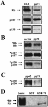
pp71 targets the hypophosphorylated forms of the Rb family. (A) U-2 OS cells were transfected with pCMV-Rb, pCMV-p107, or pCMV-p130 and either pCMVE1A or pCGN71. Lysates were harvested 48 h later, separated on 10% gels, and probed with antibodies to the indicated Rb family members. (B) U-2 OS cells were transfected with pSG5L-HApRB, pHAp107, or pHAp130 and either the empty pSG5 vector or pSG5-pp71. Lysates were harvested 48 h later, separated on 6% gels, and probed with the HA antibody. Asterisks represent phosphorylated protein species. (C) U-2 OS cells were transfected with pHAp130 and either pCGNpp65 or pCGN71. Lysates were harvested 48 h later, separated on 6% gels, and probed with the HA antibody. Asterisks represent phosphorylated protein species. (D) Glutathione-Sepharose beads preloaded with GST or a GST-pp71 fusion protein were incubated with lysates of U-2 OS cells transfected with pSG5L-HApRb and then processed for immunoblotting with an HA-specific antibody. Asterisks indicate phosphorylated Rb proteins.
FIG. 4.
FIG. 4.
pp71 degrades the Rb family. (A) U-2 OS cells cotransfected with pSG5L-HApRb, pHAp107, or pHAp130 and either pSG5 (filled squares) or pSG5-pp71 (open circles) were subjected to a pulse-chase analysis as described in Materials and Methods. The relative amounts of total protein (hypo- and hyperphosphorylated forms) are shown. (B) Phosphorimages of the 0- and 1-h time points of a representative Rb pulse-chase experiment are shown. The asterisk indicates the phosphorylated Rb species. (C) The relative amount of each individual form of Rb (lower, hypophosphorylated band; upper, hyperphosphorylated band) in the presence and absence of pp71 was determined after chase times of 0 and 1 h for two independent experiments, and the percent change during that time period is shown for each species.
FIG. 5.
FIG. 5.
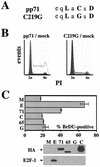
The LXCXD motif is required for the induction of S phase by pp71. (A) Sequences of wild-type pp71 and the C219G mutant in which a single cysteine residue was changed to a glycine. Conserved residues are in boldface, and acidic residues preceding the motif are underlined. (B) Quiescent Rat-1 cells were mock infected (filled histograms) or infected with ADtrack-based recombinant adenoviruses expressing either pp71 (gray) or the C219G mutant (black) and harvested at 24 h postinfection, and their DNA contents were analyzed by flow cytometry. Similar expression levels were confirmed by Western blotting (data not shown). (C) Quiescent Rat-1 cells grown on coverslips were infected with ADtrack-based recombinant adenoviruses expressing the indicated protein as described above and pulse-labeled with BrDU prior to harvesting. The coverslips were stained as described in Materials and Methods, and the number of BrDU-positive nuclei was counted by fluorescence microscopy and is presented as a percentage of the total number of nuclei counted (top). The cells remaining attached to the plates were processed for Western blot analysis of equal amounts of protein lysate with the indicated antibody (bottom). M, mock; E, E2F-1; 71, pp71; 65, pp65; G, GFP; C, C219G; HA, HA-specific antibody; E2F-1, E2F-1-specific antibody.
FIG. 6.
FIG. 6.
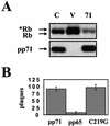
The pp71-mediated degradation of Rb requires a wild type LXCXD motif. (A) Cotransfection assays were performed as described in the legend to Fig. 3B with pSG5L-HApRb. C, pSG5-pp71-C219G; V, vector (pSG5); 71, pSG5-pp71. Asterisks indicate phosphorylated forms of the Rb protein. (B) Primary human foreskin fibroblasts were transfected with infectious HCMV DNA isolated from virions and either pCGN71, pCGNpp65, or pCGN71-C219G. The cells were overlaid with agarose, and plaques were stained and counted 18 days after transfection.
FIG. 7.
FIG. 7.
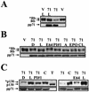
The pp71-mediated degradation of Rb and p130 requires proteasome function. (A) Cotransfection assays with pSG5L-HApRb were performed as described in the legend to Fig. 3B. Where indicated, lactacystin (L) was added 14 h before harvesting lysates. V, vector; 71, pp71. (B) Cotransfection assays were preformed as described above. Where indicated, the proteasome inhibitors lactacystin (L), proteasome inhibitor no. 1 (PI#1), ALLN (A), epoxomicin (EPO), and clasto-lactacystin β-lactone (CL), the cysteine protease inhibitor E64, or, as a control, the solvent DMSO (D) were added as described above. (C) Cotransfection assays were performed with pHAp130 as described above. The indicated additions were as described above. Also analyzed in these experiments were the pp71 C219G mutant (C) and SV40 T antigen (T).
FIG. 8.
FIG. 8.
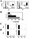
pp71 neither induces apoptosis nor transforms cells. (A) Quiescent Rat-1 cells were infected with the indicated ADIC-based recombinant adenovirus. Floating and attached cells were collected at 48 and 72 h postinfection and analyzed by flow cytometry. Quantitation of sub-G0 cells indicated by the M1 gate in the histograms was determined with CELLQuest software (Becton Dickinson). M, mock, dark gray filled histograms; 71, pp71, light gray; E, E2F-1, black. (B) Quiescent Rat-1 cells were infected with the indicated viruses and harvested at 72 h postinfection as described above. Because cells were coinfected with two viruses, the amount of each individual virus employed was half that used in the experiments for which results are shown in panel A so as to keep the total amount of input virus the same as in all other experiments. Where pp71 and E2F-1 were expressed individually, a control virus without an insert (ADIC [A]) was coinfected to maintain the proper virus level. (C) Primary rat embryo fibroblasts were transfected with the indicated plasmids and cultured for 18 to 21 days, at which time transformed colonies were counted. Error bars represent standard deviations. (D) Transformation assays were performed with baby rat kidney cells as described above.

Similar articles

Cited by

References

    1. Baldick, C. J., A. Marchini, C. E. Patterson, and T. Shenk. 1997. Human cytomegalovirus protein pp71 (ppUL82) enhances the infectivity of viral DNA and accelerates the infectious cycle. J. Virol. 71:4400-4408. - PMC - PubMed
    1. Berezutskaya, E., B. Yu, A. Morozov, P. Raychaudhuri, and S. Bagchi. 1997. Differential regulation of the pocket domains of the retinoblastoma family proteins by the HPV16 E7 oncoprotein. Cell Growth Differ. 8:1277-1286. - PubMed
    1. Boyer, S. N., D. E. Wazer, and V. Band. 1996. E7 protein of human papillomavirus-16 induces degradation of retinoblastoma protein through the ubiquitin-proteasome pathway. Cancer Res. 56:4620-4624. - PubMed
    1. Bresnahan, W. A., I. Boldogh, E. A. Thompson, and T. Albrecht. 1996. Human cytomegalovirus inhibits cellular DNA synthesis and arrests productively infected cells in G1. Virology 224:150-160. - PubMed
    1. Chellappan, S., V. B. Kraus, B. Kroger, K. Munger, P. M. Howley, W. C. Phelps, and J. R. Nevins. 1992. Adenovirus E1A, simian virus 40 tumor antigen, and human papillomavirus E7 protein share the capacity to disrupt the interaction between transcription factor E2F and the retinoblastoma gene product. Proc. Natl. Acad. Sci. USA 89:4549-4553. - PMC - PubMed

Publication types

MeSH terms

Substances