. 2003 Mar;77(6):3418-29.
doi: 10.1128/jvi.77.6.3418-3429.2003.

Vaccinia virus G7L protein Interacts with the A30L protein and is required for association of viral membranes with dense viroplasm to form immature virions

Affiliations

Vaccinia virus G7L protein Interacts with the A30L protein and is required for association of viral membranes with dense viroplasm to form immature virions

Patricia Szajner et al. J Virol. 2003 Mar.

Abstract

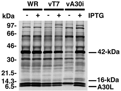
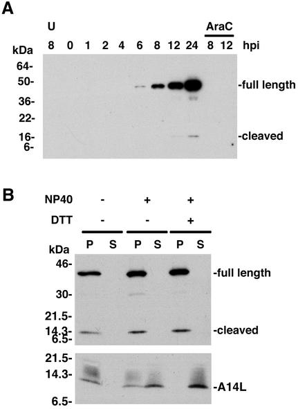
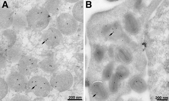
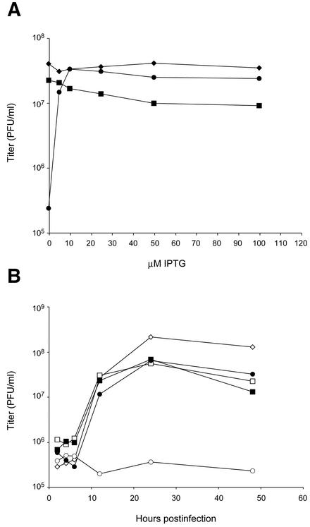
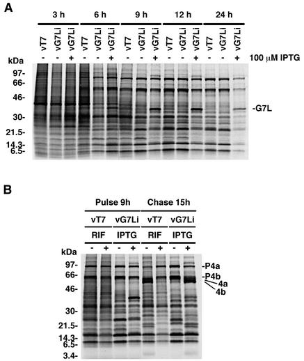
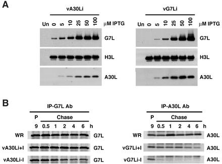
The vaccinia virus A30L protein is required for the association of electron-dense, granular, proteinaceous material with the concave surfaces of crescent membranes, an early step in viral morphogenesis. For the identification of additional proteins involved in this process, we used an antibody to the A30L protein, or to an epitope appended to its C terminus, to capture complexes from infected cells. A prominent 42-kDa protein was resolved and identified by mass spectrometry as the vaccinia virus G7L protein. This previously uncharacterized protein was expressed late in infection and was associated with immature virions and the cores of mature particles. In order to study the role of the G7L protein, a conditional lethal mutant was made by replacing the G7L gene with an inducible copy. Expression of G7L and formation of infectious virus was dependent on the addition of inducer. Under nonpermissive conditions, morphogenesis was blocked and viral crescent membranes and immature virions containing tubular elements were separated from the electron-dense granular viroplasm, which accumulated in large spherical masses. This phenotype was identical to that previously obtained with an inducible, conditional lethal A30L mutant. Additional in vivo and in vitro experiments provided evidence for the direct interaction of the A30L and G7L proteins and demonstrated that the stability of each one was dependent on its association with the other.


Figures

FIG. 1.
FIG. 1.
Coimmunoprecipitation of a 42-kDa protein with the A30L protein from VV-infected cells. BS-C-1 cells were infected with wild-type VV (WR), vT7LacOI (vT7), or vA30Li (vA30i) in the presence (+) or absence (−) of 50 μM IPTG as indicated. At 6 h after infection, the cells were labeled with a mixture of [35S]methionine and [35S]cysteine and incubated for 18 h at 37°C. Cell extracts were prepared and incubated with antiserum directed to the C-terminal 11 amino acids of the A30L protein. The immunoprecipitated products were resolved by SDS-PAGE and visualized by autoradiography. Bands corresponding to the A30L protein (which is the lower band of a doublet) and proteins with masses of 42 and 16 kDa are indicated on the right. Numbers on the left correspond to molecular masses of the marker proteins.
FIG. 2.
FIG. 2.
Identification of the 42-kDa protein as the product of the G7L ORF. (A) Affinity purification of the A30L protein complex. Proteins extracted from cells infected with vA30LiHA in the presence of IPTG were purified using a high-affinity anti-HA affinity matrix. The F.T (Flowthrough) lane contains material that did not bind to the HA matrix. The HA matrix was washed with lysis buffer (wash lanes, fractions 1 to 3), and the proteins were eluted with the HA peptide (HApep lanes, fractions 1 to 3) followed by glycine (glycine lanes, fractions 1 to 4). Proteins from each fraction were analyzed by electrophoresis on an SDS-10 to 20% polyacrylamide gel in Tricine buffer followed by silver staining. Bands corresponding to the A30L and 42-kDa proteins are indicated. Numbers on the left indicate the positions and molecular masses in kilodaltons of marker proteins. (B) Predicted amino acid sequence of the VV G7L ORF. Peptides identified by MS are underlined. Amino acids comprising the peptide used to produce polyclonal antibody are in italics, and consensus proteolysis cleavage sites are indicated by arrows. (C) Reactivity of the 42-kDa protein with G7L protein antiserum. (Left) Lysates of uninfected (lane U) and VV WR-infected BS-C-1 cells were resolved by SDS-PAGE, reacted with G7L peptide antiserum by Western blotting, and detected by chemiluminescence. (Right) Extracts of cells infected with the vA30LiHA virus in the presence (+) or absence (−) of 100 μM IPTG were incubated with the high-affinity anti-HA affinity matrix, and bound proteins were analyzed by Western blotting with G7L protein antiserum. The positions of migrations and molecular masses of marker proteins are indicated on the left.
FIG. 3.
FIG. 3.
Synthesis and virion localization of the G7L protein. (A) Temporal synthesis of the G7L protein. BS-C-1 cells were mock infected for 8 h (lane U) or infected with VV at a multiplicity of infection of 10 in the absence or presence of AraC and harvested between 0 and 24 h postinfection (hpi). Proteins from total cell extracts were resolved by electrophoresis on an SDS-4 to 20% gradient polyacrylamide gel and analyzed by Western blotting using rabbit G7L peptide antiserum. Proteins were detected by chemiluminescence. The bands corresponding to the full-length and cleaved forms of the G7L protein are indicated on the right. The positions of migrations and molecular masses of marker proteins are indicated on the left. (B) Association of the G7L protein with purified virions. Sucrose gradient-purified VV was incubated in buffer containing 1% NP-40 with or without 50 mM dithiothreitol. After centrifugation, the soluble (S lanes) and insoluble (P lanes) fractions were analyzed by Western blotting using G7L or A14L peptide antiserum. Bands corresponding to the A14L protein or to the full-length and cleaved forms of the G7L protein are indicated on the right. The numbers on the left correspond to the molecular masses of the marker proteins. DTT, dithiothreitol.
FIG. 4.
FIG. 4.
Localization of the G7L protein by immunoelectron microscopy. BS-C-1 cells were infected with VV WR at a multiplicity of infection of 2 PFU per cell. At 22 h, the cells were fixed in paraformaldehyde, cryosectioned, and incubated with an antibody to the peptide corresponding to amino acids 26 to 38 of the G7L protein and then with 10-nm-diameter gold particles conjugated to protein A. Electron micrographs are shown, with the scale indicated by the bars. (A) Field containing large numbers of IV; (B) field containing mature virions. Arrows point to representative gold particles.
FIG. 5.
FIG. 5.
Construction of a conditional lethal inducible G7L VV. (A) Schematic diagram representing the genome of vG7Li. The J2R (thymidine kinase [TK]), G7L, and A56R (HA) loci are shown. Insertions into these loci are displayed below the line. Additional abbreviations: P11, a VV late promoter, P7.5, a VV early late promoter; lacO, E. coli lac operator; lacI, E. coli lac repressor gene; T7 pol, bacteriophage T7 RNA polymerase gene; PT7, bacteriophage T7 promoter; EMC, encephalomyocarditis virus mRNA cap-independent translation enhancer element; gus, E. coli β-glucuronidase gene; gpt, E. coli guanine phosphoribosyltransferase gene. (B) Effect of IPTG on virus plaque formation. BS-C-1 cell monolayers were infected with vT7lacOI, vG7L/G7Li, or vG7Li in the presence (+) or absence (−) of 50 μM IPTG in the methylcellulose overlay. Cells were stained with crystal violet 48 h after infection.
FIG. 6.
FIG. 6.
Effect of IPTG on yields of infectious vG7Li. (A) BS-C-1 cells were infected with VV WR (♦), vG7L/G7Li (▪), or vG7Li (•) at a multiplicity of infection of 5 and incubated in the presence of 0 to 100 μM IPTG for 24 h. Virus titers were determined by plaque assay in the presence of 50 μM IPTG. (B) BS-C-1 cells were infected with VV WR (⋄), vG7L/G7Li (□, ▪), or vG7Li (○, •) in the absence (open symbols) or presence (filled symbols) of 50 μM IPTG. Cells were harvested at the indicated times after infection, and virus titers were determined as described for panel A.
FIG. 7.
FIG. 7.
Synthesis and processing of viral proteins. (A) Pulse-labeling of viral proteins. BS-C-1 cells were infected with vT7lacOI (vT7) or vG7Li at a multiplicity of infection of 10 in the presence (+) or absence (−) of 100 μM IPTG and labeled with a mixture of [35S]methionine and [35S]cysteine for 30-min periods starting at 3, 6, 9, 12, or 24 h after infection. Immediately after being labeled, the cells were washed and lysed and the labeled proteins were analyzed by electrophoresis on an SDS-4 to 20% gradient polyacrylamide gel. The gel was dried, and the proteins were visualized by autoradiography. The numbers on the left correspond to the molecular masses of the marker proteins. (B) Proteolytic processing of viral late proteins. BS-C-1 cells were infected either with vT7lacOI in the presence (+) or absence (−) of 100 μg of rifampin (RIF) per ml or with vG7Li in the presence (+) or absence (−) of 100 μM IPTG. At 9 h after infection, the cells were pulse-labeled with a mixture of [35S]methionine and [35S]cysteine for 30 min. Cells were either harvested immediately (pulse) or incubated with excess unlabeled methionine for an additional 15 h (chase). The proteins were denatured with SDS and mercaptoethanol, analyzed by electrophoresis on an SDS-4 to 20% gradient polyacrylamide gel, and visualized by autoradiography. The positions of migration of proteolytically processed structural proteins (4a and 4b) and their precursors (P4a and P4b) are shown on the right.
FIG. 8.
FIG. 8.
Electron microscopy of cells infected with vG7Li in the presence or absence of IPTG. BS-C-1 cells were infected with vG7Li in the presence (A) or absence (B to D) of 50 μM IPTG. After 24 h, the cells were fixed and prepared for transmission electron microscopy. The arrow in panel B points to electron-dense viroplasm, which is separated from IV membranes. Arrows in panel D point to cross-sections of tubules in which the membrane bilayer is clearly discerned. Electron micrographs are shown, with the scale indicated by the bars. Abbreviations: C, crescents; nu, nucleoid within an IV.
FIG. 9.
FIG. 9.
The interaction of the G7L and A30L proteins is direct. (A) The interaction of the G7L and A30L proteins does not depend on VV morphogenesis. BS-C-1 cells were infected with vA30Li (vA30i) or vA17Li (vA17i) in the presence (+) or absence (−) of IPTG or with the vT7lacOI (vT7) virus in the presence (+) or absence (−) of rifampin (RIF). At 6 h after infection, cells were labeled with a mixture of [35S]methionine and [35S]cysteine. Labeled cells were harvested at 24 h after infection, and cell extracts were incubated with the A30L peptide antiserum and then protein A-Sepharose beads. The bound proteins were resolved in an SDS-4 to 20% polyacrylamide gel and visualized by autoradiography. The G7L protein and A30L protein (lower band of a doublet) are indicted on the right. (B) The interaction of the A30L and G7L proteins is not dependent on other VV late proteins. BS-C-1 cells were infected with vTF7-3 in the presence (+) or absence (−) of AraC and transfected (+) or not transfected (−) with plasmids containing the A30L or the G7L ORF regulated by the bacteriophage T7 promoter. After 6 h, the cells were labeled with a mixture of [35S]methionine and [35S]cysteine for 18 h. Cell extracts were immunoprecipitated with A30L peptide antiserum as described for panel A, and the bound proteins were resolved on an SDS-10 to 20% polyacrylamide gel in Tricine buffer and visualized by autoradiography. (C) The A30L and G7L proteins interact in the absence of other viral proteins. Templates containing (+) or not containing (−) the A30L or G7L ORF under the control of the bacteriophage T7 promoter were transcribed and translated in a reticulocyte lysate in the presence of [35S]methionine. The in vitro-translated proteins were immunoprecipitated with A30L peptide antiserum and analyzed by SDS-PAGE as described for panel A. In all panels, the migration positions and masses of marker proteins are indicated on the left and the positions of the G7L and A30L proteins are indicated on the right.
FIG. 10.
FIG. 10.
Stability of the A30L and G7L proteins. (A) BS-C-1 cells were either mock infected (lanes Un) or infected with vA30Li (left panel) or vG7Li (right panel) in the absence or presence of increasing concentrations of IPTG. After 24 h, cells were harvested and total cell lysates were analyzed by electrophoresis on an SDS-10 to 20% gradient polyacrylamide gel in Tricine buffer followed by Western blotting using antisera to the A30L, G7L, or H3L protein as indicated. (B) BS-C-1 cells were infected with VV WR, vA30Li, or vG7Li in the presence or absence of 50 μM IPTG. After 9 h, the cells were pulse-labeled with a mixture of [35S]methionine and [35S]cysteine for 15 min. Cells were either harvested immediately (lanes P) or incubated with excess unlabeled methionine for 0.5, 1, 2, 4, and 6 h (Chase lanes). Extracts of cells infected with vA30Li or vG7Li were immunoprecipitated with the G7L (IP-G7L Ab; left panel) or A30L (IP-A30C Ab; right panel) protein antiserum, respectively. The extracts from WR-infected cells were immunoprecipitated with G7L or A30L protein antiserum as indicated. The immunoprecipitated products were analyzed by SDS-PAGE and visualized by autoradiography.

Similar articles

Cited by

References

    1. Alexander, W. A., B. Moss, and T. R. Fuerst. 1992. Regulated expression of foreign genes in vaccinia virus under the control of bacteriophage T7 RNA polymerase and the Escherichia coli lac repressor. J. Virol. 66:2934-2942. - PMC - PubMed
    1. Appleyard, G., A. J. Hapel, and E. A. Boulter. 1971. An antigenic difference between intracellular and extracellular rabbitpox virus. J. Gen. Virol. 13:9-17. - PubMed
    1. Betakova, T., E. J. Wolffe, and B. Moss. 1999. Regulation of vaccinia virus morphogenesis: phosphorylation of the A14L and A17L membrane proteins and C-terminal truncation of the A17L protein are dependent on the F10L protein kinase. J. Virol. 73:3534-3543. - PMC - PubMed
    1. Blasco, R., and B. Moss. 1992. Role of cell-associated enveloped vaccinia virus in cell-to-cell spread. J. Virol. 66:4170-4179. - PMC - PubMed
    1. Blasco, R., J. R. Sisler, and B. Moss. 1993. Dissociation of progeny vaccinia virus from the cell membrane is regulated by a viral envelope glycoprotein: effect of a point mutation in the lectin homology domain of the A34R gene. J. Virol. 67:3319-3325. - PMC - PubMed

Substances

LinkOut - more resources