Isolation and characterization of Staufen-containing ribonucleoprotein particles from rat brain
- PMID: 12592035
- PMCID: PMC149965
- DOI: 10.1073/pnas.0334355100
Isolation and characterization of Staufen-containing ribonucleoprotein particles from rat brain
Abstract
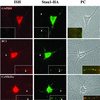
Localized mRNAs are thought to be transported in defined particles to their final destination. These particles represent large protein complexes that may be involved in recognizing, transporting, and anchoring localized messages. Few components of these ribonucleoparticles, however, have been identified yet. We chose the strategy to biochemically enrich native RNA-protein complexes involved in RNA transport to identify the associated RNAs and proteins. Because Staufen proteins were implicated in intracellular RNA transport, we chose mammalian Staufen proteins as markers for the purification of RNA transport particles. Here, we present evidence that Staufen proteins exist in two different complexes: (i) distinct large, ribosome- and endoplasmic reticulum-containing granules preferentially found in the membrane pellets during differential centrifugation and (ii) smaller particles in the S100 from rat brain homogenates. On gel filtration of the S100, we identified soluble 670-kDa Staufen1-containing and 440-kDa Staufen2-containing particles. They do not cofractionate with ribosomes and endoplasmic reticulum but rather coenrich with kinesin heavy chain. Furthermore, the fractions containing the Staufen1 particles show a 15-fold enrichment of mRNAs compared with control fractions. Most importantly, these fractions are highly enriched in BC1, and, to a lesser extent, in the alpha-subunit of the Ca(2+)/calmodulin-dependent kinase II, two dendritically localized RNAs. Finally, both RNAs colocalize with Staufen1-hemagglutinin in particles in dendrites of transfected hippocampal neurons. We therefore propose that these Staufen1-containing particles may represent RNA transport intermediates that are in transit to their final destination within neurons.
Figures


Comment in
-
Insights into mRNA transport in neurons.Proc Natl Acad Sci U S A. 2003 Feb 18;100(4):1465-6. doi: 10.1073/pnas.0630376100. Epub 2003 Feb 10. Proc Natl Acad Sci U S A. 2003. PMID: 12578967 Free PMC article. No abstract available.
Similar articles
-
Barentsz, a new component of the Staufen-containing ribonucleoprotein particles in mammalian cells, interacts with Staufen in an RNA-dependent manner.J Neurosci. 2003 Jul 2;23(13):5778-88. doi: 10.1523/JNEUROSCI.23-13-05778.2003. J Neurosci. 2003. PMID: 12843282 Free PMC article.
-
The transport of Staufen2-containing ribonucleoprotein complexes involves kinesin motor protein and is modulated by mitogen-activated protein kinase pathway.J Neurochem. 2007 Sep;102(6):2073-2084. doi: 10.1111/j.1471-4159.2007.04697.x. Epub 2007 Jun 22. J Neurochem. 2007. PMID: 17587311
-
Characterization of Staufen 1 ribonucleoprotein complexes.Biochem J. 2004 Dec 1;384(Pt 2):239-46. doi: 10.1042/BJ20040812. Biochem J. 2004. PMID: 15303970 Free PMC article.
-
Staufen: a common component of mRNA transport in oocytes and neurons?Trends Cell Biol. 2000 Jun;10(6):220-4. doi: 10.1016/s0962-8924(00)01767-0. Trends Cell Biol. 2000. PMID: 10802537 Review.
-
The role of mammalian Staufen on mRNA traffic: a view from its nucleocytoplasmic shuttling function.Cell Struct Funct. 2005;30(2):51-6. doi: 10.1247/csf.30.51. Cell Struct Funct. 2005. PMID: 16377940 Review.
Cited by
-
The long noncoding RNA mimi scaffolds neuronal granules to maintain nervous system maturity.Sci Adv. 2022 Sep 30;8(39):eabo5578. doi: 10.1126/sciadv.abo5578. Epub 2022 Sep 28. Sci Adv. 2022. PMID: 36170367 Free PMC article.
-
Staufen- and FMRP-containing neuronal RNPs are structurally and functionally related to somatic P bodies.Neuron. 2006 Dec 21;52(6):997-1009. doi: 10.1016/j.neuron.2006.10.028. Neuron. 2006. PMID: 17178403 Free PMC article.
-
Knockdown of p180 eliminates the terminal differentiation of a secretory cell line.Mol Biol Cell. 2009 Jan;20(2):732-44. doi: 10.1091/mbc.e08-07-0682. Epub 2008 Nov 26. Mol Biol Cell. 2009. PMID: 19037105 Free PMC article.
-
Matrix-screening reveals a vast potential for direct protein-protein interactions among RNA binding proteins.Nucleic Acids Res. 2021 Jul 9;49(12):6702-6721. doi: 10.1093/nar/gkab490. Nucleic Acids Res. 2021. PMID: 34133714 Free PMC article.
-
mRNA transport in dendrites: RNA granules, motors, and tracks.J Neurosci. 2006 Jul 5;26(27):7139-42. doi: 10.1523/JNEUROSCI.1821-06.2006. J Neurosci. 2006. PMID: 16822968 Free PMC article. Review.
References
-
- St. Johnston D. Cell. 1995;81:161–170. - PubMed
-
- Bashirullah A, Cooperstock R L, Lipshitz H D. Annu Rev Biochem. 1998;67:335–394. - PubMed
-
- Bassell G J, Oleynikov Y, Singer R H. FASEB J. 1999;13:447–454. - PubMed
-
- Kiebler M A, DesGroseillers L. Neuron. 2000;25:19–28. - PubMed
-
- Jansen R P. Nat Rev Mol Cell Biol. 2001;2:247–256. - PubMed
Publication types
MeSH terms
Substances
LinkOut - more resources
Full Text Sources
Other Literature Sources
Molecular Biology Databases
Miscellaneous

