. 2003 Feb;111(4):469-78.
doi: 10.1172/JCI16491.

The suppressor of cytokine signaling-1 (SOCS1) is a novel therapeutic target for enterovirus-induced cardiac injury

Affiliations

The suppressor of cytokine signaling-1 (SOCS1) is a novel therapeutic target for enterovirus-induced cardiac injury

Hideo Yasukawa et al. J Clin Invest. 2003 Feb.

Abstract

Enteroviral infections of the heart are among the most commonly identified causes of acute myocarditis in children and adults and have been implicated in dilated cardiomyopathy. Although there is considerable information regarding the cellular immune response in myocarditis, little is known about innate signaling mechanisms within the infected cardiac myocyte that contribute to the host defense against viral infection. Here we show the essential role of Janus kinase (JAK) signaling in cardiac myocyte antiviral defense and a negative role of an intrinsic JAK inhibitor, the suppressor of cytokine signaling (SOCS), in the early disease process. Cardiac myocyte-specific transgenic expression of SOCS1 inhibited enterovirus-induced signaling of JAK and the signal transducers and activators of transcription (STAT), with accompanying increases in viral replication, cardiomyopathy, and mortality in coxsackievirus-infected mice. Furthermore, the inhibition of SOCS in the cardiac myocyte through adeno-associated virus-mediated (AAV-mediated) expression of a dominant-negative SOCS1 increased the myocyte resistance to the acute cardiac injury caused by enteroviral infection. These results indicate that strategies directed at inhibition of SOCS in the heart and perhaps other organs can augment the host-cell antiviral system, thus preventing viral-mediated end-organ damage during the early stages of infection.


Figures
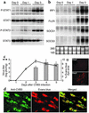
Figure 1
Figure 1
Correlation of CVB3-induced cardiac injury and JAK-STAT activation. (a) Mice were infected with CVB3. Protein lysate from the heart was blotted at the indicated days after CVB3 inoculation and probed with the antibodies indicated. (b) Northern blot of total RNA from the heart after CVB3 inoculation was probed for IRF1, FcγRI, SOCS1, and SOCS3 expression. 28S and 18S RNA are shown as controls. (c) Virus titer and the disruption of cardiac cell membrane within 5 days after CVB3 inoculation. Four-week-old male Balb/c mice were inoculated with 103 PFUs of CVB3 intraperitoneally and sacrificed at the day indicated. Evans blue dye was injected intraperitoneally 4 hours before the sacrifice (4). The left panel shows the time course of virus titer (solid line) and the percentage of Evans blue dye–positive area in the heart (gray bars). The Evans blue dye–positive areas were quantitated using NIH image software (NIH, Bethesda, Maryland, USA). The right panels show Evans blue dye–negative (Day 0) and positive (Day 3) sections (the brighter red staining areas). The data were collected from three mice for each time point and expressed as means ± SE. (d) Dual immunostaining of the infected heart demonstrates that the cells that are positive for Evans blue dye are also positive for viral capsid proteins. The left panel shows immunofluorescent staining with anti-CVB3 antibody (green), and the center panel shows Evans blue dye (red) uptake in the same field. The right panel is a merged image. Scale bars: 1 mm (c); 50 μm (d). P-STAT1, phospho-STAT1; P-STAT3, phospho-STAT3.
Figure 2
Figure 2
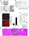
Increased myocardial injury, virus replication, and mortality in SOCS1 transgenic mice. (a) Expression of Myc-tagged SOCS1 in the heart was compared among four SOCS1 transgenic lines (A–D) by immunoblotting with anti-Myc antibody (upper panel). Heart tissue extracts for protein (lower left panel) and RNA (lower right panel) from two lines of wild-type and two lines of transgenic mice (A and B) were prepared three days after CVB3 inoculation and probed as shown. (b) Wild-type and SOCS1 transgenic mice (4-week-old males) were inoculated with 103 PFUs of virus (n = 15). The survival rate in infected, SOCS1 transgenic mice was significantly lower than in infected, wild-type littermates (P < 0.0001). (c) Evans blue dye uptake in the heart was markedly increased in surviving SOCS1 transgenic mice on day 4 after infection (red stain, left panels). The percent area of Evans blue staining in the hearts is shown (right panel; mean ± SE, n = 3, *P < 0.01). (d) Increased viral titer in the heart but not the liver of SOCS1 transgenic mice. Virus titers in SOCS1 transgenic and wild-type hearts and livers from 3–5 days after infection (mean ± SE, n = 3, *P < 0.01 comparing SOCS1 transgenic mice with wild-type littermates). (e) Hematoxylin and eosin stains of representative wild-type and SOCS1 transgenic mouse hearts 4 days after infection. Note the thrombus in the center of the ventricle (right panel). Scale bars: 1 mm (c); 100 μm (e, middle); 200 μm (e, right). Tg, transgenic; P-STAT1, phospho-STAT1; P-STAT3, phospho-STAT3.
Figure 3
Figure 3
Deterioration of cardiac function after CVB3 inoculation in SOCS1 transgenic mice. Echocardiography was performed 3 days after virus inoculation (n = 3 mice per group). Upper panels show that the LVEDD and LVESD were significantly elevated in transgenic mice (black bars) as compared with wild-type (white bars). The %FS, a parameter of cardiac function, was significantly decreased in transgenic mice as compared with wild-type mice. The lower panel shows the typical M-mode image of the two groups. Results are shown as means ± SE. *P < 0.01 for the comparison of SOCS1 transgenic mice with wild-type littermates. Tg, transgenic.
Figure 4
Figure 4
Effect of SOCS1 and SOCS3 on the cytoprotective effect of IFNs and CT-1 in cultured cardiomyocytes. (a) Myc-tagged SOCS1 or SOCS3 was expressed in neonatal rat cardiomyocytes with the use of recombinant adenovirus vectors (20) (black or gray bars, respectively). The vector containing the LacZ gene was used as a control for adenoviral vector infection (white bars). After transduction with the adenoviral vectors, the cells were stimulated with IFN-γ, IFN-β, or CT-1 for 24 hours. Cells were then infected with CVB3 (+) or maintained without virus (–) for another 30 hours. The number of cells that remained on the plate after CVB3 infection was quantitated and reported as a percentage of cells in the wells not infected with CVB3. The data are from five independent experiments and are expressed as means ± SE. *P < 0.01 for the comparison with cells transduced with adenovirus LacZ, stimulated with cytokines, and infected with CVB3. **P < 0.01 for the comparison with cells transduced with adenovirus LacZ, not stimulated with cytokines, and infected with CVB3. (b) Myocytes were incubated with adenovirus LacZ, adenovirus SOCS1, or adenovirus SOCS3, serum depleted for 24 hours, and then stimulated with 1000 ng/ml IFN-γ for 5 hours or 1 nM CT-1 for 10 minutes. Total cell extracts were prepared and blotted with phospho-STAT1, STAT1, phospho-STAT3, and STAT3 antibodies. SOCS1 and SOCS3 expression were confirmed with an anti-Myc antibody. Representative Western blots from three independent experiments are shown. SOCS1, adenovirus containing Myc-tagged SOCS1 gene; SOCS3, adenovirus containing Myc-tagged SOCS3 gene; LacZ, adenovirus containing LacZ; Stim, stimulated, P-STAT1, phospho-STAT1; P-STAT3, phospho-STAT3.
Figure 5
Figure 5
Augmentation of the JAK/STAT pathway by dnSOCS1 in cardiac myocytes. (a and b) Cardiomyocytes were transfected with a plasmid mixture containing the APRE-luciferase reporter gene (200 ng), the NF-κB–luciferase reporter gene (200 ng), the β-galactosidase gene (100 ng), the AAV shuttle plasmid, or the indicated concentrations of dnSOCS1 plasmid. After transfection, cells were incubated in the presence or absence of 1 nM CT-1 or 20 ng/ml TNF-α for 6 hours, and cell extracts were prepared. Data normalized with the β-galactosidase activity are shown. The experiments were repeated three times. Results are expressed as means ± SD. *P < 0.01 for the comparison of CT-1 with the AAV shuttle. **P < 0.01 for the comparison of CT-1 with pcDNA3-SOCS1. (c) Time course of IFN-γ–induced STAT1 activation and CT-1–induced STAT3 activation in cardiac myocytes. Myocytes were treated with adenovirus LacZ or adenovirus dnSOCS1, were serum depleted for 24 hours, and were then stimulated with 1000 ng/ml IFN-γ or 1 nM CT-1 for the indicated period, respectively. Total cell extracts were prepared and blotted with phospho-STAT1, STAT1, phospho-STAT3, and STAT3 antibodies. The dnSOCS1 expression was confirmed with an anti-Myc antibody. Data from one experiment are presented; two additional experiments yielded comparable results. P-STAT1, phospho-STAT1; P-STAT3, phospho-STAT3; APRE, acute-phase response element.
Figure 6
Figure 6
AAV-directed expression of dnSOCS1 inhibits the CVB3-mediated cytopathic effect. Hearts of mice were directly injected with AAV Myc-tagged dnSOCS1 or AAV LacZ. Two weeks later, the mice were infected with CVB3. Five days after infection, myocardial sections were stained with anti-Myc (a) or anti–β-galactosidase antibodies (d). Evans blue uptake was examined in the same section (b and e). The merged images (c and f) show that in the areas where there is expression of dnSOCS1, there is a lack of membrane disruption due to the virus infection, as compared with areas of the myocardium that express LacZ. The percent area of Evans blue dye staining in the regions of the myocardium that stained positive for dnSOCS1 or LacZ was quantitated from three mice per group (g) and was significantly decreased in dnSOCS1-expressing areas as compared with LacZ-expressing areas. Results are shown as means ± SE. *P < 0.01 for the comparison with LacZ-transduced areas. Scale bars: 1 mm (af).
Figure 7
Figure 7
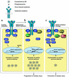
JAK-STAT antiviral defense in the cardiac myocyte and SOCS inhibition to limit early virus-induced cardiac injury. (a) Virus infection of the heart stimulates cytokine-receptor signaling through the JAK-STAT pathway. Activation of the JAK-STAT pathway induces antiviral target gene transcription stimulating the innate viral defense in the cardiac myocyte. (b) While activation of the JAK-STAT pathway has an important role in antiviral defense, phosphorylated STAT also induces SOCS expression that attenuates the innate antiviral defense by inhibiting JAK signaling. As in SOCS1-transgenic mice, increased expression of SOCS in cardiac myocytes results in robust virus replication and cardiac injury. (c) The strategies aimed at inhibition of SOCS potentiate the innate antiviral actions of cytokines that utilize the JAK-STAT pathway, resulting in the prevention of virus-mediated myocardial injury. P, phosphorylated.

Similar articles

Cited by

References

    1. Rose NR. Viral damage or “molecular mimicry” — placing the blame in myocarditis. Nat. Med. 2000;6:631–632. - PubMed
    1. Liu P, Martino T, Opavsky MA, Penninger J. Viral myocarditis: balance between viral infection and immune response. Can. J. Cardiol. 1996;12:935–943. - PubMed
    1. Baboonian C, Davies MJ, Booth JC, McKenna WJ. Coxsackie B viruses and human heart disease. Curr. Top. Microbiol. Immunol. 1997;223:31–52. - PubMed
    1. Badorff C, et al. Enteroviral protease 2A cleaves dystrophin: evidence of cytoskeletal disruption in an acquired cardiomyopathy. Nat. Med. 1999;5:320–326. - PubMed
    1. Knowlton KU, Badorff C. The immune system in viral myocarditis: maintaining the balance. Circ. Res. 1999;85:559–561. - PubMed

Publication types

MeSH terms