. 2003 Feb 6;22(5):710-21.
doi: 10.1038/sj.onc.1206151.

Selective PDZ protein-dependent stimulation of phosphatidylinositol 3-kinase by the adenovirus E4-ORF1 oncoprotein

Affiliations

Selective PDZ protein-dependent stimulation of phosphatidylinositol 3-kinase by the adenovirus E4-ORF1 oncoprotein

Kristopher K Frese et al. Oncogene. .

Abstract

While PDZ domain-containing proteins represent cellular targets for several different viral oncoproteins, including human papillomavirus E6, human T-cell leukemia virus type 1 Tax, and human adenovirus E4-ORF1, the functional consequences for such interactions have not been elucidated. Here we report that, at the plasma membrane of cells, the adenovirus E4-ORF1 oncoprotein selectively and potently stimulates phosphatidylinositol 3-kinase (PI3K), triggering a downstream cascade of events that includes activation of both protein kinase B and p70S6-kinase. This activity of E4-ORF1 could be abrogated by overexpression of its PDZ-protein targets or by disruption of its PDZ domain-binding motif, which was shown to mediate complex formation between E4-ORF1 and PDZ proteins at the plasma membrane of cells. Furthermore, E4-ORF1 mutants unable to activate the PI3K pathway failed to transform cells in culture or to promote tumors in animals, and drugs that block either PI3K or p70S6-kinase inhibited E4-ORF1-induced transformation of cells. From these results, we propose that the transforming and tumorigenic potentials of the adenovirus E4-ORF1 oncoprotein depend on its capacity to activate PI3K through a novel PDZ protein-dependent mechanism of action.


Figures

Figure 1
Figure 1
E4-ORF1 activates PI3K. (a) Elevated PI3K activity in E4-ORF1-expressing cells. An equivalent amount of extract from serum-starved CREF or CREF-E4-ORF1 cells was subjected to in vitro PI3K assays with PI substrate (bottom) as described in Materials and methods or to immunoblot analyses with anti-p85 antibodies (top). Indicated samples were untreated (−) or treated with 10 ng/ml PDGF and/or 100 nM wortmannin (wort). Results were compiled from three independent experiments. (b) PI3K lipid products accumulate at the plasma membrane of E4-ORF1-expressing cells. NIH 3T3 cells on coverslips in 6-cm dishes were lipofected with pGFP-AH or -AHR25C (0.5 μg each) and pGW1 encoding wt or the indicated mutant E4-ORF1 (50 ng each). Confluent serum-starved cells were visualized by fluorescence microscopy. Indicated cells were pretreated with 100 nM wortmannin (wort). For cells expressing E4-ORF1 mutants unable to activate PI3K, GFP-AH accumulated at variable levels in NIH 3T3 cell nuclei. Therefore, the brighter nuclear GFP-AH signal observed in the cell expressing T123D was not a general finding in these experiments. (c) Focus formation by wt and mutant E4-ORF1 proteins. CREF cells in 10-cm dishes were transfected with empty pJ4Ω (−) or pJ4Ω encoding wt or the indicated mutant E4-ORF1 (20 μg each) in triplicate. Foci were quantified 3 weeks post-transfection
Figure 2
Figure 2
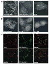
E4-ORF1 and PDZ proteins localize to the plasma membrane. (a) E4-ORF1 localizes to the plasma membrane. TE85-E4-ORF1 cells, CREF-9ORF1-low cells (CREF-E4-ORF1 cells), or 20-8 mammary tumor cells were subjected to indirect IF assays with E4-ORF1 antibodies. In the middle and right panels, cell nuclei are circumscribed by dashed lines to aid visualization of discontinuous E4-ORF1 staining present along the membrane at regions of cell–cell contact. (b) The PDZ domain-binding motif of E4-ORF1 promotes its recruitment to the plasma membrane. CREF cells stably expressing GFP-tagged wt or mutant E4-ORF1 proteins were visualized by fluorescence microscopy. Results with GFP-IA or GFP-IIIA are representative of findings with region I/II mutants IIA and IIB or region III mutants T123D and V125A, respectively. (c) E4-ORF1 colocalizes with PDZ proteins at the plasma membrane. TE85 cells stably expressing GFP-tagged wt E4-ORF1 were subjected to indirect IF assays with either ZO-2 (top three panels) or MUPP1 (bottom three panels) antibodies and visualized by deconvolution fluorescence microscopy
Figure 3
Figure 3
E4-ORF1 selectively activates PKB in a PI3K-dependent manner. (a) Stimulation of PKB activity by E4-ORF1. COS7 cells on 10-cm dishes were lipofected with pCDNA3-HA-PKB (1.6 μg), pGW1 encoding wt E4-ORF1 (6.4 μg), and pSG5-RasV12 (6.4 μg). Extracts (200 μg protein) from serum-starved cells were immunoprecipitated with anti-HA antibodies, and immunocomplexes were subjected to in vitro kinase assays with H2B substrate or to immunoblot analyses with anti-HA antibodies (inset panel). Indicated cells were treated with 100 nM wortmannin (wort). One representative experiment is shown. (b) Overexpression of p85 inhibits E4-ORF1-mediated PKB activation. NIH 3T3 cells on 6-cm dishes were lipofected with pGW1-HA-PKB (0.5 μg), pGW1 encoding wt E4-ORF1 (20 ng), and pCG-HA-p85 (2 μg). Serum-starved cells were lysed in RIPA buffer, and extracts (75 μg protein) were immunoblotted with anti-(P)Thr308 PKB or anti–HA antibodies. Indicated cells were treated with 50 μM LY294002 (LY). (c) E4-ORF1 fails to activate ERK2 in COS7 cells. COS7 cells on 10-cm dishes were lipofected with pCEP4-HA-ERK2 (1.6 μg), pGW1 encoding wt E4-ORF1 (6.4 μg), and pSG5–RasV12 (6.4 μg). Extracts (200 μg protein) from serum–starved cells were immunoprecipitated with anti-HA antibodies, and immunocomplexes were subjected to in vitro kinase assays with MBP substrate (bottom panel) or to immunoblot analyses with anti–HA antibodies (top panel). One representative experiment is shown. (d) Transformation by E4-ORF1 is intimately linked to its ability to activate PKB. CREF cells on 6-cm dishes were lipofected with pGW1 (−) and pGW1 encoding wt or the indicated mutant E4-ORF1 (right anf left panels) (2 μg each) or pCMVBam3–Neo (−) and pCMVBam3–Neo encoding wt or mutant T108S E4-ORF1 (center panel) (2 μg each). Serum-starved cells were lysed in sample buffer, and extracts (100 μg protein) were immunoblotted with anti-(P)Ser473 PKB, anti-PKB, or anti-E4-ORF1 antibodies. Indicated cells were treated with 100 nM wortmannin (wort) or 50 μM LY294002 (LY). (e) Stimulation of PKB by E4-ORF1 proteins derived from representative subgroup A–D human Ads. COS7 cells on 6–cm dishes were lipofected with pCMVBam3-Neo (−) and pCMVBam3-Neo plasmids encoding the indicated HA epitope-tagged E4-ORF1 (1.5 μg each). Serum-starved cells were lysed in RIPA buffer, and extracts (100 μg protein) were immunoblotted with anti-(P)Thr308 PKB, anti–PKB, or anti-HA antibodies. (f) E4-ORF1 activates PKB in Ad9-infected permissive cells. Serum-starved A549 cells were mock infected or infected at a multiplicity of 10 with wt Ad9 or mutant Ad9–IIIA. At the indicated times postinfection, extracts (75 μg protein) in sample buffer were immunoblotted with anti-(P)Ser473 PKB, anti-PKB, or anti–E4-ORF1 antibodies. (g) Constitutive activation of PKB in an Ad9-induced tumor cell line. Confluent serum-starved 20–8 cells or control CREF cells were lysed in RIPA buffer, and extracts (100 μg protein) were immunoblotted with anti-(P)Ser473 PKB or anti-PKB antibodies (top panels). CREF and 20-8 extracts were also immunoprecipitated with anti-E4-ORF1 antibodies, and recovered proteins were immunoblotted with the same antibodies (bottom panel). Indicated cells were untreated (−) or treated with 100 nM wortmannin (wort)
Figure 4
Figure 4
Overexpression of PDZ proteins inhibits E4-ORF1-mediated PKB activation. NIH 3T3 cells on 6-cm dishes were lipofected with pGW1–HA-PKB or pGW1-myc-PKB (0.5 μg), pGW1 encoding wt E4-ORF1 (20 ng), and pGW1 encoding HA epitope-tagged or untagged wt or indicated mutant (a) MUPP1 (2.5 μg), (b) MAGI-1 (0.5 μg), (c) ZO-2 (0.5 μg), or (d) DLG (1 μg). Serum-starved cells were lysed in RIPA buffer, and extracts (60 μg protein) were immunoblotted with anti-HA, anti-myc, and/or DLG antibodies and also with anti-(P)Thr308 PKB or anti-(P)Ser473 PKB antibodies
Figure 5
Figure 5
E4-ORF1 activates S6K. (a) Activation of S6K enzymatic activity by E4-ORF1. COS7 cells on 6-cm dishes were lipofected with PMT2-HA-p70 S6K (0.15 μg), pGW1 encoding wt E4-ORF1 (50 ng), and RasV12 (0.1 μg). Extracts (200 μg protein) from serum-starved cells were immunoprecipitated with anti-HA antibodies. Recovered proteins were subjected to in vitro kinase assays with an RRRLSSLRA peptide substrate or immunoblotted with anti-HA antibodies (inset panel). Indicated cells were treated with 100 nM wortmannin (wort) or 20 ng/ml rapamycin (rapa). One representative experiment is shown. (b) The transforming potential of E4-ORF1 is linked to its ability to activate S6K. Serum-starved CREF cells stably expressing wt E4-ORF1, the indicated mutant E4-ORF1, or no E4-ORF1 (−) were lysed in RIPA buffer, and extracts (85 μg protein) were immunoblotted with anti-(P)Thr389 S6K, anti-(P)Thr421/Ser424 S6K, anti-S6K, or anti-E4-ORF1 antibodies. Indicated cells were treated with 20 ng/ml PDGF, 50 μM LY294002 (LY), or 20 ng/ml rapamycin (rapa). (c) Constitutive activation of S6K in an Ad9-induced mammary tumor cell line. Extracts (100 μg protein) from 20-8 mammary tumor cells or control CREF cells in RIPA buffer were immunoblotted with anti-(P)Thr389 S6K or anti-S6K antibodies. Indicated cells were untreated (−) or treated with either 100 nM wortmannin (wort) or 20 ng/ml rapamycin (rapa)
Figure 6
Figure 6
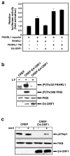
E4-ORF1 inactivates FKHRL1 and downregulates p27kip1. (a) Inactivation of FKHRL1 by E4-ORF1. NIH 3T3 cells on 6-cm dishes were lipofected with pGL3-3xFHRE (1 μg), pEF-lacZ (0.25 μg), pECE–HA–FKHRL1 wt or TM (1 μg each), and pGW1 encoding wt E4-ORF1 (0.75 μg). Extracts of serum–starved cells were prepared and assayed for luciferase activity. Results are compiled from three independent experiments. (b) E4-ORF1 promotes phosphorylation of FKHRL1. Confluent serum-starved CREF or CREF-E4-ORF1 cells were lysed in RIPA buffer, and extracts (60 μg protein) were immunoblotted with anti-(P)Thr32 FKHRL1, anti-(P)Thr308 PKB, anti-PKB, or anti–E4-ORF1 antibodies. Indicated cells were untreated (−) or treated with 50 μM LY294002 (c) Decreased p27kip1 levels in E4-ORF1-expressing cells. Confluent serum-starved CREF lines described above in (b) were lysed in RIPA buffer, and 20 μg or 100 μg of protein from cell extracts was immunoblotted with anti-p27kip1 or anti–PKB antibodies, respectively. Indicated cells were untreated (−) or treated with 100 nM wortmannin (wort)
Figure 7
Figure 7
LY294002 or rapamycin blocks transformation by E4-ORF1. (a) Soft-agar growth inhibition of E4-ORF1-expressing CREF cells by LY294002 and rapamycin. An equal number of CREF-E4-ORF1 cells or control CREF cells was suspended in soft agar without (−) or with the indicated amount of LY294002 (LY) or rapamycin (rapa) for 2 weeks and then photographed. (b) Inhibition of E4-ORF1-mediated focus formation by LY294002 and rapamycin. CREF cells on 10-cm dishes were lipofected with pJ4Ω or pJ4Ω plasmid encoding wt E4-ORF1 (8 μg each). At 1-week posttransfection, indicated cells were cultured in a medium containing either 10 μM LY294002 (LY) or 5 ng/ml rapamycin (rapa). Foci were quantified 3-weeks post–transfection. Error bars indicate range of focus numbers obtained on duplicate dishes. (c) Soft-agar growth inhibition of Ad9-induced mammary tumor cells by LY294002 and rapamycin. An equal number of 20-8 mammary tumor cells or control CREF cells was analysed as described above in (a)

Similar articles

Cited by

References

    1. Aoki M, Blazek E, Vogt PK. Proc Natl Acad Sci USA. 2001;98:136–141. - PMC - PubMed
    1. Blume-Jensen P, Hunter T. Nature. 2001;411:355–365. - PubMed
    1. Bowman T, Garcia R, Turkson J, Jove R. Oncogene. 2000;19:2474–2488. - PubMed
    1. Bradford MM. Anal Biochem. 1976;72:248–254. - PubMed
    1. Brunet A, Bonni A, Zigmond MJ, Lin MZ, Juo P, Hu LS, Anderson MJ, Arden KC, Blenis J, Greenberg ME. Cell. 1999;96:857–868. - PubMed

Publication types

MeSH terms