. 2003 Feb 3;22(3):515-28.
doi: 10.1093/emboj/cdg050.

The Helicobacter pylori CagA protein induces cortactin dephosphorylation and actin rearrangement by c-Src inactivation

Affiliations

The Helicobacter pylori CagA protein induces cortactin dephosphorylation and actin rearrangement by c-Src inactivation

Matthias Selbach et al. EMBO J. .

Abstract

The gastric pathogen Helicobacter pylori translocates the CagA protein into epithelial cells by a type IV secretion process. Translocated CagA is tyrosine phosphorylated (CagA(P-Tyr)) on specific EPIYA sequence repeats by Src family tyrosine kinases. Phos phorylation of CagA induces the dephosphorylation of as yet unidentified cellular proteins, rearrangements of the host cell actin cytoskeleton and cell scattering. We show here that CagA(P-Tyr) inhibits the catalytic activity of c-Src in vivo and in vitro. c-Src inactivation leads to tyrosine dephosphorylation of the actin binding protein cortactin. Concomitantly, cortactin is specifically redistributed to actin-rich cellular protrusions. c-Src inactivation and cortactin dephosphorylation are required for rearrangements of the actin cytoskeleton. Moreover, CagA(P-Tyr)-mediated c-Src inhibition downregulates further CagA phosphorylation through a negative feedback loop. This is the first report of a bacterial virulence factor that inhibits signalling of a eukaryotic tyrosine kinase and on a role of c-Src inactivation in host cell cytoskeletal rearrangements.


Figures
None
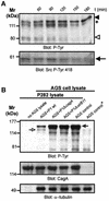
Fig. 1. Helicobacter pylori-induced cytoskeletal rearrangements depend on CagA tyrosine phosphorylation and are associated with dephosphorylation of p80. (A) Live cell imaging of an AGS cell cluster infected by wild-type H.pylori reveals cell elongation and scattering. (B) An isogenic cagA gene mutant does not stimulate this response. Complementation with wild-type cagA (P1ΔcagA/cagA) but not cagA lacking the tyrosine phosphorylation site (P1ΔcagA/cagAY972F) restores the phenotype. (C) HGF (20 pM) induced scattering of MDCK but not AGS cells. Treatment of cells with cycloheximide (10 µg/ml) prevents HGF-induced MDCK cell scattering but not the H.pylori-induced phenotype in AGS cells. (D) The time course of CagA tyrosine phosphorylation (filled arrowhead) parallels dephosphorylation of p80 (open arrowhead). In order to distinguish CagAP-Tyr from a 120 kDa host cell protein (asterisk) CagA was also immunoprecipitated (lower panels). (E) Effect of different H.pylori mutants on the tyrosine phosphorylation pattern of AGS cells. Dephosphorylation of p80 strictly correlates with CagA phosphorylation. α-tubulin blots served as loading controls. Scale bars: 20 µm.
None
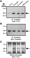
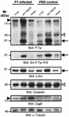
Fig. 2. Cortactin is specifically dephosphorylated upon infection with wild-type H.pylori. Cortactin was immunoprecipitated from infected AGS cells with a monoclonal anti-cortactin antibody. Probing with a phosphotyrosine-specific antibody reveals dephosphorylation of cortactin (A). The anti-cortactin blot shows that equal amounts of cortactin were precipitated (B). Tyrosine phosphorylation pattern of whole cell lysates are shown as control (C). Arrows indicate the position of cortactin on the gels.
None
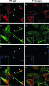
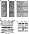
Fig. 3. Cytoskeletal rearrangements are associated with cortactin re-localization. AGS cells infected with wild-type H.pylori (A, C, E and G) or the isogenic cagA mutant (B, D, F and H) were stained with anti-cortactin antibody (A and B), phalloidin (C and D) and anti-H.pylori antiserum (E and F). Confocal laser scanning microscopy reveals cortactin co-localization with F-actin at the tip and the base of the protrusions (G, arrowheads). In cells infected with the cagA mutant, cortactin has a spot-like distribution throughout the cytoplasm (H, arrows). Scale bar: 10 µm.
None
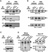
Fig. 4. Cortactin dephosphorylation is independent of the PTPase Shp-2. Cells were transfected with a shp-2 construct deleted in the phosphatase domain (shp-2ΔPTPase) that acts in a dominant-negative manner. (A) Shp-2ΔPTPase is expressed in AGS cells (upper panel, open arrowhead) and migrates below endogenous Shp-2 (filled arrowhead). Shp-2ΔPTPase does not significantly affect CagA tyrosine phosphorylation (lower panels, arrow). (BHelicobacter pylori induces cortactin dephosphorylation (arrow) irrespective of Shp-2ΔPTPase expression. (C) As a control for dominant-negative function Shp-2ΔPTPase-induced repression of basal MAPK activity was assayed with an activation specific antibody. α-tubulin blots served as loading controls. (D) Cells were infected with H.pylori, transfected with a CagA expression construct or transfected and infected simultaneously. Only CagAP-Tyr translocated by live bacteria (open arrowhead), but not transfected CagAP-Tyr (closed arrowhead), induced cortactin dephosphorylation (arrow). Conversely, only transfected but not translocated CagAP-Tyr co-immunoprecipitated with Shp-2 (right panels).
None
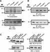
Fig. 5. CagAP-Tyr-specific inactivation of c-Src. (A) The catalytic activity of immunoprecipitated c-Src was determined by an in vitro kinase assay with [γ-32P]ATP. Helicobacter pylori wild-type infection strongly reduces c-Src autophosphorylation (upper panel, arrow). Blotting with a c-Src-specific antibody shows that similar amounts of c-Src were precipitated (lower panel, arrow). The arrowhead marks the immunoglobulin heavy chain. (B) Western blotting with phosphospecific anti-c-Src antibodies revealed that H.pylori induces c-Src inactivation by dephosphorylation of Y418 (upper panel) and phosphorylation of Y527 (lower panel). (C) Effect of CagA phosphorylation (filled arrowhead) on c-Src inactivation. Only bacteria complemented with wild-type cagA (P1ΔcagΑ/cagA), but not cagA mutated in the phosphorylation site (P1ΔcagΑ/cagAY972F) induced Y418 dephosphorylation in c-Src (open arrowhead).
None
Fig. 6. CagAP-Tyr-induced c-Src inactivation inhibits succeeding CagA phosphorylation. (A) A time course reveals that accumulation of CagAP-Tyr (filled arrowhead) in the host cell correlates with the degree of cortactin dephosphorylation (open arrowhead) and c-Src inactivation (arrow). The asterisk denotes a 120 kDa host cell protein. (B) CagAP-Tyr inhibits succeeding CagA phosphorylation. Cells infected with the P1 strains were lysed, combined with an excess of lysate derived from the H.pylori strain P282 and in vitro phosphorylation reactions were performed. Tyrosine phosphorylation of CagA from the strain P282 (filled arrow) and P1 (open arrow) was analysed. P1 wild-type infection strongly reduced phosphorylation of CagA from strain P282 (second lane). The AGS cell control marked with an asterisk was not incubated with P282 lysate (last lane). Similar amounts of CagA and host cell lysates were present in anti-CagA and anti-α-tubulin blots (lower panels).
None
Fig. 7. Purified CagA inhibits recombinant c-Src activity in vitro. (A) Purified CagA significantly reduced autophosphorylation of recombinant c-Src (open arrowhead). (B) Purified CagA was phosphorylated (CagAP-Tyr) with nonradioactive ATP before addition of [γ-32P]ATP (first lane). CagAP-Tyr had a marked inhibitory effect on c-Src activity. The lower panels show Coomassie Blue stains of the exposed gels to ensure equal amounts of CagA were loaded.
None
Fig. 8. CagAP-Tyr induces cortactin dephosphorylation through inactivation of c-Src. AGS cells were transiently transfected with c-src or activated src (src Y527F) constructs. The cells were either infected with the strain P1 or left uninfected. Activated src (i) induced the hyperphosphorylation of CagA (filled arrowhead) and Src (arrow); (ii) prevented cortactin dephosphorylation (open arrowhead); and (iii) prevented Src inactivation (second panel, arrow). c-Src, cortactin, CagA and α-tubulin blots were performed as controls.
None
Fig. 9. c-Src inactivation is essential for cytoskeletal rearrangements. AGS cells were transiently transfected with wild-type c-Src or constitutively active SrcY527F. Subsequently, the cells were infected with H.pylori, stained for F-actin (B and E) and Src expression (C and F) followed by epifluorescence microscopy. While cells expressing c-Src showed characteristic actin-rich protrusions (A, B and C), constitutively active Src prevented these H.pylori- induced cytoskeletal rearrangements (D, E and F, arrow). Arrowheads indicate neighbouring non-transfected cells as internal control. Scale bar: 10 µm.
None
Fig. 10. Model for CagA-induced signalling leading to the cytoskeletal rearrangements of gastric epithelial cells. Helicobacter pylori translocates CagA by a type IV secretion dependent process. CagA is tyrosine-phosphorylated by Src which also phosphorylates cortactin. CagAP-Tyr inactivates c-Src by a mechanism involving phosphorylation of Y527 and dephosphorylation of Y418. Src inactivation prevents succeeding CagA phosphorylation and leads to cortactin dephosphorylation. Dephosphorylated cortactin has enhanced actin cross-linking and/or nucleation activity and may induce the characteristic rearrangements of the actin cytoskeleton involved in cell scattering, designated as the hummingbird phenotype.

Similar articles

Cited by

References

    1. Akopyants N.S. et al. (1998) Analyses of the cag pathogenicity island of Helicobacter pylori. Mol. Microbiol., 28, 37–53. - PubMed
    1. Asahi M. et al. (2000) The Helicobacter pylori CagA protein can be tyrosine phosphorylated in gastric epithelial cells. J. Exp. Med., 191, 593–602. - PMC - PubMed
    1. Backert S., Ziska,E., Brinkmann,V., Zimny-Arndt,U., Fauconnier,A., Jungblut,P.R., Naumann,M. and Meyer,T.F. (2000) Translocation of the Helicobacter pylori CagA protein in gastric epithelial cells by a type IV secretion apparatus. Cell Microbiol., 2, 155–164. - PubMed
    1. Backert S., Moese,S., Selbach,M., Brinkmann,V. and Meyer,T.F. (2001) Phosphorylation of tyrosine 972 of the Helicobacter pylori CagA protein is essential for induction of a scattering phenotype in gastric epithelial cells. Mol. Microbiol., 42, 631–644. - PubMed
    1. Bliska J.B., Clemens,J.C., Dixon,J.E. and Falkow,S. (1992) The Yersinia tyrosine phosphatase: specificity of a bacterial virulence determinant for phosphoproteins in the J774A.1 macrophage. J. Exp. Med., 176, 1625–1630. - PMC - PubMed

Publication types

MeSH terms