A critical role for PPARalpha-mediated lipotoxicity in the pathogenesis of diabetic cardiomyopathy: modulation by dietary fat content
- PMID: 12552126
- PMCID: PMC298755
- DOI: 10.1073/pnas.0336724100
A critical role for PPARalpha-mediated lipotoxicity in the pathogenesis of diabetic cardiomyopathy: modulation by dietary fat content
Abstract
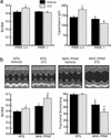
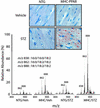
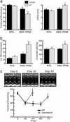
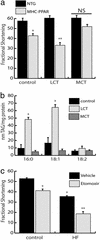
To explore the role of peroxisome proliferator-activated receptor alpha (PPARalpha)-mediated derangements in myocardial metabolism in the pathogenesis of diabetic cardiomyopathy, insulinopenic mice with PPARalpha deficiency (PPARalpha(-/-)) or cardiac-restricted overexpression [myosin heavy chain (MHC)-PPAR] were characterized. Whereas PPARalpha(-/-) mice were protected from the development of diabetes-induced cardiac hypertrophy, the combination of diabetes and the MHC-PPAR genotype resulted in a more severe cardiomyopathic phenotype than either did alone. Cardiomyopathy in diabetic MHC-PPAR mice was accompanied by myocardial long-chain triglyceride accumulation. The cardiomyopathic phenotype was exacerbated in MHC-PPAR mice fed a diet enriched in triglyceride containing long-chain fatty acid, an effect that was reversed by discontinuing the high-fat diet and absent in mice given a medium-chain triglyceride-enriched diet. Reactive oxygen intermediates were identified as candidate mediators of cardiomyopathic effects in MHC-PPAR mice. These results link dysregulation of the PPARalpha gene regulatory pathway to cardiac dysfunction in the diabetic and provide a rationale for serum lipid-lowering strategies in the treatment of diabetic cardiomyopathy.
Figures
Similar articles
-
The cardiac phenotype induced by PPARalpha overexpression mimics that caused by diabetes mellitus.J Clin Invest. 2002 Jan;109(1):121-30. doi: 10.1172/JCI14080. J Clin Invest. 2002. PMID: 11781357 Free PMC article.
-
Rescue of cardiomyopathy in peroxisome proliferator-activated receptor-alpha transgenic mice by deletion of lipoprotein lipase identifies sources of cardiac lipids and peroxisome proliferator-activated receptor-alpha activators.Circulation. 2010 Jan 26;121(3):426-35. doi: 10.1161/CIRCULATIONAHA.109.888735. Epub 2010 Jan 11. Circulation. 2010. PMID: 20065164 Free PMC article.
-
CD36 deficiency rescues lipotoxic cardiomyopathy.Circ Res. 2007 Apr 27;100(8):1208-17. doi: 10.1161/01.RES.0000264104.25265.b6. Epub 2007 Mar 15. Circ Res. 2007. PMID: 17363697
-
The role of the peroxisome proliferator-activated receptor alpha pathway in pathological remodeling of the diabetic heart.Curr Opin Clin Nutr Metab Care. 2004 Jul;7(4):391-6. doi: 10.1097/01.mco.0000134371.70815.32. Curr Opin Clin Nutr Metab Care. 2004. PMID: 15192440 Review.
-
PPAR signaling in the control of cardiac energy metabolism.Trends Cardiovasc Med. 2000 Aug;10(6):238-45. doi: 10.1016/s1050-1738(00)00077-3. Trends Cardiovasc Med. 2000. PMID: 11282301 Review.
Cited by
-
Role of ceramide in diabetes mellitus: evidence and mechanisms.Lipids Health Dis. 2013 Jul 8;12:98. doi: 10.1186/1476-511X-12-98. Lipids Health Dis. 2013. PMID: 23835113 Free PMC article. Review.
-
Metabolic stress in the myocardium: adaptations of gene expression.J Mol Cell Cardiol. 2013 Feb;55:130-8. doi: 10.1016/j.yjmcc.2012.06.008. Epub 2012 Jun 21. J Mol Cell Cardiol. 2013. PMID: 22728216 Free PMC article. Review.
-
Genome-Wide Expression Profiling of Anoxia/Reoxygenation in Rat Cardiomyocytes Uncovers the Role of MitoKATP in Energy Homeostasis.Oxid Med Cell Longev. 2015;2015:756576. doi: 10.1155/2015/756576. Epub 2015 Jun 15. Oxid Med Cell Longev. 2015. PMID: 26171116 Free PMC article.
-
A critical role for eukaryotic elongation factor 1A-1 in lipotoxic cell death.Mol Biol Cell. 2006 Feb;17(2):770-8. doi: 10.1091/mbc.e05-08-0742. Epub 2005 Nov 30. Mol Biol Cell. 2006. PMID: 16319173 Free PMC article.
-
Radionuclide imaging of myocardial metabolism.Circ Cardiovasc Imaging. 2010 Mar;3(2):211-22. doi: 10.1161/CIRCIMAGING.109.860593. Circ Cardiovasc Imaging. 2010. PMID: 20233863 Free PMC article. Review. No abstract available.
References
-
- Rubler S, Dlugash J, Yuceoglu Y Z, Kumral T, Branwood A W, Grishman A. Am J Cardiol. 1972;30:595–602. - PubMed
-
- Stone P H, Muller J E, Hartwell T, York B J, Rutherford J D, Parker C B, Turi Z G, Strauss H W, Willerson J T, Robertson T. J Am Coll Cardiol. 1989;14:49–57. - PubMed
-
- Neely J R, Rovetto M J, Oram J F. Prog Cardiovasc Dis. 1972;15:289–329. - PubMed
-
- Lopaschuk G D, Spafford M. Circ Res. 1989;65:378–387. - PubMed
-
- Belke D D, Larsen T S, Gibbs E M, Severson D L. Am J Physiol. 2000;279:E1104–E1113. - PubMed
Publication types
MeSH terms
Substances
Grants and funding
LinkOut - more resources
Full Text Sources
Other Literature Sources
Medical
Molecular Biology Databases
Research Materials

