Flexibility of mouse classical and plasmacytoid-derived dendritic cells in directing T helper type 1 and 2 cell development: dependency on antigen dose and differential toll-like receptor ligation
- PMID: 12515817
- PMCID: PMC2193804
- DOI: 10.1084/jem.20021908
Flexibility of mouse classical and plasmacytoid-derived dendritic cells in directing T helper type 1 and 2 cell development: dependency on antigen dose and differential toll-like receptor ligation
Abstract
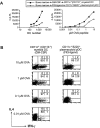
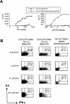
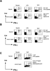
Distinct dendritic cell (DC) subsets have been suggested to be preprogrammed to direct either T helper cell (Th) type 1 or Th2 development, although more recently different pathogen products or stimuli have been shown to render these DCs more flexible. It is still unclear how distinct mouse DC subsets cultured from bone marrow precursors, blood, or their lymphoid tissue counterparts direct Th differentiation. We show that mouse myeloid and plasmacytoid precursor DCs (pDCs) cultured from bone marrow precursors and ex vivo splenic DC subsets can induce the development of both Th1 and Th2 effector cells depending on the dose of antigen. In general, high antigen doses induced Th1 cell development whereas low antigen doses induced Th2 cell development. Both cultured and ex vivo splenic plasmacytoid-derived DCs enhanced CD4(+) T cell proliferation and induced strong Th1 cell development when activated with the Toll-like receptor (TLR)9 ligand CpG, and not with the TLR4 ligand lipopolysaccharide (LPS). The responsiveness of plasmacytoid pDCs to CpG correlated with high TLR9 expression similarly to human plasmacytoid pDCs. Conversely, myeloid DCs generated with granulocyte/macrophage colony-stimulating factor enhanced Th1 cell development when stimulated with LPS as a result of their high level of TLR4 expression. Polarized Th1 responses resulting from high antigen dose were not additionally enhanced by stimulation of DCs by TLR ligands. Thus, the net effect of antigen dose, the state of maturation of the DCs together with the stimulation of DCs by pathogen-derived products, will determine whether a Th1 or Th2 response develops.
Figures


Similar articles
-
Toll-like receptor ligands modulate dendritic cells to augment cytomegalovirus- and HIV-1-specific T cell responses.J Immunol. 2003 Oct 15;171(8):4320-8. doi: 10.4049/jimmunol.171.8.4320. J Immunol. 2003. PMID: 14530357
-
TLR ligands can activate dendritic cells to provide a MyD88-dependent negative signal for Th2 cell development.J Immunol. 2005 Jan 15;174(2):742-51. doi: 10.4049/jimmunol.174.2.742. J Immunol. 2005. PMID: 15634894
-
A novel role for IL-3: human monocytes cultured in the presence of IL-3 and IL-4 differentiate into dendritic cells that produce less IL-12 and shift Th cell responses toward a Th2 cytokine pattern.J Immunol. 2002 Jun 15;168(12):6199-207. doi: 10.4049/jimmunol.168.12.6199. J Immunol. 2002. PMID: 12055233
-
Differential migration behavior and chemokine production by myeloid and plasmacytoid dendritic cells.Hum Immunol. 2002 Dec;63(12):1164-71. doi: 10.1016/s0198-8859(02)00755-3. Hum Immunol. 2002. PMID: 12480260 Review.
-
Natural type I interferon-producing cells as a link between innate and adaptive immunity.Hum Immunol. 2002 Dec;63(12):1126-32. doi: 10.1016/s0198-8859(02)00751-6. Hum Immunol. 2002. PMID: 12480256 Review.
Cited by
-
Different parasite inocula determine the modulation of the immune response and outcome of experimental Trypanosoma cruzi infection.Immunology. 2013 Feb;138(2):145-56. doi: 10.1111/imm.12022. Immunology. 2013. PMID: 23113506 Free PMC article.
-
Bone marrow plasmacytoid dendritic cells can differentiate into myeloid dendritic cells upon virus infection.Nat Immunol. 2004 Dec;5(12):1227-34. doi: 10.1038/ni1136. Epub 2004 Nov 7. Nat Immunol. 2004. PMID: 15531885 Free PMC article.
-
Immunomodulatory properties of defensins and cathelicidins.Curr Top Microbiol Immunol. 2006;306:27-66. doi: 10.1007/3-540-29916-5_2. Curr Top Microbiol Immunol. 2006. PMID: 16909917 Free PMC article. Review.
-
Efficient and versatile manipulation of the peripheral CD4+ T-cell compartment by antigen targeting to DNGR-1/CLEC9A.Eur J Immunol. 2010 May;40(5):1255-65. doi: 10.1002/eji.201040419. Eur J Immunol. 2010. PMID: 20333625 Free PMC article.
-
Gene expression analysis of dendritic cells that prevent diabetes in NOD mice: analysis of chemokines and costimulatory molecules.J Leukoc Biol. 2011 Sep;90(3):539-50. doi: 10.1189/jlb.0311126. Epub 2011 May 31. J Leukoc Biol. 2011. PMID: 21628331 Free PMC article.
References
-
- Banchereau, J., and R.M. Steinman. 1998. Dendritic cells and the control of immunity. Nature. 392:245–252. - PubMed
-
- Shortman, K., and Y.J. Liu. 2002. Mouse and human dendritic cell subtypes. Nat. Rev. Immunol. 2:151–161. - PubMed
-
- Steinman, R.M. 1991. The dendritic cell system and its role in immunogenicity. Annu. Rev. Immunol. 9:271–296. - PubMed
-
- Lanzavecchia, A., and F. Sallusto. 2001. Regulation of T cell immunity by dendritic cells. Cell. 106:263–266. - PubMed
-
- Rissoan, M.-C., V. Soumelis, N. Kadowaki, G. Grouard, F. Briere, R. de Waal Malefyt, and Y.-J. Liu. 1999. Reciprocal control of T helper cell and dendritic cell differentiation. Science. 283:1183–1186. - PubMed
Publication types
MeSH terms
Substances
LinkOut - more resources
Full Text Sources
Other Literature Sources
Research Materials

