. 2003 Jan 20;455(4):463-76.
doi: 10.1002/cne.10491.

Immunocytochemical description of five bipolar cell types of the mouse retina

Affiliations

Immunocytochemical description of five bipolar cell types of the mouse retina

Silke Haverkamp et al. J Comp Neurol. .

Abstract

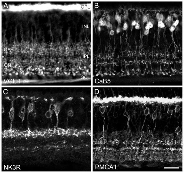
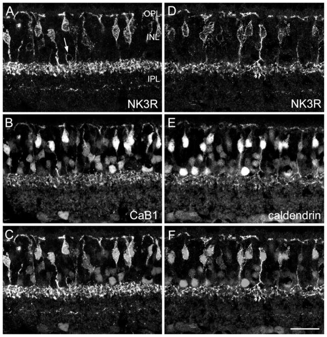
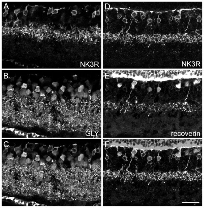
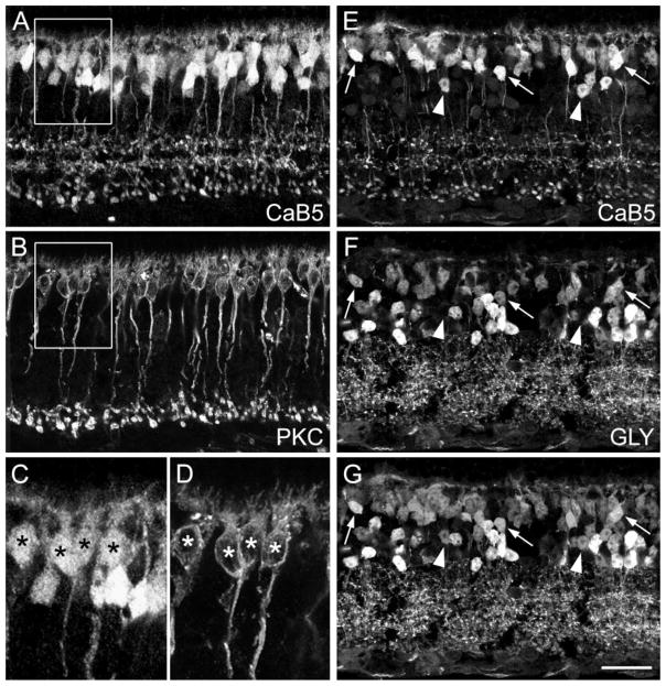
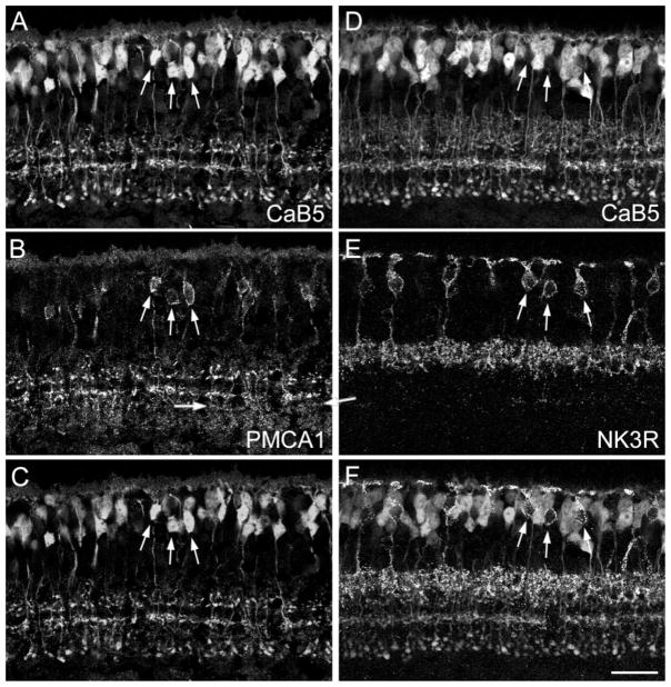
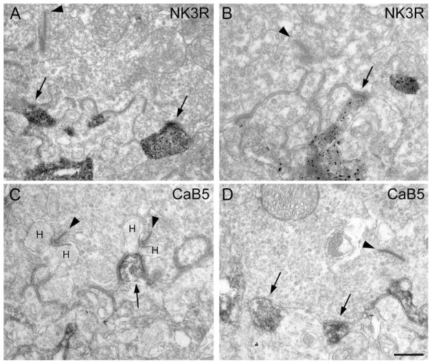
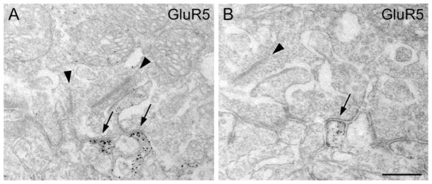
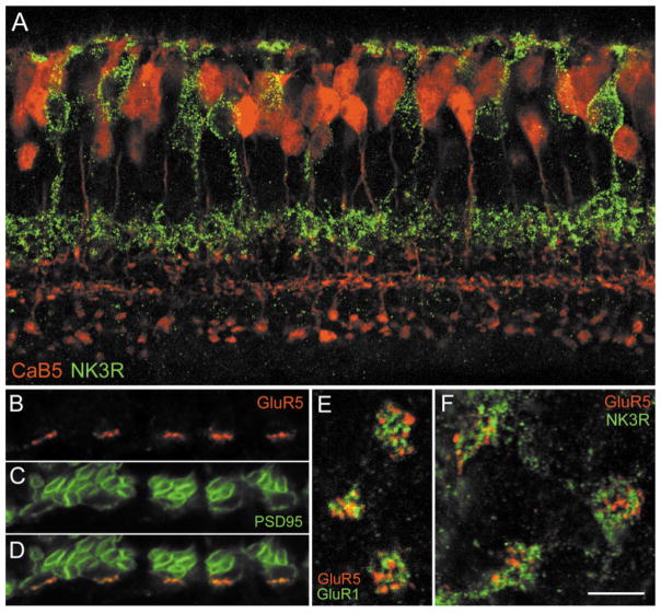
With the ever-growing number of transgenic mice being used in vision research, a precise knowledge of the cellular organization of the mouse retina is required. As with the cat, rabbit, rat, and primate retinae, as many as 10 cone bipolar types and one rod bipolar type can be expected to exist in the mouse retina; however, they still have to be defined. In the current study, several immunocytochemical markers were applied to sections of mouse retina, and the labeling of bipolar cells was studied using confocal microscopy and electron microscopy. By using antibodies against the neurokinin-3 receptor NK3R; the plasma membrane calcium ATPase1 (PMCA1); and the calcium (Ca)-binding proteins CaB1, CaB5, caldendrin, and recoverin, three different OFF-cone bipolar cells could be identified. One type of ON-cone bipolar cell was identified through its immunoreactivity for CaB5 and PMCA1. Rod bipolar cells, comparable in morphology to those of other mammalian retinae, expressed protein kinase Calpha and CaB5. It was also shown that putative OFF-cone bipolar cells receive light signals through flat contacts at the cone pedicle base, whereas ON-cone bipolar signaling involves invaginating contacts. The distribution of the kainate receptor subunit GluR5 was studied by confocal and electron microscopy. GluR5 was expressed at flat bipolar cell contacts; however, it appears to be involved with only certain types of OFF-cone bipolar cells. This suggests that different bipolar cell types receive their light signals through different sets of glutamate receptors.


Figures

Fig. 1
Fig. 1
Fluorescence photomicrographs of vertical frozen sections through mouse retina that were labeled immunocytochemically. The retinal layers are indicated. OPL, outer plexiform layer; INL, inner nuclear layer; IPL, inner plexiform layer; GCL, ganglion cell layer. A: Conventional photomicrograph showing immunolabeling for the vesicular glutamate transporter 1 (VGluT1). Photoreceptor terminals in the OPL and bipolar cell axon terminals in the IPL are labeled. In the outer half of the IPL, putative OFF-cone bipolar cells terminate; in the inner half of the IPL, putative ON-cone bipolar cells and rod bipolar cells terminate. B: Confocal photomicrograph showing immunolabeling for the calcium-binding protein CaB5. Dendrites of bipolar cells in the OPL, their perikarya in the INL, and their axons terminating in three bands in the IPL are labeled. C: Confocal micrograph of a section immunolabeled for the neurokinin-3 receptor NK3R. Bipolar cells and their axons terminating in the outer IPL are labeled. The processes labeled in the inner IPL are amacrine cell dendrites. D: Confocal micrograph showing immunolabeling for the plasma membrane calcuim ATPase 1 (PMCA1). Photoreceptor terminals in the OPL, bipolar cell bodies in the INL, and bipolar cell axon terminals in the IPL are labeled. Scale bar = 19 μm for A, 20 μm for B,D, 17.7 μm for C.
Fig. 2
Fig. 2
Confocal micrographs of vertical frozen sections through mouse retina that were doubly labeled for NK3R and for the calcium-binding proteins CaB1 (A–C) and caldendrin (D–F). A: NK3R immunofluorescence. Many bipolar cells are labeled. The arrow indicates an amacrine cell body. B: CaB1 immunolabeling can be observed in many bipolar cells, amacrine cells, and ganglion cells. C: Superimposition of A and B shows that the NK3R-labeled bipolar cells from A and the strongly CaB1-immunoreactive bipolar cells from B correspond to the same cells. D–F: NK3R-labeled bipolar cells also express caldendrin immunoreactivity. Scale bar = 25 μm.
Fig. 3
Fig. 3
Confocal micrographs of vertical frozen sections through mouse retina that were doubly labeled for NK3R and glycine (A–C) or for NK3R and recoverin (D–F). A: NK3R immunofluorescence is found in eight bipolar cell perikarya and in their axon terminals in the IPL. B: Bipolar cells, amacrine cells, and their processes in the IPL express glycine immunoreactivity. C: Superimposition of A and B shows that none of the eight NK3R-labeled bipolar cell perikarya exhibits glycine immunofluorescence. D: NK3R-labeled bipolar cells and their axon terminals in the IPL. E: Recoverin immunoreactivity is also expressed in bipolar cell perikarya and their axons. F: Superimposition of D and E shows that approximatelyonly half of the NK3R-labeled perikarya are also immunoreactive for recoverin. Scale bar = 25 μm.
Fig. 4
Fig. 4
Confocal micrographs of vertical frozen sections through mouse retina that were doubly labeled for the calcium-binding protein CaB5 and protein kinase Cα (PKCα; A–D) or for CaB5 and glycine (E–G). A: CaB5 immunoreactivity is found in many bipolar cell perikarya, in their descending axons, and in three distinct bands of axon terminals. B: PKCα labels rod bipolar (RB) cells, their descending axons, and their axon terminals in the innermost IPL. Comparison with A shows that RB axon terminals also express CaB5. C: High-power micrograph showing the CaB5-immunolabeled cell bodies from the boxed area in A. D: PKCα-immunolabeled cell bodies from the corresponding area in B. The asterisks indicate the double-labeled RB cell perikarya. E: CaB5-labeled section. F: Same section as in E labeled for glycine (GLY). The three arrows indicate bipolar cells expressing both CaB5 and glycine. The arrowheads indicate bipolar cells that express CaB5 but not glycine. G: Superimposition of E and F. Scale bar = 25 μm for A,B,E,F,G, 11.4 μm for C,D.
Fig. 5
Fig. 5
Confocal micrographs of vertical frozen sections through mouse retina that were doubly labeled for CaB5 and for PMCA1 (A–C) or for CaB5 and NK3R (D–F). A: CaB5 immunoreactivity is found in many bipolar cell perikarya, in their descending axons, and in three distinct bands of axon terminals. B: PMCA1 immunoreactivity decorates the perikaryal membrane of a few bipolar cells in the INL and their axons, which form two bands in the IPL. Weaker immunoreactivity is also present in additional bands of putative axon terminals in the IPL (horizontal arrows). C: Superimposition of A and B shows that the PMCA1-immunolabeled bipolar cells and their axon terminals represent a subgroup of the CaB5-labeled bipolar cells (vertical arrows). D: CaB5-immunolabeled section. E: Same section labeled for NK3R. F: Superimposition of D and E. The arrows show that NK3R-immunoreactive bipolar cells are not immunoreactive for CaB5. Scale bar = 25 μm.
Fig. 6
Fig. 6
Electron micrograph showing the cone contacts of bipolar cell dendrites. A: Cone pedicle base containing one presynaptic ribbon (arrowhead). Two NK3R-immunoreactive processes (arrows) make flat contacts at the cone pedicle base. B: Another cone pedicle base, containing a presynaptic ribbon (arrowhead) engaged with a NK3R-labeled flat bipolar cell contact (arrow). C: Cone pedicle base containing two presynaptic ribbons (arrowheads) and their lateral horizontal cell dendrites (H). The CaB5-immunoreactive invaginating process (arrow) represents the central element opposed to the ribbon. D: Cone pedicle base containing one presynaptic ribbon (arrowhead). Two CaB5-immunoreactive processes (arrows) make flat contacts at the cone pedicle base. Scale bar = 0.57 μm for A, 0.37 μm for B, 0.5 μm for C,D.
Fig. 7
Fig. 7
Electron micrographs showing the expression of the glutamate receptor subunit GluR5 at the cone pedicle base. A: Cone pedicle base containing two presynaptic ribbons. Two GluR5-immunoreactive processes make flat contacts at the cone pedicle base (arrows). B: Another cone pedicle containing a presynaptic ribbon (arrowhead) is engaged with a flat bipolar cell contact expressing GluR5. Scale bar = 0.5 μm for A, 0.4 μm for B.
Fig. 8
Fig. 8
A: Confocal fluorescence micrograph of a vertical section through the mouse retina doubly labeled for CaB5 (shown in red) and the NK3 receptor (shown in green). B–F: Confocal fluorescence micrographs showing the expression of the GluR5 subunit at cone pedicles of the mouse retina. B–D: Vertical section through the OPL doubly labeled for GluR5 and PSD95. B: GluR5 immunofluorescence (red) decorates the base of 5 cone pedicles. C: PSD95 immunofluorecence (green) reveals many rod spherules and the five cone pedicles. D: Superimposition of B and C shows the coincidence of GluR5 and PSD95 at the cone pedicle bases. E: Horizontal confocal section through three cone pedicles of the mouse retina doubly labeled for GluR5 (red) and GluR1 (green). The GluR1 and GluR5 clusters are not in register. F: Horizontal confocal section through three cone pedicles of the mouse retina doubly labeled for GluR5 (red) and NK3R (green). The GluR5 clusters and the NK3R-labeled bipolar cell dendritic tips are not in register. Scale bar = 15 μm for A, 6 μm for B–D, 5 μm for E,F.

Similar articles

Cited by

References

    1. Andressen C, Mai JK. Localization of the CD15 carbohydrate epitope in the vertebrate retina. Vis Neurosci. 1997;14:253–262. - PubMed
    1. Berrebi AS, Oberdick J, Sangameswaran L, Christakos S, Morgan JI, Mugnaini E. Cerebellar Purkinje cell markers are expressed in retinal bipolar neurons. J Comp Neurol. 1991;308:630–649. - PubMed
    1. Blumauer N, Brecha NC. Off-type of cone bipolar cells express neurokinin-3 (NK-3) receptors in the rat retina. Soc Neurosci Abstr. 1998:2091.
    1. Boycott BB, Wässle H. Morphological classification of bipolar cells in the macaque monkey retina. Eur J Neurosci. 1991;3:1069–1088. - PubMed
    1. Brandstätter JH, Koulen P, Wässle H. Selective synaptic distribution of kainate receptor subunits in the two plexiform layers of the rat retina. J Neurosci. 1997;17:9298–9307. - PMC - PubMed

Publication types

MeSH terms