. 2002 Dec 23;159(6):957-69.
doi: 10.1083/jcb.200207028. Epub 2002 Dec 16.

Symmetrical dimethylarginine methylation is required for the localization of SMN in Cajal bodies and pre-mRNA splicing

Affiliations

Symmetrical dimethylarginine methylation is required for the localization of SMN in Cajal bodies and pre-mRNA splicing

Francois-Michel Boisvert et al. J Cell Biol. .

Abstract

The nuclear structures that contain symmetrical dimethylated arginine (sDMA)-modified proteins and the role of this posttranslational modification is unknown. Here we report that the Cajal body is a major epitope in HeLa cells for an sDMA-specific antibody and that coilin is an sDMA-containing protein as analyzed by using the sDMA-specific antibody and matrix-assisted laser desorption ionization time of flight mass spectrometry. The methylation inhibitor 5'-deoxy-5'-methylthioadenosine reduces the levels of coilin methylation and causes the appearance of SMN-positive gems. In cells devoid of Cajal bodies, such as primary fibroblasts, sDMA-containing proteins concentrated in speckles. Cells from a patient with spinal muscular atrophy, containing low levels of the methyl-binding protein SMN, localized sDMA-containing proteins in the nucleoplasm as a discrete granular pattern. Splicing reactions are efficiently inhibited by using the sDMA-specific antibody or by using hypomethylated nuclear extracts, showing that active spliceosomes contain sDMA polypeptides and suggesting that arginine methylation is important for efficient pre-mRNA splicing. Our findings support a model in which arginine methylation is important for the localization of coilin and SMN in Cajal bodies.


Figures
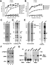
Figure 1.
Figure 1.
SYM10 recognizes many cellular proteins including coilin and the Sm proteins B, B', and D. (A and B) The specificity of SYM10 was examined using an ELISA. The quantity of peptide is indicated on the abscissa and the absorbance on the ordinate. (C) HeLa cells were metabolically labeled with (methyl-3H)-l-methionine in the presence of translation inhibitors, lysed, and immunoprecipitated with the indicated antibodies. The 3H-labeled proteins were visualized by fluorography. The migration of SmB, B', and D proteins is indicated. (D) HeLa cells were metabolically labeled with [35S]methionine, lysed, and immunoprecipitated with the SYM10 antibody, Y12, or normal rabbit serum. Proteins were separated using high TEMED SDS-PAGE. The 35S-labeled proteins were visualized by autoradiography. The migration of Sm proteins and U1A is indicated. (E) HeLa cell lysates were immunoblotted with the NRS or the SYM10 antibody (1:1,000). (F) The methylosome and SMN complexes are immunoprecipitated by SYM10. Immunoprecipitation was performed in HeLa cells using SYM10, and the proteins were immunoblotted with PRMT5, SMN, and Sm B/B' (ANA128) antibodies. (G) Coilin is recognized directly by SYM10. Immunoprecipitation was performed in HeLa cells using α-coilin antibodies followed by immunoblotting with SYM10 (1:1,000; left). Coilin is immunoprecipitated by SYM10. Immunoprecipitation was performed with the SYM10 antibody using HeLa cells transfected with myc-coilin followed by immunoblotting with α-Myc antibodies to detect transfected myc-coilin (right).
Figure 2.
Figure 2.
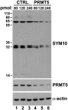
Down-regulation of PRMT5 by siRNA results in a loss of recognized proteins by SYM10. Control siRNAs (lane 1–3) or siRNAs engineered to specifically target PRMT5 mRNAs (lane 4–6) were introduced in Jurkat cells using electroporation. Cell extracts were prepared 72 h posttransfection, and the levels of SYM10 (top), PRMT5 (middle), and actin (bottom) were assessed by immunoblotting.
Figure 3.
Figure 3.
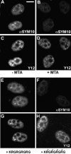
Nuclear bright foci stained with SYM10. HeLa cells treated with DMSO (−MTA) or the methylation inhibitor MTA (+MTA) for 24h were labeled for immunofluorescence using SYM10 (A and B) and the snRNP antibody Y12 (C and D). A peptide competition was performed using the immunizing peptide in a nonmethylated form (E and G) and in a fully methylated form (F and H). The antibody was preincubated with the peptide at a final concentration of 10 μM for 15 min on ice before immunofluorescence labeling using SYM10 (E and F) and the snRNP antibody Y12 (G and H). B and D, E and G, and F and H were double labeled, and the cells were visualized by fluorescence microscopy. Bar, 10 μM.
Figure 4.
Figure 4.
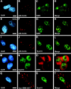
SMN colocalizes with sDMA-containing proteins in Cajal bodies. HeLa cells were labeled for immunofluorescence with a coilin antibody (B) and a SMN antibody (C). Colocalization within Cajal bodies of both proteins is shown in the merged image (D). HeLa cells treated with MTA (+MTA) for 24 h were immunostained for coilin (F) and SMN (G). HeLa cells were labeled for immunofluorescence with a coilin mAb (J) and with the SYM10 antibody (K). HeLa cells were labeled for immunofluorescence with a SMN antibody (O) and with SYM10 (N). HeLa cells were transfected with myc-SMNΔN27 and immunostained using the SYM10 antibody (S) and a myc antibody (R). The merged image is shown in T. Cell nuclei were counterstained with DAPI (A, E, I, M, and Q, blue). Bars, 10 μM.
Figure 5.
Figure 5.
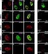
The localization of sDMA proteins is disrupted in a patient with SMA. Human fibroblasts WI-38 were doubly stained with Y12 (A) and SYM10 (B) or an anticoilin antibody (P). Fibroblasts from a patient with Werdnig-Hoffman disease SMA type I, SMN1−/−, GM03813 (SMA13), and fibroblasts from his unaffected mother SMN1−/+ GM03814 (SMA14) were either mock treated (DMSO, D–F and J–L) or treated with 750 μM MTA (G–I and M–O) for 24 h and stained using Y12 (D, G, J and M) and SYM10 (E, H, K and N). Fibroblasts from the patient with SMA (SMA13) or his unaffected mother (SMA14) were treated with DMSO (Q and S) or treated with 750 μM MTA (MTA, R and T) for 24 h and stained using anti-SMN antibodies (Q–S). Bars, 5 μm.
Figure 6.
Figure 6.
The level of PRMT5 and sDMA-containing proteins is not affected in the SMA cell line. (A) Cell lysates from the indicated cell lines were immunoblotted with PRMT5, SMN, SmB/B' (ANA128), or actin antibodies. The asterisk indicates a nonspecific band. (B) PRMT5 immunoprecipitations were performed using the indicated cell lines. Immunoprecipitated proteins were incubated with GST–SmB' as an exogenous substrate in the presence of (methyl-3H)–SAM. Proteins were resolved by SDS-PAGE, stained with Coomassie blue (top), and visualized by fluorography (bottom). The migration of GST–SmB' IgG H and L chains are indicated on the right. (C) SYM10 immunoblotting was performed on extracts prepared from the indicated cell lines.
Figure 7.
Figure 7.
Pre-mRNA splicing and spliceosomal formation is impaired in hypomethylated nuclear extracts, and SYM10 inhibits pre-mRNA splicing. (A) Splicing reactions were performed by using a 32P-labeled AdML transcription unit pre-mRNA substrate (2 fmoles) in the presence of increasing amount (25–250 ng) of purified antibodies. RNAs were resolved on an 8% denaturing polyacrylamide gel. The positions of pre-mRNA and splicing products and intermediates are indicated on the left. (B) DMSO (Mock) or MTA-treated extracts were stained with Coomassie blue or immunoblotted with the indicated antibodies. (C) Splicing reactions were performed by using a 32P-labeled caspase 2 pre-mRNA. The pre-mRNA transcripts were incubated with either the mock-treated or with the MTA-treated nuclear extracts for increasing amount of time. RNAs were resolved on a 6% denaturing polyacrylamide gel. (D) Splicing complexes assembly on the 32P-labeled adenovirus major late transcripts using nuclear extracts that are either mock treated or MTA treated. Heterogeneous (H) and spliceosomal complexes (A/B/C) are indicated on the left.

Similar articles

Cited by

References

    1. Almeida, F., R. Saffrich, W. Ansorge, and M. Carmo-Fonseca. 1998. Microinjection of anti-coilin antibodies affects the structure of coiled bodies. J. Cell Biol. 142:899–912. - PMC - PubMed
    1. Andrade, L.E.C., E.M. Tan, and E.K.L. Chan. 1993. Immunocytochemical analysis of the coiled body in the cell cycle and during cell proliferation. Proc. Natl. Acad. Sci. USA. 90:1947–1951. - PMC - PubMed
    1. Baldwin, G.S., and P.R. Carnegie. 1971. Specific enzymic methylation of an arginine in the experimental allergic encephalomyelitis protein from human myelin. Science. 171:579–581. - PubMed
    1. Bedford, M.T., A. Frankel, M.B. Yaffe, S. Clarke, P. Leder, and S. Richard. 2000. Arginine methylation inhibits the binding of proline-rich ligands to Src homology 3, but not WW, domains. J. Biol. Chem. 275:16030–16036. - PubMed
    1. Bohmann, K., J.A. Ferreira, and A.I. Lamond. 1995. Mutational analysis of p80 coilin indicates a functional interaction between coiled bodies and the nucleolus. J. Cell Biol. 131:817–831. - PMC - PubMed

Publication types

MeSH terms