. 2002 Dec 16;196(12):1605-15.
doi: 10.1084/jem.20021552.

The PAAD/PYRIN-family protein ASC is a dual regulator of a conserved step in nuclear factor kappaB activation pathways

Affiliations

The PAAD/PYRIN-family protein ASC is a dual regulator of a conserved step in nuclear factor kappaB activation pathways

Christian Stehlik et al. J Exp Med. .

Abstract

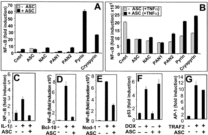
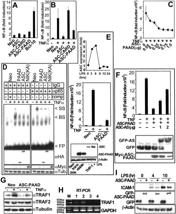
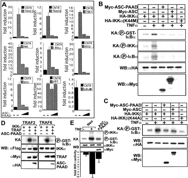
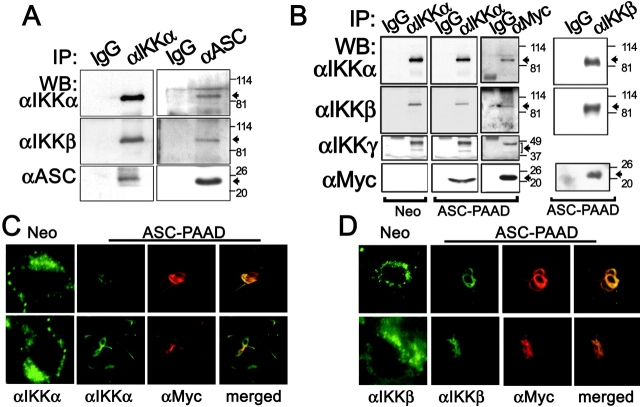
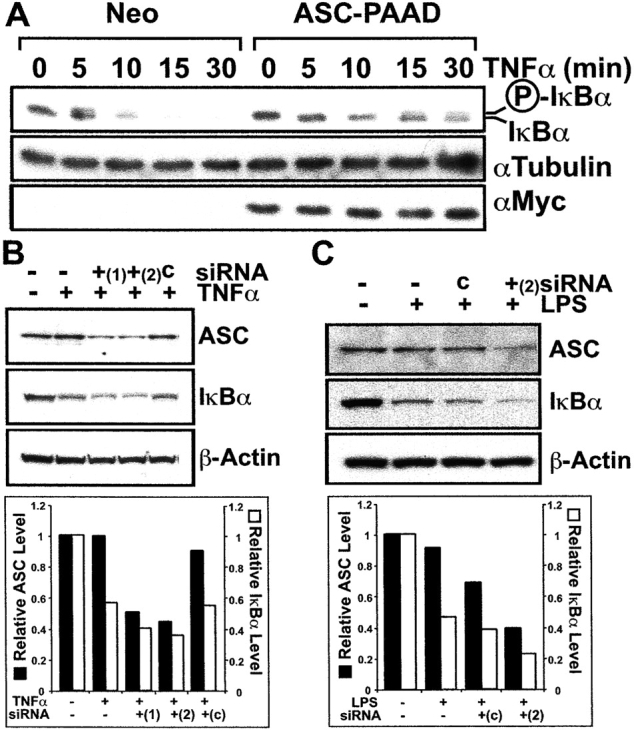
Apoptosis-associated speck-like protein containing a Caspase recruitment domain (ASC) belongs to a large family of proteins that contain a Pyrin, AIM, ASC, and death domain-like (PAAD) domain (also known as PYRIN, DAPIN, Pyk). Recent data have suggested that ASC functions as an adaptor protein linking various PAAD-family proteins to pathways involved in nuclear factor (NF)-kappaB and pro-Caspase-1 activation. We present evidence here that the role of ASC in modulating NF-kappaB activation pathways is much broader than previously suspected, as it can either inhibit or activate NF-kappaB, depending on cellular context. While coexpression of ASC with certain PAAD-family proteins such as Pyrin and Cryopyrin increases NF-kappaB activity, ASC has an inhibitory influence on NF-kappaB activation by various proinflammatory stimuli, including tumor necrosis factor (TNF)alpha, interleukin 1beta, and lipopolysaccharide (LPS). Elevations in ASC protein levels or of the PAAD domain of ASC suppressed activation of IkappaB kinases in cells exposed to pro-inflammatory stimuli. Conversely, reducing endogenous levels of ASC using siRNA enhanced TNF- and LPS-induced degradation of the IKK substrate, IkappaBalpha. Our findings suggest that ASC modulates diverse NF-kappaB induction pathways by acting upon the IKK complex, implying a broad role for this and similar proteins containing PAAD domains in regulation of inflammatory responses.


Figures

Figure 1.
Figure 1.
Differential effects of ASC on NF-κB activation. (A and B) HEK293T cells were transfected with either 200 ng of control plasmid or 200 ng of various plasmids encoding PAAD-family members (ASC, NAC, PAN1, PAN2, Pyrin, or Cryopyrin) alone (gray bars) or in combination with 200 ng of ASC-encoding plasmid (black bars). Transfections also included 100 ng of pNF-κB and 6 ng of pRL-TK, maintaining the total DNA of transfections at 1 μg. At 36 h after transfection, cells were either left untreated (A) or stimulated with TNFα for 8 h (B) and analyzed for NF-κB activity by reporter gene assay. Data represent the mean ± SD (n = 3) and are expressed as fold-induction relative to cells transfected with pcDNA3 control plasmid without TNFα stimulation, and are representative of several experiments. (C) HEK293N cells, transfected as above, were cotransfected with IL-1RI and IL-1RAcP and induced with IL-1β for 8 h. (D and E) Cells were transfected with plasmids encoding Bcl-10 (D) or Nod (E) alone or in combination with ASC, and NF-κB activity was measured. (F and G) To assess specificity of NF-κB reporter gene assays in HEK293N cells, p53 transcriptional activity was determined after transfection with p53-responsive luciferase reporter plasmid pRL-p53 and stimulation the next day with DNA-damaging drug doxorubicin (DOX) (40 ng ml−1) (F), or AP-1 transcriptional activity was determined after transfection with control or TRAF2-encoding plasmids with or without ASC and the AP-1–responsive plasmid pRL-AP1 (G). Transcriptional activity was measured 1 d later by reporter gene assay (fold induction, mean ± SD; n = 3).
Figure 2.
Figure 2.
The PAAD of ASC regulates NF-κB activity, NF-κB DNA-binding activity, and expression of endogenous NF-κB target genes. (A–C) NF-κB activity as measured by reporter gene assays. Data represent fold induction (mean ± SD; n = 3) and are representative of several experiments. (A) NF-κB activity data are presented for HEK293N cells transfected with plasmids as indicated. (B) HEK293N cells were transfected with plasmids as indicated and NF-κB activity was measured after TNFα stimulation for 8 h. (C) Dose dependence of ASC-mediated inhibition of NF-κB activity, shown by transfecting increasing amounts of ASC-PAAD plasmid into HEK293N cells and measuring TNFα-induced NF-κB activity by reporter gene assay 1 d later. (D) NF-κB DNA-binding activity was measured by EMSA in nuclear lysates prepared from HEK293T cells that had been transiently transfected with the indicated plasmids. NIK(KK429,430AA) served as a control inhibiting TNF induction of NF-κB DNA-binding activity. As indicated, either IgG or anti-p65 antibodies were added for producing “super-shifted” DNA–protein complexes, or in lane 2 unlabeled NF-κB–binding DNA probe was included as a competitor for demonstrating binding specificity: band-shift (BS); super-shift (SS); free probe (FP). (E; top panel) Expression of ASC protein was measured by immunoblotting in THP-1 cells treated for the indicated times with 600 ng ml−1 LPS. Data represent quantification of scanned bands on blots using densitometry, normalized for Tubulin expression, and presented as a fold-increase relative to untreated cells. (Middle panel) HEK293N Myc-ASC-PAAD or Neo stable transfectants were assayed for NF-κB reporter gene activity at 8 h after stimulation with TNFα (mean ± SD; n = 3). (Bottom panel) Expression of Neo, ASC-PAAD in transient (trans.) and stable transfected HEK293N cells was compared with endogenous ASC in THP-1 cells untreated (−) or treated (+) for 6 h with LPS, by immunoblotting. Expression in the stable cell line was 1.5 times that of LPS stimulated and 2.9 times that of resting THP-1 cells. (F) HEK293N-Neo and Myc-ASC-PAAD stable transfectants were transiently transfected with plasmids encoding GFP or GFP-antisense ASC together with a NF-κB-luciferase reporter plasmid and NF-κB activity was measured 8 h after TNFα stimulation. Alternatively, lysates were analyzed by immunoblotting with anti-GFP and anti-Myc antibodies. Note that the shift in molecular weight of GFP in antisense ASC-transfected cells is due to an artificial open reading frame, created by insertion of the antisense cDNA into pEGFP. (G) HEK293N cells were transiently transfected with either Neo control or Myc-ASC-PAAD plasmids, treated with TNFα for 4 h where indicated and lysates were SDS-PAGE/immunoblotted using anti-TRAF1, anti-TRAF2, and anti-Tubulin antibodies to measure expression of the endogenous proteins. (H) Alternatively, total RNA was isolated from stably transfected HEK293N cells and analyzed by RT-PCR for TRAF1 and GAPDH. Lanes represent: HEK293N-Neo cells (1) untreated and (2) treated for 4 h with TNFα; and HEK293N-ASC-PAAD cells (3) untreated and (4) treated for 4 h with TNFα; (M) molecular size marker. (I) THP-1 cells that had been stably transfected with plasmids encoding GFP or GFP-ASC-PAAD were treated with LPS as indicated and lysates were subject to SDS-PAGE/immunoblotting using anti- ICAM-1, anti-GFP, and anti-βActin antibodies.
Figure 3.
Figure 3.
ASC inhibits NF-κB induction at the level of the IKK complex. (A) HEK293N cells were transfected with 200 ng of plasmids encoding various NF-κB–inducing proteins, as indicated, and either 200 or 400 ng of ASC-encoding plasmid. NF-κB activity was measured 1 d later by reporter gene assays as described above, and data presented as fold-induction relative to cells not transfected with effector plasmids (mean ± SD; n = 3). The IκBαM (S32, 36A) mutant served as a negative control. (B) IKKα in vitro kinase activity toward GST-IκBα in transient transfected HEK293N cells in response to TNFα. Kinase dead IKKα (K44M) was used as a control for blocking TNFα induction of IKK activity. Shown are phosphorylated GST-IκBα, as well as auto-phosphorylated IKKα and phosphorylated endogenous IκBα, which associates with the IKK complex. (C) In vitro kinase assays as above were performed using HA-IKKβ and HA-IKKβ (K44A). (D) HEK293N cells were transiently transfected with plasmids encoding effectors of the TNFα (TRAF2) and IL-1β (TRAF6) pathways and in vitro kinase assays were performed, measuring IKKα kinase activity on GST-IκBα. (E) Endogenous IKKα kinase activity was measured by in vitro kinase assay in HEK293N-ASC-PAAD or -Neo stable transfectants. The IKKα kinase assay data from five experiments were quantified by scanning-densitometry analysis of the autoradiograms, and are presented below the gel as a bar graph showing fold-activity compared with uninduced HEK293N-Neo cells, normalized to IKKα expression as determined by immunoblotting (mean ± SD). KA, kinase assay; WB, Western blot.
Figure 4.
Figure 4.
ASC associates with IKKα and IKKβ. (A and B) CoIP assays were performed using THP-1 cells that had been treated with LPS for 30 min. Cleared lysates were subject to coIP using either IgG, anti-IKKα, or anti-ASC antibodies and the resulting immune-complexes were analyzed by WB using various antibodies, as indicated. (B) Cleared lysates from stable HEK293N-Neo and HEK293N-ASC-PAAD cells were subjected to coIP using either IgG, anti-IKKα, anti-IKKβ, or anti-Myc antibodies and the resulting immune-complexes were analyzed by WB. (C and D) Immunofluorescence analysis of stably transfected HEK293N-Neo and HEK293N-ASC-PAAD cells was performed, using either anti-IKKα (C) or anti-IKKβ (D) polyclonal rabbit and goat antibodies, respectively, in combination with mouse monoclonal anti-Myc epitope antibody for detection of Myc-ASC-PAAD protein. From left to right: Localization of FITC-labeled IKKα or IKKβ is shown for Neo cells and ASC-PAAD–expressing cells, followed by localization of TRITC-labeled Myc-ASC-PAAD, and then two-color fluorescence analysis (merge).
Figure 5.
Figure 5.
ASC regulates IκBα levels. (A) Endogenous IκBα expression was analyzed by immunoblotting in stably transfected HEK293N-Neo or ASC-PAAD cells at various times following stimulation with TNFα. The position of unphosphorylated and phosphorylated IκBα proteins are indicated. (B) MCF7 cells were transfected on two consecutive days with two different double-strand ASC siRNAs (ASC nucleotides 721–741 [+1]; and nucleotides 474–494 [+2]) or control (c) siRNA using Oligofectamine. After 24 h, cells were either left untreated (−) or treated with 10 ng ml−1 TNFα for 10 min (+). Cell lysates were prepared, normalized for protein content, and analyzed by immunoblotting with antibodies specific for ASC (top), IκBα (middle), or βActin (bottom). (C) TPA-differentiated THP-1 cells were transfected with ASC (ASC nucleotides 474–494 [+2]) or control (c) siRNA on two consecutive days, then stimulated the following day for 30 min with LPS. Cell lysates were analyzed by immunoblotting for ASC (top), IκBα (middle), or βActin (bottom). (B and C) Blots were also quantified by scanning-densitometry analysis of the autoradiograms, and are presented below the gel as a bar graph showing fold expression compared with uninduced cells, normalized to βActin expression as determined by immunoblotting.

Similar articles

Cited by

References

    1. Aravind, L., V.M. Dixit, and E.V. Koonin. 1999. The domains of death: evolution of the apoptosis machinery. Trends Biochem. Sci. 24:47–53. - PubMed
    1. Weber, C.H., and C. Vincenz. 2001. The death domain superfamily: a tale of two interfaces? Trends Biochem. Sci. 26:475–481. - PubMed
    1. Pawlowski, K., F. Pio, Z.-L. Chu, J.C. Reed, and A. Godzik. 2001. PAAD-a new protein domain associated with apoptosis, cancer and autoimmune diseases. Trends Biochem. Sci. 26:85–87. - PubMed
    1. Bertin, J., and P.S. DiStefano. 2000. The PYRIN domain: a novel motif found in apoptosis and inflammation proteins. Cell Death Differ. 7:1273–1274. - PubMed
    1. Fairbrother, W.J., N.C. Gordon, E.W. Humke, K.M. O'Rourke, M.A. Starovasnik, J.-P. Yin, and V.M. Dixit. 2001. The PYRIN domain: a member of the death domain-fold superfamily. Protein Sci. 10:1911–1918. - PMC - PubMed

Publication types

MeSH terms