Freshly isolated bovine coronary endothelial cells do not express the BK Ca channel gene
- PMID: 12482889
- PMCID: PMC2290710
- DOI: 10.1113/jphysiol.2002.029843
Freshly isolated bovine coronary endothelial cells do not express the BK Ca channel gene
Abstract
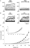
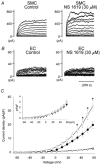
Recent reports have suggested that different types of Ca(2+)-activated K(+) channels may be selectively expressed either in the vascular endothelial cells (ECs) or smooth muscle cells (SMCs) of a single artery. In this study, we directly compared mRNA, protein and functional expression of the high-conductance Ca(2+)-activated K(+) (BK(Ca)) channel between freshly isolated ECs and SMCs from bovine coronary arteries. Fresh ECs and SMCs were enzymatically isolated, and their separation verified by immunofluorescent detection of alpha-actin and platelet/endothelium cell adhesion molecule (PECAM) proteins, respectively. Subsequently, studies using a sequence-specific antibody directed against the pore-forming alpha-subunit of the BK(Ca) channel only detected its expression in the SMCs, whereas PECAM-positive ECs were devoid of the alpha-subunit protein. Additionally, multicell RT-PCR performed using cDNA derived from either SMCs or ECs only detected mRNA encoding the BK(Ca) alpha-subunit in the SMCs. Finally, whole-cell recordings of outward K(+) current detected a prominent iberiotoxin-sensitive BK(Ca) current in SMCs that was absent in ECs, and the BK(Ca) channel opener NS 1619 only enhanced K(+) current in the SMCs. Thus, bovine coronary SMCs densely express BK(Ca) channels whereas adjacent ECs in the same artery appear to lack the expression of the BK(Ca) channel gene. These findings indicate a cell-specific distribution of Ca(2+)-activated K(+) channels in SMCs and ECs from a single arterial site.
Figures


Similar articles
-
Characterisation of K+ channels in human fetoplacental vascular smooth muscle cells.PLoS One. 2013;8(2):e57451. doi: 10.1371/journal.pone.0057451. Epub 2013 Feb 21. PLoS One. 2013. PMID: 23437391 Free PMC article.
-
Characterization of an apamin-sensitive small-conductance Ca(2+)-activated K(+) channel in porcine coronary artery endothelium: relevance to EDHF.Br J Pharmacol. 2002 Mar;135(5):1133-43. doi: 10.1038/sj.bjp.0704551. Br J Pharmacol. 2002. PMID: 11877319 Free PMC article.
-
Selective blockade of the intermediate-conductance Ca2+-activated K+ channel suppresses proliferation of microvascular and macrovascular endothelial cells and angiogenesis in vivo.Arterioscler Thromb Vasc Biol. 2005 Apr;25(4):704-9. doi: 10.1161/01.ATV.0000156399.12787.5c. Epub 2005 Jan 20. Arterioscler Thromb Vasc Biol. 2005. PMID: 15662023
-
Limits of isolation and culture: intact vascular endothelium and BKCa.Am J Physiol Heart Circ Physiol. 2009 Jul;297(1):H1-7. doi: 10.1152/ajpheart.00042.2009. Epub 2009 May 1. Am J Physiol Heart Circ Physiol. 2009. PMID: 19411289 Review.
-
Mechanisms of cellular synchronization in the vascular wall. Mechanisms of vasomotion.Dan Med Bull. 2010 Oct;57(10):B4191. Dan Med Bull. 2010. PMID: 21040688 Review.
Cited by
-
Calcium-Dependent Ion Channels and the Regulation of Arteriolar Myogenic Tone.Front Physiol. 2021 Nov 8;12:770450. doi: 10.3389/fphys.2021.770450. eCollection 2021. Front Physiol. 2021. PMID: 34819877 Free PMC article. Review.
-
Functional coupling of TRPV4, IK, and SK channels contributes to Ca(2+)-dependent endothelial injury in rodent lung.Pulm Circ. 2015 Jun;5(2):279-90. doi: 10.1086/680166. Pulm Circ. 2015. PMID: 26064452 Free PMC article.
-
BACE1 Translation: At the Crossroads Between Alzheimer's Disease Neurodegeneration and Memory Consolidation.J Alzheimers Dis Rep. 2019 May 15;3(1):113-148. doi: 10.3233/ADR-180089. J Alzheimers Dis Rep. 2019. PMID: 31259308 Free PMC article. Review.
-
Connexins and gap junctions in the EDHF phenomenon and conducted vasomotor responses.Pflugers Arch. 2010 May;459(6):897-914. doi: 10.1007/s00424-010-0830-4. Epub 2010 Apr 9. Pflugers Arch. 2010. PMID: 20379740 Review.
-
Novel role of endothelial BKCa channels in altered vasoreactivity following hypoxia.Am J Physiol Heart Circ Physiol. 2010 Nov;299(5):H1439-50. doi: 10.1152/ajpheart.00124.2010. Epub 2010 Sep 3. Am J Physiol Heart Circ Physiol. 2010. PMID: 20817829 Free PMC article.
References
-
- Campbell W, Gebremehdin D, Pratt PF, Harder DR. Identification of epoxyeicosatrienoic acid as endothelium-derived hyperpolarizing factor. Circulation Research. 1996;78:415–423. - PubMed
-
- Campbell WB, Deeter C, Gauthier KM, Ingraham RH, Falck JR, Li PL. 14,15-Dihydroxyeicosatrienoic acid relaxes bovine coronary arteries by activation of KCa channels. American Journal of Physiology – Heart and Circulatory Physiology. 2002;282:H1656–1664. - PubMed
Publication types
MeSH terms
Substances
Grants and funding
LinkOut - more resources
Full Text Sources
Miscellaneous

