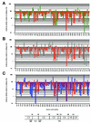
Fine mapping of the interaction of neutralizing and nonneutralizing monoclonal antibodies with the CD4 binding site of human immunodeficiency virus type 1 gp120
- PMID: 12477867
- PMCID: PMC140633
- DOI: 10.1128/jvi.77.1.642-658.2003
Fine mapping of the interaction of neutralizing and nonneutralizing monoclonal antibodies with the CD4 binding site of human immunodeficiency virus type 1 gp120
Abstract
Alanine scanning mutagenesis was performed on monomeric gp120 of human immunodeficiency virus type 1 to systematically identify residues important for gp120 recognition by neutralizing and nonneutralizing monoclonal antibodies (MAbs) to the CD4 binding site (CD4bs). Substitutions that affected the binding of broadly neutralizing antibody b12 were compared to substitutions that affected the binding of CD4 and of two nonneutralizing anti-CD4bs antibodies (b3 and b6) with affinities for monomeric gp120 comparable to that of b12. Not surprisingly, the sensitivities to a number of amino acid changes were similar for the MAbs and for CD4. However, in contrast to what was seen for the MAbs, no enhancing mutations were observed for CD4, suggesting that the virus has evolved toward an optimal gp120-CD4 interaction. Although the epitope maps of the MAbs overlapped, a number of key differences between b12 and the other two antibodies were observed. These differences may explain why b12, in contrast to nonneutralizing antibodies, is able to interact not only with monomeric gp120 but also with functional oligomeric gp120 at the virion surface. Neutralization assays performed with pseudovirions bearing envelopes from a selection of alanine mutants mostly showed a reasonable correlation between the effects of the mutations on b12 binding to monomeric gp120 and neutralization efficacy. However, some mutations produced an effect on b12 neutralization counter to that predicted from gp120 binding data. It appears that these mutations have different effects on the b12 epitope on monomeric gp120 and functional oligomeric gp120. To determine whether monomeric gp120 can be engineered to preferentially bind MAb b12, recombinant gp120s were generated containing combinations of alanine substitutions shown to uniquely enhance b12 binding. Whereas b12 binding was maintained or increased, binding by five nonneutralizing anti-CD4bs MAbs (b3, b6, F105, 15e, and F91) was reduced or completely abolished. These reengineered gp120s are prospective immunogens that may prove capable of eliciting broadly neutralizing antibodies.
Figures


Similar articles
-
Nonneutralizing antibodies to the CD4-binding site on the gp120 subunit of human immunodeficiency virus type 1 do not interfere with the activity of a neutralizing antibody against the same site.J Virol. 2003 Jan;77(2):1084-91. doi: 10.1128/jvi.77.2.1084-1091.2003. J Virol. 2003. PMID: 12502824 Free PMC article.
-
A novel human antibody against human immunodeficiency virus type 1 gp120 is V1, V2, and V3 loop dependent and helps delimit the epitope of the broadly neutralizing antibody immunoglobulin G1 b12.J Virol. 2003 Jun;77(12):6965-78. doi: 10.1128/jvi.77.12.6965-6978.2003. J Virol. 2003. PMID: 12768015 Free PMC article.
-
PGV04, an HIV-1 gp120 CD4 binding site antibody, is broad and potent in neutralization but does not induce conformational changes characteristic of CD4.J Virol. 2012 Apr;86(8):4394-403. doi: 10.1128/JVI.06973-11. Epub 2012 Feb 15. J Virol. 2012. PMID: 22345481 Free PMC article.
-
CD4-gp120 interactions.Curr Opin Immunol. 1991 Aug;3(4):552-8. doi: 10.1016/0952-7915(91)90020-2. Curr Opin Immunol. 1991. PMID: 1721822 Review.
-
CD4 monoclonal antibodies in organ transplantation--a review of progress.Transplantation. 1991 Oct;52(4):579-89. doi: 10.1097/00007890-199110000-00001. Transplantation. 1991. PMID: 1926337 Review. No abstract available.
Cited by
-
Rapid conformational epitope mapping of anti-gp120 antibodies with a designed mutant panel displayed on yeast.J Mol Biol. 2013 Jan 23;425(2):444-56. doi: 10.1016/j.jmb.2012.11.010. Epub 2012 Nov 15. J Mol Biol. 2013. PMID: 23159556 Free PMC article.
-
Specific amino acids in the N-terminus of the gp41 ectodomain contribute to the stabilization of a soluble, cleaved gp140 envelope glycoprotein from human immunodeficiency virus type 1.Virology. 2007 Mar 30;360(1):199-208. doi: 10.1016/j.virol.2006.09.046. Epub 2006 Nov 7. Virology. 2007. PMID: 17092531 Free PMC article.
-
Molecular features of the broadly neutralizing immunoglobulin G1 b12 required for recognition of human immunodeficiency virus type 1 gp120.J Virol. 2003 May;77(10):5863-76. doi: 10.1128/jvi.77.10.5863-5876.2003. J Virol. 2003. PMID: 12719580 Free PMC article.
-
Punica granatum (Pomegranate) juice provides an HIV-1 entry inhibitor and candidate topical microbicide.BMC Infect Dis. 2004 Oct 14;4:41. doi: 10.1186/1471-2334-4-41. BMC Infect Dis. 2004. PMID: 15485580 Free PMC article.
-
Antibody responses elicited in macaques immunized with human immunodeficiency virus type 1 (HIV-1) SF162-derived gp140 envelope immunogens: comparison with those elicited during homologous simian/human immunodeficiency virus SHIVSF162P4 and heterologous HIV-1 infection.J Virol. 2006 Sep;80(17):8745-62. doi: 10.1128/JVI.00956-06. J Virol. 2006. PMID: 16912322 Free PMC article.
References
-
- Allaway, G. P., K. L. Davis-Bruno, G. A. Beaudry, E. B. Garcia, E. L. Wong, A. M. Ryder, K. W. Hasel, M.-C. Gauduin, R. A. Koup, J. S. McDougal, and P. J. Maddon. 1995. Expression and characterization of CD4-IgG2, a novel heterotetramer that neutralizes primary HIV type 1 isolates. AIDS Res. Hum. Retrovir. 11:533-539. - PubMed
-
- Baba, T. W., V. Liska, R. Hofmann-Lehmann, J. Vlasak, W. Xu, S. Ayehunie, L. A. Cavacini, M. R. Posner, H. Katinger, G. Stiegler, B. J. Bernacky, T. A. Rizvi, R. Schmidt, L. R. Hill, M. E. Keeling, Y. Lu, J. E. Wright, T. C. Chou, and R. M. Ruprecht. 2000. Human neutralizing monoclonal antibodies of the IgG1 subtype protect against mucosal simian-human immunodeficiency virus infection. Nat. Med. 6:200-206. - PubMed
-
- Barbas, C. F., III, E. Bjorling, F. Chiodi, N. Dunlop, D. Cababa, T. M. Jones, S. L. Zebedee, M. A. Persson, P. L. Nara, E. Norrby, and D. R. Burton. 1992. Recombinant human Fab fragments neutralize human type 1 immunodeficiency virus in vitro. Proc. Natl. Acad. Sci. USA 89:9339-9343. - PMC - PubMed
-
- Barbas, C. F., III, T. A. Collet, W. Amberg, P. Roben, J. M. Binley, D. Hoekstra, D. Cababa, T. M. Jones, R. A. Williamson, G. R. Pilkington, N. L. Haigwood, E. Cabezas, A. C. Satterthwait, I. Sanz, and D. R. Burton. 1993. Molecular profile of an antibody response to HIV-1 as probed by combinatorial libraries. J. Mol. Biol. 230:812-823. - PubMed
Publication types
MeSH terms
Substances
Grants and funding
LinkOut - more resources
Full Text Sources
Other Literature Sources
Research Materials

