. 2002 Dec 1;30(23):5074-86.
doi: 10.1093/nar/gkf647.

Localisation of the human hSuv3p helicase in the mitochondrial matrix and its preferential unwinding of dsDNA

Affiliations

Localisation of the human hSuv3p helicase in the mitochondrial matrix and its preferential unwinding of dsDNA

Michal Minczuk et al. Nucleic Acids Res. .

Abstract

We characterised the human hSuv3p protein belonging to the family of NTPases/helicases. In yeast mitochondria the hSUV3 orthologue is a component of the degradosome complex and participates in mtRNA turnover and processing, while in Caenorhabditis elegans the hSUV3 orthologue is necessary for viability of early embryos. Using immunofluorescence analysis, an in vitro mitochondrial uptake assay and sub-fractionation of human mitochondria we show hSuv3p to be a soluble protein localised in the mitochondrial matrix. We expressed and purified recombinant hSuv3p protein from a bacterial expression system. The purified enzyme was capable of hydrolysing ATP with a K(m) of 41.9 micro M and the activity was only modestly stimulated by polynucleotides. hSuv3p unwound partly hybridised dsRNA and dsDNA structures with a very strong preference for the latter. The presented analysis of the hSuv3p NTPase/helicase suggests that new functions of the protein have been acquired in the course of evolution.


Figures
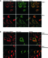
Figure 1
Figure 1
Mitochondrial localisation of hSuv3p in mammalian cells. (A) COS-1 and HeLa cells were grown on coverslips, incubated with the mitochondria-specific dye MitoTracker CMXRos (300 nM), fixed with 4% formaldehyde and permeabilised with 1% Triton X-100. The cells were immunostained with anti-hSuv3p serum and visualised with FITC-conjugated secondary antibody. Fluorescent images of MitoTracker (red) and hSuv3p specific signal (green) were taken by a confocal microscope. Co-localisation of hSuv3p and mitochondria appear yellow in digitally overlaid images. (B) COS-1 and HeLa cells were grown on coverslips and transiently transfected with cDNA encoding c-myc-tagged hSuv3p (pchSUV3myc). After incubation with MitoTracker, fixation and permeabilisation as described in (A), the cells were immunostained with anti-c-myc monoclonal antibody, which was then visualised with FITC-conjugated antibody. Fluorescent images of MitoTracker (red) and c-myc-tagged hSuv3p (green) were taken by a confocal microscope. Co-localisation of hSuv3p and mitochondria appear yellow in digitally overlaid images.
Figure 2
Figure 2
N-terminal sequence is necessary for the mitochondrial import of the hSuv3p protein. COS-1 and HeLa cells were grown on coverslips and transiently transfected with cDNA encoding N-terminally truncated (the first 18 amino acids) hSuv3p with a c-myc tag at the C-terminus (pcΔmthSUV3myc). After incubation with the mitochondria-specific label MitoTracker CMXRos (300 nM), fixation with 4% formaldehyde and permeabilisation with 1% Triton X-100 the cells were immunostained with anti-c-myc monoclonal antibody. The c-myc-tagged N-terminally truncated hSuv3p was visualised with FITC-conjugated antibody (green); the mitochondria stained with MitoTracker are red. The hSuv3p protein devoid of the 18 N-terminal amino acids did not co-localise with the mitochondria in either COS-1 or HeLa cells.
Figure 3
Figure 3
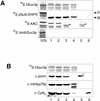
The hSuv3p protein imported into isolated yeast mitochondria is localised in the mitochondrial matrix. (A) In vitro synthesised radiolabelled proteins, hSuv3p (wild-type), pSu9–DHFR (localised in the mitochondrial matrix), AAC (localised in the mitochondrial inner membrane) and ΔmthSuv3p (hSuv3p protein devoid of the 18 N-terminal amino acids), were incubated with isolated yeast mitochondria. After import, mitochondria were treated with protease (trypsin) to remove non-imported precursor (lane 1). In the next step the mitoplasts were prepared by osmotic shock (lane 2) and treated with proteinase K (lane 3). After alkali extraction of the mitoplasts with Na2CO3 the soluble fraction (lane 4) and the membrane pellets (lane 5) were analysed. The inter-membrane space fraction released after osmotic swelling was also examined (lane 6). All the protein samples were separated by SDS–PAGE, transferred onto nitrocellulose membranes and visualised by autoradiography. For pSu9–DHFR arrows indicate the precursor (P) and mature peptide cleaved by mitochondrial MPP (M). Asterisks show the protease-resistant fragment of the protein AAC inserted into the mitochondrial inner membrane. The 10% of the input is also shown. (B) The mitochondrial fractions transferred onto nitrocellulose membranes after the import of hSuv3p from (A) were verified by immunoblotting with antibodies specific to the endogenous proteins localised in different mitochondrial subcompartments: anti-porin for the outer membrane (α porin), anti-mtHsp70p for the matrix (α mtHsp70p) and anti-Cytb2 for the inter-membrane space (α Cytb2).
Figure 4
Figure 4
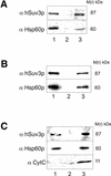
Localisation of the hSuv3p protein in the mitochondrial matrix of human mitochondria. (A) Crude mitochondrial fraction (lane 1), cytosolic fraction (lane 2) and total cell extract (lane 3) of HeLa cells were separated by SDS–PAGE, immunoblotted and probed with the anti-hSuv3p serum (α hSuv3p). (B) Soluble submitochondrial fraction (lane 1), membrane submitochondrial fraction (lane 2) and total extract of purified mitochondria of HeLa cells (lane 3) were immunoprobed with the anti-hSuv3p serum (α hSuv3p) as above. (C) Mitoplasts (lane 1), post-mitoplast supernatant (lane 2) and total extract of purified mitochondria of HeLa cells (lane 3) were separated by SDS–PAGE, immunoblotted and probed with the anti-hSuv3p serum (α hSuv3p). Subcellular and submitochondrial fractions were verified by immunoblotting with antibodies specific to the endogenous proteins localised in different mitochondrial subcompartments: anti-Hsp60p for the matrix (α Hsp60p) and in (C) anti-CytC for the intermembrane space (α CytC). Molecular masses are indicated on the left-hand side of the blot images.
Figure 5
Figure 5
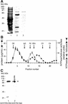
Purification of hSuv3p from E.coli. (A) SDS–PAGE analysis of purification steps. Triton X-100 soluble fraction (lane 1); Ni2+ affinity chromatography (lane 2); Superdex-200 column (lane 3). The samples were separated by SDS–PAGE and the proteins stained with Coomassie Blue. M(r) kDa, molecular mass markers in kilodaltons. (B) Gel filtration purification of hSuv3p. The distribution of protein and of NTPase and helicase activities of the hSuv3p preparation obtained by Ni2+ affinity chromatography on an analytical Superdex-200 column is shown. The NTPase and the helicase assays were performed as described in Materials and Methods. In the case of the helicase assay a dsDNA substrate was used. The column was calibrated with dextran blue (DB) (2000 kDa) and with the following marker proteins: IgG (160 kDa); PB, phosphorylase b (97 kDa); BSA (66 kDa); OV, ovalbumin (45 kDa); CA, carbonic anhydrase (30 kDa). (C) Aliquots of pooled fractions from the Superdex-200 column displaying ATPase and helicase activity were removed and precipitated with TCA. Samples were separated by SDS–PAGE followed by immunoblotting with anti-hSuv3p antiserum (lane 1) and with anti-his tag antibody (lane 2). The nitrocellulose filters were autoradiographed for 24 h. M(r) kDa, molecular mass markers in kilodaltons. The arrow indicates the position of hSuv3p.
Figure 6
Figure 6
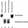
Lack of the NTPase and helicase activity of hSuv3p mutated in the Walker A motif. An aliquot of 2 pmol of homogeneously purified hSuv3p wild-type enzyme (lane 3) and its mutated form hSuv3p(G207V) (lane 4) was allowed to react with a dsDNA substrate (4.7 pM) as described in Materials and Methods. The substrate and released strand were separated in a TBE polyacrylamide gel and visualised by exposure of the dried gel to X-ray film for 24 h.
Figure 7
Figure 7
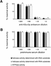
Depletion of enzymatic activities of hSuv3p NTPase/helicase by anti-hSuv3p antibodies. Aliquots of the purified hSuv3p NTPase/helicase (2 pmol) were incubated with anti-hSuv3p serum (A) or with preimmune serum (B) at 30°C for 30 min, then adjusted to the concentrations indicated in the figure. Thereafter the unwinding or ATPase reactions were started and the samples were processed as described in Materials and Methods. Values for the enzymatic activities are shown as means ± SEMs for representative experiments performed in triplicate.
Figure 8
Figure 8
Modulation of the ATPase activity of hSuv3p by AMP-PNP with variations in ATP concentration. (A) Investigation of the ATPase activity as a function of increasing concentrations of AMP-PNP in the presence of different ATP concentrations. The demonstrated assays were performed at ATP concentrations of hSuv3p ATPase (41.9 µM) equal to (diamond) 100 × Km; (triangle) 1 × Km; (square) 0.001 × Km; and (circle) 0.00001 × Km. The activities of the enzyme measured for each ATP concentration in the absence of AMP-PNP was referred to as 100%. (B) Kinetic analysis of the inhibition of the hSuv3p ATPase at lower ATP concentrations. Data obtained at ATP concentrations equal to (circle) 0.00001 × Km, (open triangle) 0.00003 × Km and (open square) 0.0001 × Km were replotted according to the method of Dixon (37).
Figure 9
Figure 9
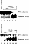
Comparison of efficacy of RNA and DNA unwinding reaction mediated by hSuv3p as a function of decreasing enzyme concentration. The reaction mixture was composed as described in Materials and Methods with various hSuv3p enzyme concentrations used: 0.66 fM (lanes 2 and 8); 66 fM (lanes 3 and 9); 6.6 pM (lanes 4 and 10); 0.66 nM (lanes 5 and 11); 66 nM (lanes 6 and 12). The substrate and released strand were separated in a TBE polyacrylamide gel and visualised by exposure of the dried gel to X-ray film for 24 h. The results shown are representative of three independent experiments.

Similar articles

Cited by

References

    1. Margossian S.P., Li,H., Zassenhaus,H.P. and Butow,R.A. (1996) The DexH box protein Suv3p is a component of a yeast mitochondrial 3′-to-5′ exoribonuclease that suppresses group I intron toxicity. Cell, 84, 199–209. - PubMed
    1. Dmochowska A., Golik,P. and Stepien,P.P. (1995) The novel nuclear gene DSS-1 of Saccharomyces cerevisiae is necessary for mitochondrial biogenesis. Curr. Genet., 28, 108–112. - PubMed
    1. Stepien P.P., Margossian,S.P., Landsman,D. and Butow,R.A. (1992) The yeast nuclear gene suv3 affecting mitochondrial post-transcriptional processes encodes a putative ATP-dependent RNA helicase. Proc. Natl Acad. Sci. USA, 89, 6813–6817. - PMC - PubMed
    1. Conrad-Webb H., Perlman,P.S., Zhu,H. and Butow,R.A. (1990) The nuclear SUV3-1 mutation affects a variety of post-transcriptional processes in yeast mitochondria. Nucleic Acids Res., 18, 1369–1376. - PMC - PubMed
    1. Stepien P.P., Kokot,L., Leski,T. and Bartnik,E. (1995) The suv3 nuclear gene product is required for the in vivo processing of the yeast mitochondrial 21s rRNA transcripts containing the r1 intron. Curr. Genet., 27, 234–238. - PubMed

Publication types

MeSH terms