. 2002 Dec 2;21(23):6590-602.
doi: 10.1093/emboj/cdf652.

A novel function for human factor C1 (HCF-1), a host protein required for herpes simplex virus infection, in pre-mRNA splicing

Affiliations

A novel function for human factor C1 (HCF-1), a host protein required for herpes simplex virus infection, in pre-mRNA splicing

Paul Ajuh et al. EMBO J. .

Abstract

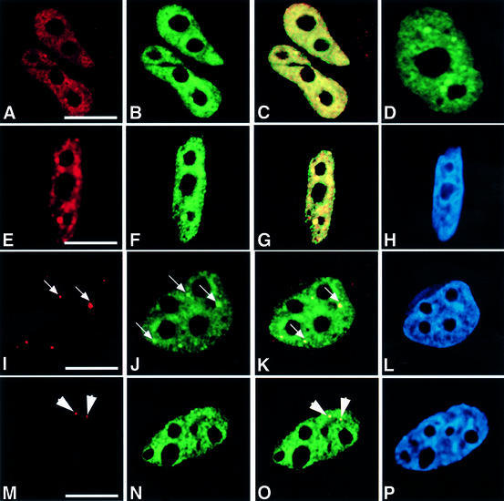
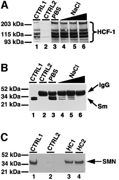
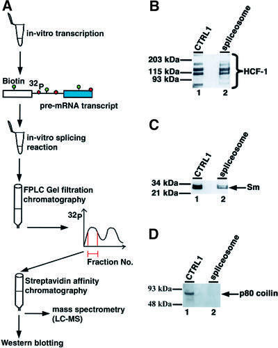
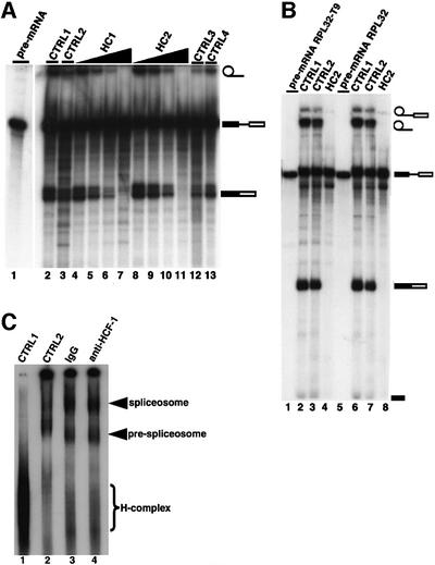
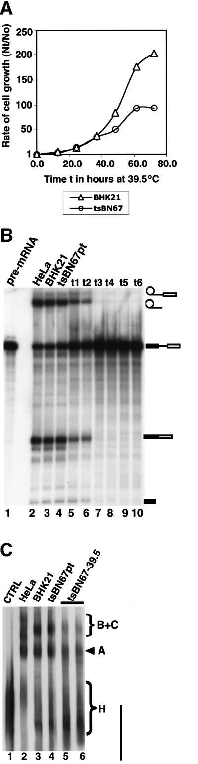
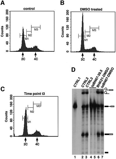
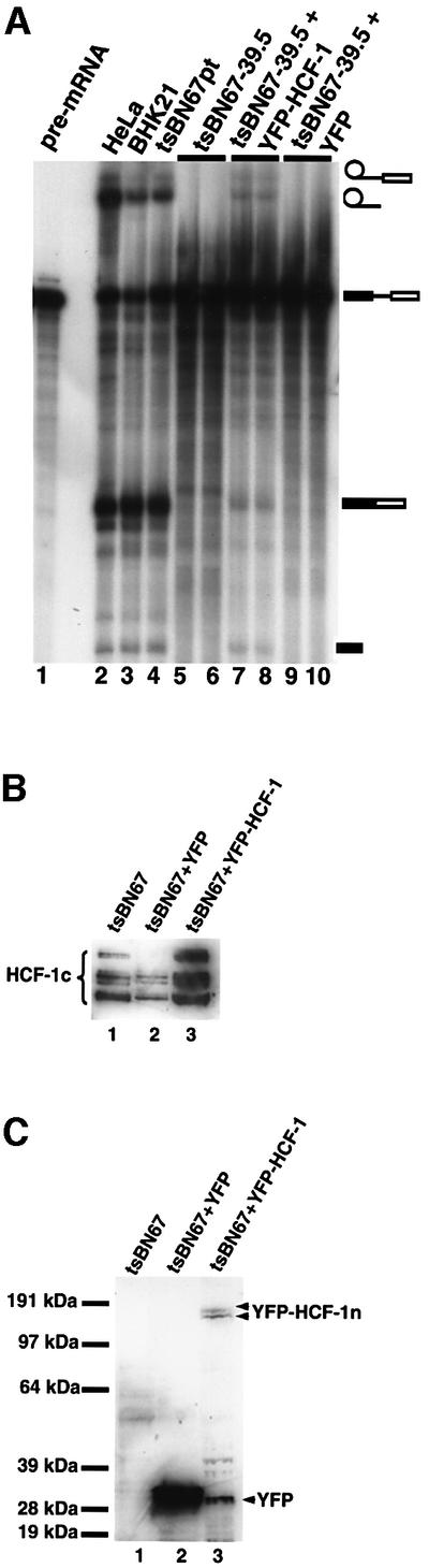
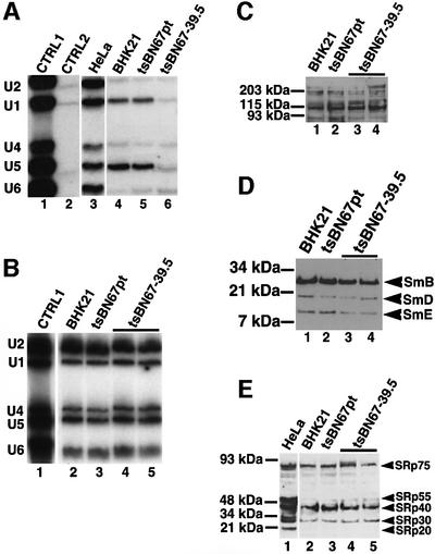
Human factor C1 (HCF-1) is needed for the expression of herpes simplex virus 1 (HSV-1) immediate-early genes in infected mammalian cells. Here, we provide evidence that HCF-1 is required for spliceosome assembly and splicing in mammalian nuclear extracts. HCF-1 interacts with complexes containing splicing snRNPs in uninfected mammalian cells and is a stable component of the spliceosome complex. We show that a missense mutation in HCF-1 in the BHK21 hamster cell line tsBN67, at the non-permissive temperature, inhibits the protein's interaction with U1 and U5 splicing snRNPs, causes inefficient spliceosome assembly and inhibits splicing. Transient expression of wild-type HCF-1 in tsBN67 cells restores splicing at the non-permissive temperature. The inhibition of splicing in tsBN67 cells correlates with the temperature-sensitive cell cycle arrest phenotype, suggesting that HCF-1-dependent splicing events may be required for cell cycle progression.


Figures

None
Fig. 1. HCF-1 co-localizes with nuclear structures that contain pre-mRNA splicing factors. HeLa cells were transfected with pEYFP-HCF, and indirect immunofluorescence experiments were performed on the transfected cells using various antibodies. The arrows point to gems, while the arrowheads point to Cajal bodies. (A) Light microscopic image of an indirect immunofluorescence experiment on HeLa cells using the polyclonal HC2 antibody. (B) A microscopic image obtained from the direct fluorescence of YFP–HCF-1 in the same cells as in (A). (C) Superimposed optical sections from (A) and (B). Co-localization of green and red results in the yellow colour. Note the presence of some green fluorescence that does not co-localize with the antibody staining. This may be due to the stronger fluorescence signal produced by the YFP–HCF-1 fusion protein. Secondly, YFP–HCF-1/cellular HCF-1 may exist in some complexes that, although visible by direct YFP fluorescence, are inaccessible to the anti-HCF-1 peptide antibodies used for cell staining. (D) Light microscopic image of a live HeLa cell expressing YFP–HCF-1 (green). (E, I and M) Microscopic images of indirect immunofluorescence experiments on HeLa cells using anti-Sm antibody (Y12), anti-SMN mAb (MANSMA1) and anti-p80 coilin mAb (5P10), respectively. (F, J and N) Microscopic images obtained from the direct fluorescence of YFP–HCF-1 in the same cells as in (E), (I) and (M), respectively. (G, K and O) Superimposed optical sections from (E) and (F), (I) and (J), and (M) and (N), respectively. (H, L and P) Microscopic images obtained from DAPI staining of the HeLa cells shown in (E), (I) and (M), respectively. Bar = 5 µm.
None
Fig. 2. HCF-1 interacts with complexes that contain splicing snRNPs and SMN. (A) Immunoprecipitation using anti-HCF-1 antibody (HC2). Lane 1 (marked CTRL1) is the positive control and contained HeLa nuclear extract. CTRL2 (lane 2) is a control immunoprecipitation using pre-immune IgG. Lanes 3–6 contained immunoprecipitates using HC2 antibodies, except that the immunoprecipitate in lane 3 was washed with PBS containing 0.01% Triton X-100 whereas the immunoprecipitates in lanes 4–6 were washed with PBS/Triton X-100 as above but with increasing concentrations of NaCl (i.e. 200, 300 and 400 mM), respectively. The beads containing immunoprecipitated proteins were loaded onto a 4–12% SDS–polyacrylamide gradient gel (Novex) and probed by western blotting using anti-HCF-1 antibodies. HCF-1 bands on the blot were revealed by ECL (Amersham-Pharmacia) and are shown by the bracket on the right of (A). (B) The experiment and the lane markings of the panel are identical to (A) except that the immunoblot was probed with the anti-Sm antibody Y12 instead of anti-HCF-1 antibodies. (C) A 10 µg aliquot of HC1 (lane 3) or HC2 (lane 4) was used to immunoprecipitate HCF-1 from ∼0.2 mg of HeLa nuclear extract. CTRL1 is a positive control lane containing ∼25 µg of HeLa nuclear extract. CTRL2 (lane 2) is a negative control immunoprecipitation reaction using 10 µg of pre-immune IgG. The immunoblot was probed with anti-SMN mAbs (MANSMA1), and the SMN bands revealed by ECL.
None
Fig. 3. HCF-1 is a component of the spliceosome complex in HeLa nuclear extract. Spliceosomes purified as described in Materials and methods were separated on a 10 or 4–12% SDS–polyacrylamide gradient gel. (A) Cartoon showing the assembly and purification scheme used to purify the human spliceosome complex. The pre-mRNA transcript was labelled with both biotin-UTP and [32P]GTP. (B) The spliceosomal proteins were blotted onto nitrocellulose filters and probed with anti-HCF-1 antibodies. Lane 1 is a control lane containing ∼25–30 µg of nuclear extract. Lane 2 contained the purified spliceosome complex. (C and D) The contents of the lanes are identical to those in (A) except that the blots were probed with the anti-Sm antibody (Y12) and anti-p80 coilin mAb (5P10), respectively.
None
Fig. 4. Inhibition of pre-mRNA splicing by anti-HCF-1 antibodies. (A) HeLa nuclear extracts (∼50 µg) were pre-incubated with 0.15–1.2 pmol anti-HCF-1 antibodies before addition to the splicing reaction. Lane 2 (CTRL1) is a control splicing reaction to which no antibodies were added. Lane 3 (CTRL2) is a control splicing assay containing 1.2 pmol of rabbit pre-immune IgG. Lanes 4–7 indicate splicing reactions that contained 0.15, 0.30, 0.6 and 1.2 pmol of the HC1 antibody, respectively. Lanes 8–11 represent splicing assays that contained 0.15, 0.30, 0.6 and 1.2 pmol of the HC2 antibody, respectively. Lanes 12 and 13 (CTRL3 and CTRL4) are control reactions that contained 1.2 pmol each of HC1 and HC2, respectively, except that here each anti-HCF-1 antibody had been pre-incubated with its cognate peptide before addition to the splicing reaction. The symbols on the right represent the input pre-mRNA and splicing products. (B) Inhibition of splicing of the RPL32 pre-mRNAs by anti-HCF-1 antibodies. Pre-mRNAs transcribed in vitro from the pGEM-RPL32 and pGEM-RPL32:T9 plasmids (Vilardell and Warner, 1997; and references therein) were used in splicing reactions containing 1.2 pmol of the HC2 antibody or pre- immune IgG. RPL32 pre-mRNA was used in the reactions in lanes 1–4, while RPL32:T9 pre-mRNA was used in splicing reactions in lanes 5–8. Lanes 3 and 7 are control reactions that contained pre- immune IgG. HC2 antibody was added to the reactions in lanes 4 and 8. No antibodies were added to samples in lanes 2 and 6. (C) Spliceosome assembly is unaffected by anti-HCF-1 antibodies in splicing reactions. Pre-mRNA splicing reactions were performed in vitro for 45–60 min using HeLa nuclear extract that had been pre-incubated for 15–20 min at 30°C with 1.2 pmol of the HC2 antibody or pre-immune IgG. The splicing complexes were separated on a native agarose–polyacrylamide composite gel. Lane 1 is a control reaction performed on ice. No antibodies were added to the reaction in lane 2. The reactions in lanes 3 and 4 contained pre-immune IgG and HC2 antibodies, respectively.
None
Fig. 5. Nuclear extracts from the hamster BHK21 and tsBN67 cell line containing a missense mutation in HCF-1 are incompetent in splicing. (A) Analysis of the growth rates of BHK21 and tsBN67 cells at 39.5°C. A total of 2 × 104 tsBN67 and BHK21 cells were seeded into 10 cm dishes and grown at 37°C. After 48 h, the temperature was shifted to 39.5°C. The cells were harvested at 12 h intervals by trypsin ization and counted using a haemocytometer. The cell counts obtained at each time point (Nt) were divided by the cell count (N0) at the time the cells were shifted to the non-permissive temperature, and plotted against the time in hours. (B) Nuclear extracts were prepared from the BHK21 and tsBN67 cells maintained at 39.5°C at 18–24 h intervals (t1–t6; lanes 5–10). About 45–50 µg of nuclear extract from each time point were used in each splicing experiment. Lane 1 contained the input pre-mRNA. Lane 2 is a positive control of a splicing reaction using HeLa nuclear extract. The lane marked BHK21 (lane 3) contained nuclear extract from BHK21 cells maintained indefinitely at 39.5°C. Lane 4 contained nuclear extract from tsBN67 cells maintained at the permissive temperature. (C) Nuclear extracts from tsBN67 cells are inefficient in spliceosome assembly. Pre-mRNA splicing reactions were performed in vitro for 45–60 min. The splicing complexes were then separated on a native agarose–polyacrylamide composite gel. Lane 1 is a control reaction using BHK21 nuclear extract and performed on ice. Lane 2 contained a splicing reaction using HeLa nuclear extract. Lanes 3 and 4 contained BHK21 and tsBN67 nuclear extracts prepared as in lanes 3 and 4 of (B) above. Lanes 5 and 6 contained tsBN67 nuclear extracts from time points t3 and t4.
None
Fig. 6. Nuclear extracts from BHK21 and tsBN67 cells arrested at G0/G1 using DMSO can splice adeno-pre-mRNA efficiently. BHK21 and tsBN67 cells were maintained for 48 h at 37°C in culture medium containing 1% (v/v) DMSO. No DMSO was added to control cells. (AC) Cell cycle analysis of tsBN67 cells. The labels M1, M2 and M3 represent cells at the G0/G1, S and G2/M phases of the cell cycle, respectively. (A) DNA content FACS analysis of untreated cells. (B) DNA content FACS analysis of DMSO-treated cells. (C) DNA content FACS analysis of tsBN67 cells incubated at 39.5°C up to time point t3. (D) Nuclear extracts were prepared from treated and untreated cells for use in splicing experiments. Lane 1 contained 30% of the input pre-mRNA transcript. Lanes 2 and 3 contained control nuclear extracts from untreated BHK21 and tsBN67 cells, respectively. Lanes 4 and 5 contained a splicing reaction using nuclear extract from tsBN67 cells maintained at the non-permissive temperature (time point t3), whereas lanes 6 and 7 contained nuclear extracts from DMSO-treated BHK21 and tsBN67 cells, respectively.
None
Fig. 7. Transfection of tsBN67 cells maintained at the non-permissive temperature restores splicing to the tsBN67 extracts. (A) tsBN67 cells were transfected with wild-type HCF-1 in the plasmid pEYFP-HCF and the transfection efficiency monitored by fluorescence microscopy (data not shown). Transfected cells were maintained at 39.5°C up to the time point t3 above. Nuclear extracts were then prepared from the cells and used for in vitro splicing assays. Lane 1 contained input pre-mRNA. Lane 2 contained a splicing reaction using HeLa nuclear extract. The lane marked BHK21 (lane 3) contained nuclear extract from BHK21 cells maintained at 39.5°C. Lane 4 contained nuclear extract from tsBN67 cells maintained at the permissive temperature. Lanes 5 and 6 represent duplicate splicing reactions using tsBN67 nuclear extracts (from cells maintained at 39.5°C) prepared at t3. The lanes marked 7 and 8 contained splicing reactions using nuclear extracts from tsBN67 cells transfected with wild-type HCF-1 plasmid, maintained at 39.5°C, and extracts prepared at time t3 (10–12 h after lag period). Lanes 9 and 10 contained control reactions using extracts from tsBN76 cells treated identically to the cells used for the reactions in lanes 7 and 8 except that the tsBN67 cells used here were transfected with a plasmid that contained only YFP. (B and C) Nuclear extracts from transfected and untransfected cells were probed with (B) anti-HCF-1 and (C) anti-YFP antibodies (BD Biosciences, UK). The samples in (B) and (C) are identical. The lanes marked 1 contained nuclear extracts from untransfected cells while the lanes marked 2 and 3 contained tsBN67 cells transfected with pEYFP-C1 and pEYFP-HCF, respectively. Note that ∼3- to 4-fold less nuclear extract was loaded for the lanes marked 2 in order to compensate for the high signal from the YFP. Note also that only the doublet of bands representing the N-terminal HCF-1 fragments was detected in (C, lane 3) because the YFP protein is fused to the N-terminus of HCF-1 in the pEYFP-HCF construct, whereas the antibodies HC1 and HC2 recognize the C-terminal fragments of the processed HCF-1 polypeptides.
None
Fig. 8. The association between HCF-1 and U1 or U5 splicing snRNPs is inhibited in tsBN67 nuclear extracts. (A) Anti-HCF-1 antibody immunoprecipitates using ∼10 µg of antibody and ∼0.25 mg of nuclear extract were probed with 32P-labelled snRNA probes. Lane 1 is a positive control containing snRNAs in HeLa nuclear extract. Lane 2 contained immunoprecipitates using pre-immune IgG, whereas lanes 3–6 contained anti-HCF-1 antibody immunoprecipitates from HeLa cells, BHK21 cells (maintained at 39.5°C), tsBN67 cells maintained at the permissive temperature and tsBN67 cells maintained at 39.5°C, respectively. (B) Total nuclear RNA was extracted from ∼100 µg of nuclear extracts and the snRNAs in the extracts separated on an 8 M urea– polyacrylamide gel and transferred onto a nylon membrane by northern blotting. The snRNA bands were visualized by probing the membrane with [32P]GTP-labelled snRNA probes and autoradiography. Lanes 1–3 contained snRNAs in nuclear extracts from HeLa cells, BHK21 cells maintained at 39.5°C and tsBN67 cells maintained at the permissive temperature, respectively. Lanes 4 and 5 contained duplicate samples of RNA prepared from non-splicing-competent nuclear extracts of tsBN67 cells maintained at 39.5°C. (CE) About 30 µg of nuclear extracts from BHK21 cells maintained at 39.5°C (lane 1), tsBN67 cells maintained at the permissive temperature (lane 2) and tsBN67 cells maintained at 39.5°C (lanes 3 and 4, i.e. duplicate), respectively, were probed with anti-HCF-1 antibodies (C), anti-Sm antibodies (D) and anti-SR protein antibodies (E).

Similar articles

Cited by

References

    1. Adams J., Kelso,R. and Cooley,L. (2000) The kelch superfamily of proteins: propellers of cell function. Trends Cell Biol., 10, 17–24. - PubMed
    1. Ajuh P.M., Browne,G.J., Hawkes,N.A., Cohen,P.T., Roberts,S.G. and Lamond,A.I. (2000) Association of a protein phosphatase 1 activity with the human factor C1 (HCF) complex. Nucleic Acids Res., 28, 678–686. - PMC - PubMed
    1. Bach M., Krol,A. and Luhrmann,R. (1990) Structure-probing of U1 snRNPs gradually depleted of the U1-specific proteins A, C and 70k. Evidence that A interacts differentially with developmentally regulated mouse U1 snRNA variants. Nucleic Acids Res., 18, 449–457. - PMC - PubMed
    1. Beales M., Flay,N., McKinney,R., Habara,Y., Ohshima,Y., Tani,T. and Potashkin,J. (2000) Mutations in the large subunit of U2AF disrupt pre-mRNA splicing, cell cycle progression and nuclear structure. Yeast, 16, 1001–1013. - PubMed
    1. Bentley D. (2002) The mRNA assembly line: transcription and processing machines in the same factory. Curr. Opin. Cell Biol., 14, 336–342. - PubMed

Publication types